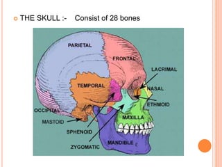
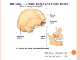
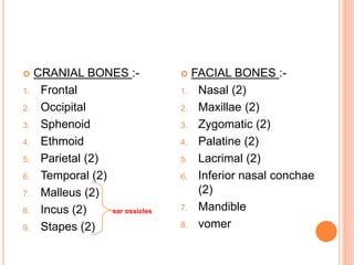
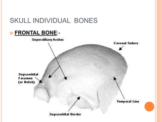
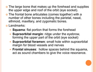
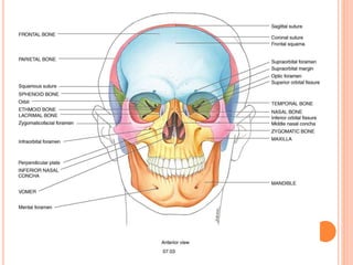
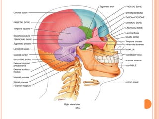
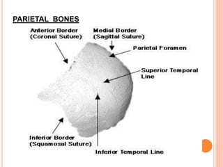
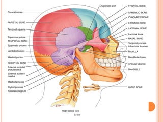
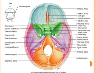
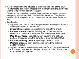
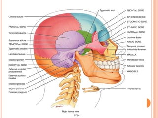
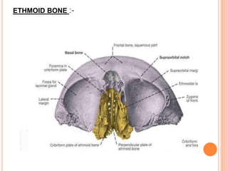
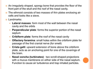
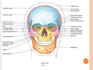
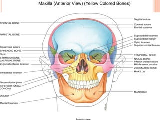
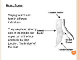
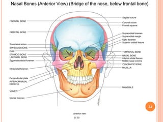
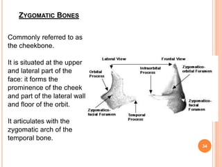
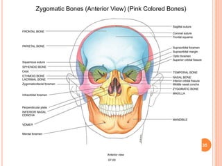
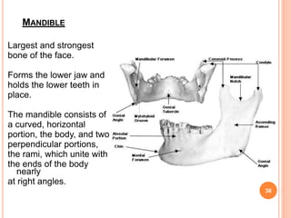
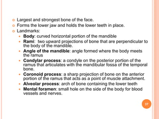
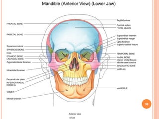
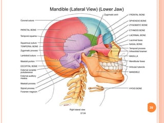
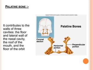
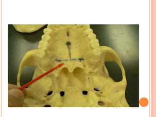
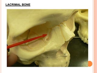
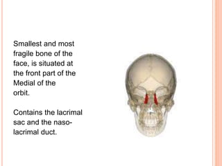
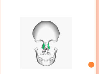
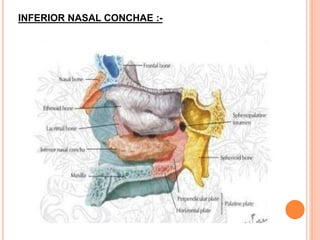
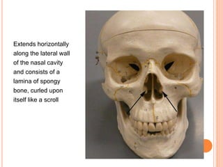
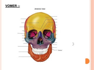
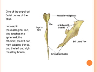
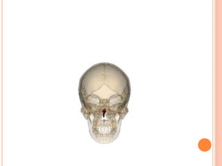
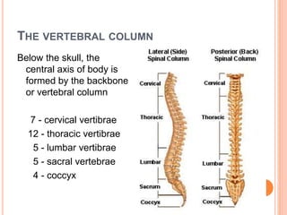
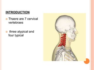
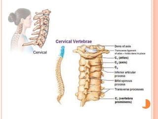
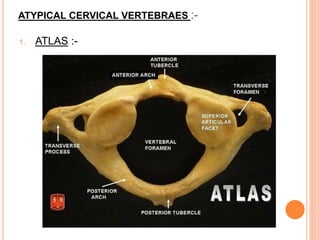
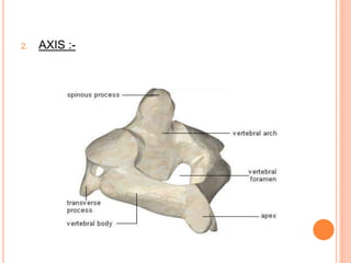
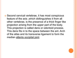
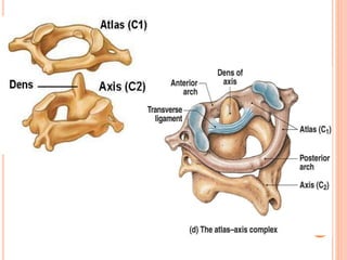
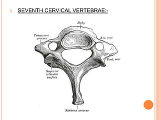
BY: ABDUL AKBAR
1ST YEAR PG OMR
CONTENTS
 INTRODUCTION
 SKULL INDIVIDUAL BONES
FRONTAL
PAREITAL
OCCIPITAL
TEMPORAL
SPHENOID
ETHMOID
MAXILLAE
ZYGOMATIC
NASAL
PALATINE
LACRIMAL
INFERIOR NASAL CONCHAE
VOMER
MANDIBLE
 CERVICAL VERTEBRAES
ATLAS
AXIS
TYPICAL CERVICAL VERTEBRAE
SEVENTH CERVICAL VERTEBRAE
INTRODUCTION
 THE SKULL :- Consist of 28 bones
CRANIAL BONES -14
FACIAL BONES -14
 CRANIAL BONES :-
1. Frontal
2. Occipital
3. Sphenoid
4. Ethmoid
5. Parietal (2)
6. Temporal (2)
7. Malleus (2)
8. Incus (2) ear ossicles
9. Stapes (2)
 FACIAL BONES :-
1. Nasal (2)
2. Maxillae (2)
3. Zygomatic (2)
4. Palatine (2)
5. Lacrimal (2)
6. Inferior nasal conchae
(2)
7. Mandible
8. vomer
SKULL INDIVIDUAL BONES
 FRONTAL BONE:-
 The large bone that makes up the forehead and supplies
the upper edge and roof of the orbit (eye socket).
 The frontal bone articulates (comes together) with a
number of other bones including the parietal, nasal,
ethmoid, maxillary, and zygomatic bones.
 Landmarks:
 Squama: flat portion that forms the forehead
 Supraorbital margin: ridge under the eyebrow,
forming the upper part of the orbit (eye socket)
 Supraorbital foramen: small hole within supraorbital
margin for blood vessels and nerves
 Frontal sinuses: hollow spaces behind the squama,
act as sound chambers to give the voice resonance.
PARIETAL BONES
 The main bone on the side of the skull.
 The word "parietal" comes from the Latin "parietalis"
meaning "belonging to the wall."
 It articulates (joins) with the other parietal bone in the
midline (top of the head), with the frontal bone in front of
it, with the occipital bone behind it, and with the sphenoid
and temporal bones lower down on the side of the skull.
OCCIPITAL BONE
 From the Latin, meaning the part of the head opposite
the front.
 The bone that forms the rear and the rear bottom of the
skull.
 The occipital bone articulates (joins) with the parietal and
temporal bones of the skull, the sphenoid bone in front of
it, and the first cervical vertebra (the atlas) beneath it
 Landmarks:
 Foramen magnum: large hole, allowing passage of
the spinal cord
 External occipital protuberance (EOP): prominent
projection on back of occiput
 Nuchal lines: a superior and inferior line running
laterally from the midline, serve as a point of muscle
attachment
SPHENOID BONE
 A prominent, irregular, wedge-shaped bone at the base of the
skull. The sphenoid bone has been called the "keystone" of
the cranial floor since it is in contact with all of the other cranial
bones.
 The Greek physician Galan wrote that the sphenoid bone was
"like a wedge thrust between the skull and the superior
maxilla."
 Landmarks
 Greater wings: large lateral projections of bone that help to
form the lateral border of the skull
 Lesser wings: smaller lateral projections of bone above
the greater wings
 Pterygoid processes: two long downward projections from
the greater wings that act as a point of muscle attachment.
 Sella turcica: known as the Turkish Saddle which cradles
the pituitary gland.
TEMPORAL BONE
 A large irregular bone situated at the base and side of the skull.
The temporal bone is connected with the mandible (the jaw bone)
via the temporomandibular (TM) joint.
 The temporal bone is formed of three parts (squamous, tympanic
and petrous) that are distinct at birth but then fuse. The petrous
portion of the temporal bone contains the structures of the inner
ear.
 Landmarks:
 Squama: flat portion of the temporal bone forming the anterior
and superior part of the temple
 Zygomatic process: process forming part of the cheek
 Petrous portion: internal, forming part of the floor of the
cranium. Contains the ear canal and internal ear structures.
 Mandibular fossa: socket between squama and petrous
portion, articulates with the condyle of the mandible (TMJ)
 External auditory meatus: opening to the ear canal
 Mastoid process: bony prominence behind the external
auditory meatus
 Styloid process: looks like an elephant’s tusk located between
the mastoid process and the jaw. Acts as a point of attachment
for muscles and ligaments.
ETHMOID BONE :-
 An irregularly shaped, spongy bone that provides the floor of the
front part of the skull and the roof of the nasal cavity.
 The ethmoid consists of two masses of thin plates enclosing air
cells and looks like a sieve.
 Landmarks:
 Lateral masses: form most of the wall between the nasal
cavity and the orbits
 Perpendicular plate: forms the superior portion of the nasal
septum
 Cribiform plate: forms the roof of the nasal cavity
 Olfactory foramina: small holes within the cribiform plate for
passage of the first cranial nerve (for smell)
 Crista galli: upward extension of bone above the cribiform
plate, acts as an anchoring point for one of the coverings of
the brain.
 Nasal concha (turbinates): two scroll-shaped projections
with a mucus membrane on either side of the nasal septum.
Function to cause air turbulence and trap inhaled particles.
MAXILLARY BONES
29
 The largest bones of the face, except for the mandible
and form, by their union, the whole of the upper jaw.
 They hold the upper teeth, and connect on the left and
right to the zygomatic bones (cheek bones).
 Each assists in forming the boundaries of three
cavities, namely, the roof of the mouth, the floor and
lateral wall of the nose, and the floor of the orbit.
 Landmarks:
 Infra Orbital foramen: hole below the orbit, for
blood vessels and nerves
 Alveolar process: arch of the maxilla containing the
upper teeth
 Palatine process: horizontal projection of the
maxilla forming the anterior ¾ of the hard palate.
31
Maxilla (Anterior View) (Yellow Colored Bones)
NASAL BONES
Varying in size and
form in different
individuals
They are placed side by
side at the middle and
upper part of the face
and form, by their
junction, "the bridge" of
the nose
32
33
Nasal Bones (Anterior View) (Bridge of the nose, below frontal bone)
ZYGOMATIC BONES
Commonly referred to as
the cheekbone.
It is situated at the upper
and lateral part of the
face: it forms the
prominence of the cheek
and part of the lateral wall
and floor of the orbit.
It articulates with the
zygomatic arch of the
temporal bone.
34
35
Zygomatic Bones (Anterior View) (Pink Colored Bones)
MANDIBLE
Largest and strongest
bone of the face.
Forms the lower jaw and
holds the lower teeth in
place.
The mandible consists of
a curved, horizontal
portion, the body, and two
perpendicular portions,
the rami, which unite with
the ends of the body
nearly
at right angles.
36
 Largest and strongest bone of the face.
 Forms the lower jaw and holds the lower teeth in place.
 Landmarks:
 Body: curved horizontal portion of the mandible
 Rami: two upward projections of bone that are perpendicular to
the body of the mandible.
 Angle of the mandible: angle formed where the body meets
the ramus
 Condylar process: a condyle on the posterior portion of the
ramus that articulates with the mandibular fossa of the temporal
bone.
 Coronoid process: a sharp projection of bone on the anterior
portion of the ramus that acts as a point of muscle attachment.
 Alveolar process: arch of bone containing the lower teeth
 Mental foramen: small hole on the side of the body for blood
vessels and nerves.
37
38
Mandible (Anterior View) (Lower Jaw)
39
Mandible (Lateral View) (Lower Jaw)
PALATINE BONE :-
It contributes to the
walls of three
cavities: the floor
and lateral wall of
the nasal cavity,
the roof of the
mouth, and the
floor of the orbit
LACRIMAL BONE
Smallest and most
fragile bone of the
face, is situated at
the front part of the
Medial of the
orbit.
Contains the lacrimal
sac and the naso-
lacrimal duct.
INFERIOR NASAL CONCHAE :-
Extends horizontally
along the lateral wall
of the nasal cavity
and consists of a
lamina of spongy
bone, curled upon
itself like a scroll
VOMER :-
One of the unpaired
facial bones of the
skull.
Located in
the midsagittal line,
and touches the
sphenoid, the
ethmoid, the left and
right palatine bones,
and the left and right
maxillary bones.
THE VERTEBRAL COLUMN
Below the skull, the
central axis of body is
formed by the backbone
or vertebral column
7 - cervical vertibrae
12 - thoracic vertibrae
5 - lumbar vertibrae
5 - sacral vertebrae
4 - coccyx
CERVICAL VERTIBRAES
INTRODUCTION
 Thaere are 7 cervical
vertebraes
 three atypical and
four typical
ATYPICAL CERVICAL VERTEBRAES :-
1. ATLAS :-
 Atlas is the first cervical vertibrae,it look very defferent
from a typical vertebre ,it has no body and no spine.
 It has two lateral masses ,joined togother anteriorly by
anterior arch and posteriorly by posterior arch.
 Two large transverse process seen on lateral sides ,and
pierced by a foramen called foramen transversarium.
 Superior aspect of each lateral masses show elongated
concave facets which articulate with the corresponding
condyle of occipital bone and form a atlanto-occipital
joint.
 The inferior aspect each lateral masses large oval facet
for articulation with corresponding superior articular
aspect of axis to form atlanto-axis joint.
2. AXIS :-
 Second cervical vertebrae, it has most conspicous
feature of the axis ,which didtinguishes it from all
other vertebrae, is the presence of a thick finger like
projection arising from the upper part of the body.
This projection is called dens or odontoid process.
This dens fits in to the space between the ant. Arch
of the atlas and its transverse ligament to form the
median atlanto occipital joint.
3. SEVENTH CERVICAL VERTEBRAE:-
 Seventh cervical vertebrae deffers from typical
vertebrae in having a long thick spinous process
which ends in a single tubercle.
 The tip of the process forms a prominent surface
land marks and becase of this it reffered to as
vertibrae prominence.
 The transverse process are also large and have
prominent posterior tubercles.
TYPICAL CERVICAL VERTEBRAES:-
STRUCTURE OF A TYPICAL VERTEBRAE:-
PARTS :-
1. body:- lies anteriorly,having flat upper and lower
surfaces. That attached to adjoining vertebrae
through intervertebral discs.
2. Pedicles:- (right and left) are short rounded bars
projects backwards from posterior part of body.
3. Lamina:- each pedicle is continous posterio
medially and form a vertical plate of bone called
the lamina.
4. The pedicle and lamina togother constitute the vertebral
arch.
5. Spine:-passing backwards from the junction of teo
laminae , there is spinous process.
6. Vertebral foramen:-bounded anteriorly by posterior
aspect of body . On sides of pedicles and behind the
lamina.
7. Transverse process:-passing laterally from the junction
of each pedicle and the corresponding lamina there is a
transverse process
8. Superior articular process:-projecting upwords from the
juntion of the pedicle and the laminae and it directed
posteriorly and some what laterally.
9. Inferior articular process:- projecting down words from
junction of the pedicle and laminae and it is directed
forwards and some what medially.
Osteology

Osteology

  • 1.
  • 2.
    CONTENTS  INTRODUCTION  SKULLINDIVIDUAL BONES FRONTAL PAREITAL OCCIPITAL TEMPORAL SPHENOID ETHMOID MAXILLAE ZYGOMATIC NASAL PALATINE LACRIMAL INFERIOR NASAL CONCHAE VOMER MANDIBLE  CERVICAL VERTEBRAES ATLAS AXIS TYPICAL CERVICAL VERTEBRAE SEVENTH CERVICAL VERTEBRAE
  • 3.
  • 5.
     THE SKULL:- Consist of 28 bones
  • 6.
  • 7.
     CRANIAL BONES:- 1. Frontal 2. Occipital 3. Sphenoid 4. Ethmoid 5. Parietal (2) 6. Temporal (2) 7. Malleus (2) 8. Incus (2) ear ossicles 9. Stapes (2)  FACIAL BONES :- 1. Nasal (2) 2. Maxillae (2) 3. Zygomatic (2) 4. Palatine (2) 5. Lacrimal (2) 6. Inferior nasal conchae (2) 7. Mandible 8. vomer
  • 8.
  • 9.
     The largebone that makes up the forehead and supplies the upper edge and roof of the orbit (eye socket).  The frontal bone articulates (comes together) with a number of other bones including the parietal, nasal, ethmoid, maxillary, and zygomatic bones.  Landmarks:  Squama: flat portion that forms the forehead  Supraorbital margin: ridge under the eyebrow, forming the upper part of the orbit (eye socket)  Supraorbital foramen: small hole within supraorbital margin for blood vessels and nerves  Frontal sinuses: hollow spaces behind the squama, act as sound chambers to give the voice resonance.
  • 12.
  • 13.
     The mainbone on the side of the skull.  The word "parietal" comes from the Latin "parietalis" meaning "belonging to the wall."  It articulates (joins) with the other parietal bone in the midline (top of the head), with the frontal bone in front of it, with the occipital bone behind it, and with the sphenoid and temporal bones lower down on the side of the skull.
  • 15.
  • 16.
     From theLatin, meaning the part of the head opposite the front.  The bone that forms the rear and the rear bottom of the skull.  The occipital bone articulates (joins) with the parietal and temporal bones of the skull, the sphenoid bone in front of it, and the first cervical vertebra (the atlas) beneath it  Landmarks:  Foramen magnum: large hole, allowing passage of the spinal cord  External occipital protuberance (EOP): prominent projection on back of occiput  Nuchal lines: a superior and inferior line running laterally from the midline, serve as a point of muscle attachment
  • 19.
  • 20.
     A prominent,irregular, wedge-shaped bone at the base of the skull. The sphenoid bone has been called the "keystone" of the cranial floor since it is in contact with all of the other cranial bones.  The Greek physician Galan wrote that the sphenoid bone was "like a wedge thrust between the skull and the superior maxilla."  Landmarks  Greater wings: large lateral projections of bone that help to form the lateral border of the skull  Lesser wings: smaller lateral projections of bone above the greater wings  Pterygoid processes: two long downward projections from the greater wings that act as a point of muscle attachment.  Sella turcica: known as the Turkish Saddle which cradles the pituitary gland.
  • 23.
  • 24.
     A largeirregular bone situated at the base and side of the skull. The temporal bone is connected with the mandible (the jaw bone) via the temporomandibular (TM) joint.  The temporal bone is formed of three parts (squamous, tympanic and petrous) that are distinct at birth but then fuse. The petrous portion of the temporal bone contains the structures of the inner ear.  Landmarks:  Squama: flat portion of the temporal bone forming the anterior and superior part of the temple  Zygomatic process: process forming part of the cheek  Petrous portion: internal, forming part of the floor of the cranium. Contains the ear canal and internal ear structures.  Mandibular fossa: socket between squama and petrous portion, articulates with the condyle of the mandible (TMJ)  External auditory meatus: opening to the ear canal  Mastoid process: bony prominence behind the external auditory meatus  Styloid process: looks like an elephant’s tusk located between the mastoid process and the jaw. Acts as a point of attachment for muscles and ligaments.
  • 26.
  • 27.
     An irregularlyshaped, spongy bone that provides the floor of the front part of the skull and the roof of the nasal cavity.  The ethmoid consists of two masses of thin plates enclosing air cells and looks like a sieve.  Landmarks:  Lateral masses: form most of the wall between the nasal cavity and the orbits  Perpendicular plate: forms the superior portion of the nasal septum  Cribiform plate: forms the roof of the nasal cavity  Olfactory foramina: small holes within the cribiform plate for passage of the first cranial nerve (for smell)  Crista galli: upward extension of bone above the cribiform plate, acts as an anchoring point for one of the coverings of the brain.  Nasal concha (turbinates): two scroll-shaped projections with a mucus membrane on either side of the nasal septum. Function to cause air turbulence and trap inhaled particles.
  • 29.
  • 30.
     The largestbones of the face, except for the mandible and form, by their union, the whole of the upper jaw.  They hold the upper teeth, and connect on the left and right to the zygomatic bones (cheek bones).  Each assists in forming the boundaries of three cavities, namely, the roof of the mouth, the floor and lateral wall of the nose, and the floor of the orbit.  Landmarks:  Infra Orbital foramen: hole below the orbit, for blood vessels and nerves  Alveolar process: arch of the maxilla containing the upper teeth  Palatine process: horizontal projection of the maxilla forming the anterior ¾ of the hard palate.
  • 31.
    31 Maxilla (Anterior View)(Yellow Colored Bones)
  • 32.
    NASAL BONES Varying insize and form in different individuals They are placed side by side at the middle and upper part of the face and form, by their junction, "the bridge" of the nose 32
  • 33.
    33 Nasal Bones (AnteriorView) (Bridge of the nose, below frontal bone)
  • 34.
    ZYGOMATIC BONES Commonly referredto as the cheekbone. It is situated at the upper and lateral part of the face: it forms the prominence of the cheek and part of the lateral wall and floor of the orbit. It articulates with the zygomatic arch of the temporal bone. 34
  • 35.
    35 Zygomatic Bones (AnteriorView) (Pink Colored Bones)
  • 36.
    MANDIBLE Largest and strongest boneof the face. Forms the lower jaw and holds the lower teeth in place. The mandible consists of a curved, horizontal portion, the body, and two perpendicular portions, the rami, which unite with the ends of the body nearly at right angles. 36
  • 37.
     Largest andstrongest bone of the face.  Forms the lower jaw and holds the lower teeth in place.  Landmarks:  Body: curved horizontal portion of the mandible  Rami: two upward projections of bone that are perpendicular to the body of the mandible.  Angle of the mandible: angle formed where the body meets the ramus  Condylar process: a condyle on the posterior portion of the ramus that articulates with the mandibular fossa of the temporal bone.  Coronoid process: a sharp projection of bone on the anterior portion of the ramus that acts as a point of muscle attachment.  Alveolar process: arch of bone containing the lower teeth  Mental foramen: small hole on the side of the body for blood vessels and nerves. 37
  • 38.
  • 39.
  • 40.
    PALATINE BONE :- Itcontributes to the walls of three cavities: the floor and lateral wall of the nasal cavity, the roof of the mouth, and the floor of the orbit
  • 42.
  • 43.
    Smallest and most fragilebone of the face, is situated at the front part of the Medial of the orbit. Contains the lacrimal sac and the naso- lacrimal duct.
  • 45.
  • 46.
    Extends horizontally along thelateral wall of the nasal cavity and consists of a lamina of spongy bone, curled upon itself like a scroll
  • 47.
  • 48.
    One of theunpaired facial bones of the skull. Located in the midsagittal line, and touches the sphenoid, the ethmoid, the left and right palatine bones, and the left and right maxillary bones.
  • 50.
    THE VERTEBRAL COLUMN Belowthe skull, the central axis of body is formed by the backbone or vertebral column 7 - cervical vertibrae 12 - thoracic vertibrae 5 - lumbar vertibrae 5 - sacral vertebrae 4 - coccyx
  • 51.
  • 52.
    INTRODUCTION  Thaere are7 cervical vertebraes  three atypical and four typical
  • 54.
  • 55.
     Atlas isthe first cervical vertibrae,it look very defferent from a typical vertebre ,it has no body and no spine.  It has two lateral masses ,joined togother anteriorly by anterior arch and posteriorly by posterior arch.  Two large transverse process seen on lateral sides ,and pierced by a foramen called foramen transversarium.  Superior aspect of each lateral masses show elongated concave facets which articulate with the corresponding condyle of occipital bone and form a atlanto-occipital joint.  The inferior aspect each lateral masses large oval facet for articulation with corresponding superior articular aspect of axis to form atlanto-axis joint.
  • 56.
  • 57.
     Second cervicalvertebrae, it has most conspicous feature of the axis ,which didtinguishes it from all other vertebrae, is the presence of a thick finger like projection arising from the upper part of the body. This projection is called dens or odontoid process. This dens fits in to the space between the ant. Arch of the atlas and its transverse ligament to form the median atlanto occipital joint.
  • 59.
  • 60.
     Seventh cervicalvertebrae deffers from typical vertebrae in having a long thick spinous process which ends in a single tubercle.  The tip of the process forms a prominent surface land marks and becase of this it reffered to as vertibrae prominence.  The transverse process are also large and have prominent posterior tubercles.
  • 62.
  • 63.
    STRUCTURE OF ATYPICAL VERTEBRAE:- PARTS :- 1. body:- lies anteriorly,having flat upper and lower surfaces. That attached to adjoining vertebrae through intervertebral discs. 2. Pedicles:- (right and left) are short rounded bars projects backwards from posterior part of body. 3. Lamina:- each pedicle is continous posterio medially and form a vertical plate of bone called the lamina.
  • 64.
    4. The pedicleand lamina togother constitute the vertebral arch. 5. Spine:-passing backwards from the junction of teo laminae , there is spinous process. 6. Vertebral foramen:-bounded anteriorly by posterior aspect of body . On sides of pedicles and behind the lamina. 7. Transverse process:-passing laterally from the junction of each pedicle and the corresponding lamina there is a transverse process 8. Superior articular process:-projecting upwords from the juntion of the pedicle and the laminae and it directed posteriorly and some what laterally. 9. Inferior articular process:- projecting down words from junction of the pedicle and laminae and it is directed forwards and some what medially.