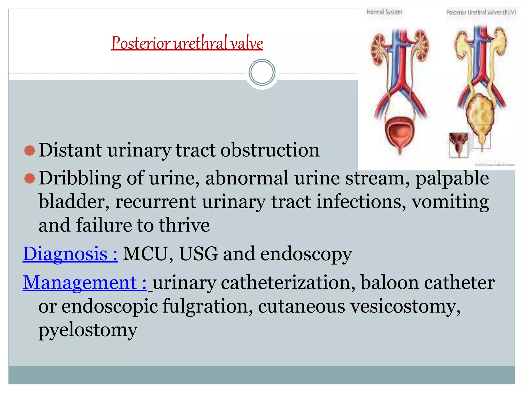
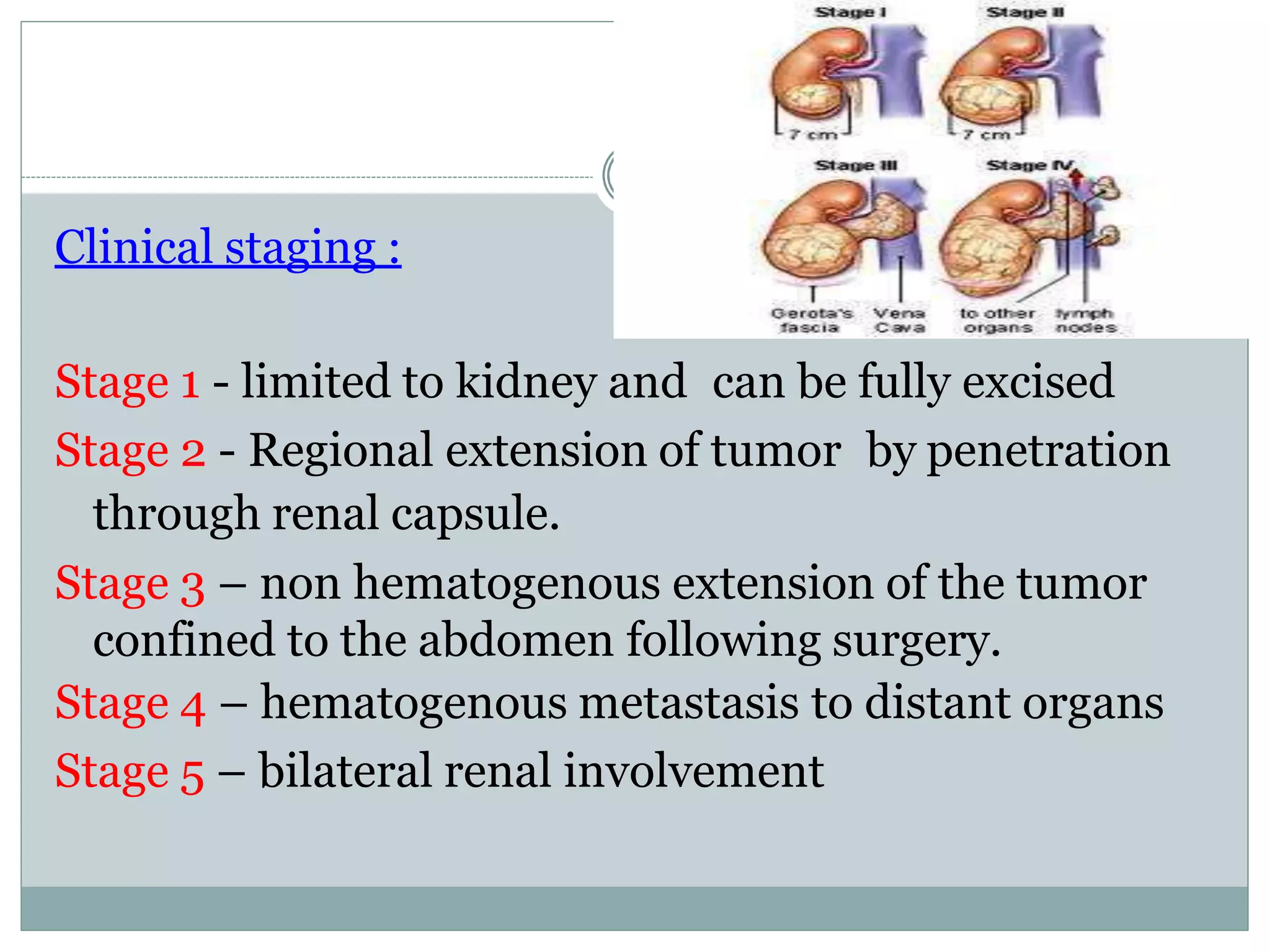
CONGENITAL GENITO URINARYDISORDERS
introduction
Disorders of kidney and urinary tract are commonly
seen in pediatric units as medical and surgical
problems.
Incidence: 3 to 6 per 1000 live births
Congenital abnormalities of the kidney and urinary tract
⚫These problems usually required surgical correction.
⚫Some of them are producing no clinical symptoms.
⚫25% cases of CRF are due to congenital anomalies
⚫8 to 10 % of children affected with congenital
anomalies of urinary tract.
en
Kidney and Ureter Bladder and Urethra
•Renal agenesis
•Renal hypoplasia
•Horse-shoe kidney
•Poly cystic renal disease
•Ectopic kidney
•Duplication of renal
pelvis and ureter
•Hydronephrosis
•Pelvi-ureteric
obstruction
•Congenital renal
neoplasm
•ureterocele
•Ectopia vesicaehus
•Patent urachus
•Bladder and neck
obstruction
•Posterior urethral valves
•Neurogenic bladder
•Hypospadias
•Epispadias
•Phimosis
•Urethral stenosis
•Meatal stenosis
Renal agenesis
Definition :It is the absence of kidney due to failure of
ureteric bud formation.
Type: Bilateral
Unilateral
Renal hypoplasia and dysplasia
⚫it occurs due to reduction of renal mass affecting the
nephron.
⚫It may be unilateral or bilateral
⚫It may be segmental, simple
RENAL DYSPLASIA :
⚫Disorganization of lung parenchyma with immature
nephron and ductal elements resulting in large or
small kidney.
⚫It may be multicystic , hypo plastic or aplastic
Horse – shoe kidney
⚫It develops when lower poles of the kidneys are fused
in the midline due to fusion of ureteric buds during
fetal development.
⚫Child presents with pyuria, albuminuria, vomiting
⚫Surgery is indicated
Polycystic kidneys
⚫Commonest congenital anomalies as inherited
autosomal disease.
⚫It is an complex syndrome
⚫Resulting from progressive dilatation of specific
portion of the nephron.
Types : infantile
adult
Diagnosis : IVP , renal angiography
Obstructive lesions of the urinary tract
⚫It mainly caused by congenital abnormalities like
pelvi-ureteric junction obstruction and posterior
urethral valves which may lead to irreversible renal
damage.
Pelvi– uretericjunctionstenosis
⚫Unilateral or bilateral
⚫Urinary tract infections and upper abdominal pain
Diagnosis : ultrasound, IVP , renal function test
Management:
⚫Surgery is indicated for removal of obstruction
hydronephrosis
⚫ It is the dilatation of renal pelvis
⚫ Unilateral or bilateral
⚫ Due to obstruction of urine flow in the distal urinary
tract
⚫ Males > females
⚫ Abdominal pain, failure to thrive , anemia, hypertension,
hematuria, renal failure.
Diagnosis : USG, IVP ,MCU
micturating cysto-
urethrogram (
Management :
⚫ Surgical removal or pyeloplasty,percutaneous
nephrostomy
Posteriorurethralvalve
⚫Distant urinary tract obstruction
⚫Dribbling of urine, abnormal urine stream, palpable
bladder, recurrent urinary tract infections, vomiting
and failure to thrive
Diagnosis : MCU, USG and endoscopy
Management : urinary catheterization, baloon catheter
or endoscopic fulgration, cutaneous vesicostomy,
pyelostomy
Meatal stenosis
⚫ Congenital abnormality
⚫ Due to meatal ulcer and scaring
⚫Meatal dilatation or meatoplasty
Exstrophyof bladder(ectopiavesicae)
⚫It is a congenital malformation
⚫Lower portion of the abdominal wall and the anterior
wall of the bladder are missing so that bladder is
everted through the opening and may found on the
lower abdomen with continuous passage of urine to
the outside.
⚫Male are more commonly affected
Clinical manifestations:
⚫ Urinary dribbling
⚫ Skin excoriation
⚫ Infection and ulceration or the bladder mucosa
⚫ Ambiguous genitalia
⚫ UTI
⚫ Growth failure
Diagnosis :
⚫ Cystoscopic examination
⚫ X-ray
⚫ USG
⚫ IVP
Management :
⚫Surgical closure of the bladder within 48 hours
⚫Urinary conversion before reconstructive surgery
⚫Orthopedic surgery
⚫Supportive nursing care
⚫Pre operative care
⚫Post operative care
⚫Follow up
epispadias
⚫Abnormal urethral opening on the dorsal aspect of
penis.
⚫Due to abnormal development of the infraumbilical
wall and upper wall of urethra.
⚫Rare in females
Classification : anterior epispadias
posterior epispadias
female epispadias – bifid clitoris
subsymphyseal with incontinence
of urine
Management : surgical correction
1. 1.5 to 2 years of age for penile lengthening,
elongation of urethral strip and chordee correction.
2. operation done at least 6 months after first stage
for urethral reconstruction
3. 3 to 4 years of age for bladder neck reconstruction
and correction of VUR Vesicoureteral reflux
⚫ Cystoplasty
⚫ Supportive nursing care
hypospadias
⚫ it is the congenital abnormal urethral opening on
the ventral aspect( under surface ) of the penis.
⚫Common in male children.
Classification :
⚫Anterior hypospadias(65 to 70%) : it may found as
glandular or coronal or on distal penile shaft
⚫Middle (10-15%) penile shaft hypospadias.
⚫Posterior hypospadias(20%) : it may be found on
proximal penile shaft or as penoscrotal,scortal or
perineal type.
Problems related to hypospadias :
1. Painful downward curvature of penis
2. Deflected stream of urine
3.Inability void urine while standing
Management :
⚫ surgical reconstruction
⚫ Meatotomy
⚫ Chordee correction
⚫ urethroplasty
phimosis
⚫Narrow opening of the prepuce that prevents it being
drawn back over the glans penis.
Management:
⚫Circumcision
⚫ apply Betamethasone cream
paraphimosis
⚫It is the retraction of a phimotic foreskin, behind
coronal sulcus.
⚫It may develop phimotic child which also need for
surgical management by circumcision or reduction
with application of lubricant under deep sedation.
Clinical features :
⚫edematous
⚫Severe pain
Wilm’stumor(nephroblastoma)
⚫ Max wilm’s , German surgeon described this most
common renal tumor of childhood.
⚫ Chromosomal deletions 11 and 16
⚫ Highly malignant embryonal tumor
⚫ Tumor develops in kidney parenchyma ,invading the
surrounding tissues.
Clinical features :
⚫ abdominal mass
⚫ Microscopic hematuria
⚫ Fever
⚫ Pallor
⚫ Superficial vein engorgement
Clinical staging :
Stage 1 - limited to kidney and can be fully excised
Stage 2 - Regional extension of tumor by penetration
through renal capsule.
Stage 3 – non hematogenous extension of the tumor
confined to the abdomen following surgery.
Stage 4 – hematogenous metastasis to distant organs
Stage 5 – bilateral renal involvement
Diagnostic evaluation :
⚫History
⚫Physical examination
⚫X-ray abdomen ,chest
⚫USG
⚫CT scan
⚫MRI
⚫Renal function test
Management :
⚫Nephrectomy
⚫Chemotherapy
⚫radiotherapy
Undescended testis
⚫Testis cannot be made to reach the bottom of the
scrotum
⚫It is also known as Cryptochordism
⚫5% of full term male infants may have unilateral or
bilateral testis.
Types :
⚫Retractile
⚫Palpable
⚫impalpable
Diagnosis :
⚫USG
⚫Laproscopy
⚫MRI
Complications :
⚫Trauma
⚫Malignant
⚫Psychological trauma
Management :
⚫Orcheopexy
⚫Hormonal treatment
Vesico– urethral reflux
⚫ Retrograde flow of bladder urine up the ureter during
voiding.
Causes:
⚫ Insertion of ureter in to the bladder
⚫ Infection
⚫Edema
Clinical features:
⚫ Dysuria
⚫ Urinary frequency and urgency
⚫ Urine retention
⚫ Cloudy or blood tinged urine
Management :
⚫Correction of structural anomalies
⚫Administration of low dose antibiotics
Detectingkidney and urinary tract abnormalities beforebirth
⚫Ultrasound examination
⚫Antenatal screening
⚫Screening of high risk groups
Prevention of congenital genitourinary anomalies
⚫Advice to future parents
⚫To minimize exposure of pregnant women to risk
factors
⚫To prevent over weight/ obesity
⚫To promote effective information on diet
⚫To improve folate status
⚫To avoid contraindicated vaccination
⚫To include school education programmes
Nursing management of thechild withurologic surgery
⚫Promoting understanding of parents
⚫Preparing for diagnostic procedures
⚫Involving the parents in child care
⚫Monitoring intake and output
⚫Preventing infections
⚫Providing comfort
⚫Providing adequate nutrition
⚫Teaching the parents about related care
CONGENITAL GENITO URINARY DISORDERS.pptx

CONGENITAL GENITO URINARY DISORDERS.pptx

  • 1.
  • 2.
    introduction Disorders of kidneyand urinary tract are commonly seen in pediatric units as medical and surgical problems. Incidence: 3 to 6 per 1000 live births
  • 4.
    Congenital abnormalities ofthe kidney and urinary tract ⚫These problems usually required surgical correction. ⚫Some of them are producing no clinical symptoms. ⚫25% cases of CRF are due to congenital anomalies ⚫8 to 10 % of children affected with congenital anomalies of urinary tract.
  • 5.
    en Kidney and UreterBladder and Urethra •Renal agenesis •Renal hypoplasia •Horse-shoe kidney •Poly cystic renal disease •Ectopic kidney •Duplication of renal pelvis and ureter •Hydronephrosis •Pelvi-ureteric obstruction •Congenital renal neoplasm •ureterocele •Ectopia vesicaehus •Patent urachus •Bladder and neck obstruction •Posterior urethral valves •Neurogenic bladder •Hypospadias •Epispadias •Phimosis •Urethral stenosis •Meatal stenosis
  • 6.
    Renal agenesis Definition :Itis the absence of kidney due to failure of ureteric bud formation. Type: Bilateral Unilateral
  • 7.
    Renal hypoplasia anddysplasia ⚫it occurs due to reduction of renal mass affecting the nephron. ⚫It may be unilateral or bilateral ⚫It may be segmental, simple RENAL DYSPLASIA : ⚫Disorganization of lung parenchyma with immature nephron and ductal elements resulting in large or small kidney. ⚫It may be multicystic , hypo plastic or aplastic
  • 8.
    Horse – shoekidney ⚫It develops when lower poles of the kidneys are fused in the midline due to fusion of ureteric buds during fetal development. ⚫Child presents with pyuria, albuminuria, vomiting ⚫Surgery is indicated
  • 9.
    Polycystic kidneys ⚫Commonest congenitalanomalies as inherited autosomal disease. ⚫It is an complex syndrome ⚫Resulting from progressive dilatation of specific portion of the nephron. Types : infantile adult Diagnosis : IVP , renal angiography
  • 10.
    Obstructive lesions ofthe urinary tract ⚫It mainly caused by congenital abnormalities like pelvi-ureteric junction obstruction and posterior urethral valves which may lead to irreversible renal damage.
  • 11.
    Pelvi– uretericjunctionstenosis ⚫Unilateral orbilateral ⚫Urinary tract infections and upper abdominal pain Diagnosis : ultrasound, IVP , renal function test Management: ⚫Surgery is indicated for removal of obstruction
  • 12.
    hydronephrosis ⚫ It isthe dilatation of renal pelvis ⚫ Unilateral or bilateral ⚫ Due to obstruction of urine flow in the distal urinary tract ⚫ Males > females ⚫ Abdominal pain, failure to thrive , anemia, hypertension, hematuria, renal failure. Diagnosis : USG, IVP ,MCU micturating cysto- urethrogram ( Management : ⚫ Surgical removal or pyeloplasty,percutaneous nephrostomy
  • 14.
    Posteriorurethralvalve ⚫Distant urinary tractobstruction ⚫Dribbling of urine, abnormal urine stream, palpable bladder, recurrent urinary tract infections, vomiting and failure to thrive Diagnosis : MCU, USG and endoscopy Management : urinary catheterization, baloon catheter or endoscopic fulgration, cutaneous vesicostomy, pyelostomy
  • 15.
    Meatal stenosis ⚫ Congenitalabnormality ⚫ Due to meatal ulcer and scaring ⚫Meatal dilatation or meatoplasty
  • 16.
    Exstrophyof bladder(ectopiavesicae) ⚫It isa congenital malformation ⚫Lower portion of the abdominal wall and the anterior wall of the bladder are missing so that bladder is everted through the opening and may found on the lower abdomen with continuous passage of urine to the outside. ⚫Male are more commonly affected
  • 17.
    Clinical manifestations: ⚫ Urinarydribbling ⚫ Skin excoriation ⚫ Infection and ulceration or the bladder mucosa ⚫ Ambiguous genitalia ⚫ UTI ⚫ Growth failure Diagnosis : ⚫ Cystoscopic examination ⚫ X-ray ⚫ USG ⚫ IVP
  • 18.
    Management : ⚫Surgical closureof the bladder within 48 hours ⚫Urinary conversion before reconstructive surgery ⚫Orthopedic surgery ⚫Supportive nursing care ⚫Pre operative care ⚫Post operative care ⚫Follow up
  • 19.
    epispadias ⚫Abnormal urethral openingon the dorsal aspect of penis. ⚫Due to abnormal development of the infraumbilical wall and upper wall of urethra. ⚫Rare in females Classification : anterior epispadias posterior epispadias female epispadias – bifid clitoris subsymphyseal with incontinence of urine
  • 21.
    Management : surgicalcorrection 1. 1.5 to 2 years of age for penile lengthening, elongation of urethral strip and chordee correction. 2. operation done at least 6 months after first stage for urethral reconstruction 3. 3 to 4 years of age for bladder neck reconstruction and correction of VUR Vesicoureteral reflux ⚫ Cystoplasty ⚫ Supportive nursing care
  • 22.
    hypospadias ⚫ it isthe congenital abnormal urethral opening on the ventral aspect( under surface ) of the penis. ⚫Common in male children. Classification : ⚫Anterior hypospadias(65 to 70%) : it may found as glandular or coronal or on distal penile shaft ⚫Middle (10-15%) penile shaft hypospadias. ⚫Posterior hypospadias(20%) : it may be found on proximal penile shaft or as penoscrotal,scortal or perineal type.
  • 23.
    Problems related tohypospadias : 1. Painful downward curvature of penis 2. Deflected stream of urine 3.Inability void urine while standing Management : ⚫ surgical reconstruction ⚫ Meatotomy ⚫ Chordee correction ⚫ urethroplasty
  • 24.
    phimosis ⚫Narrow opening ofthe prepuce that prevents it being drawn back over the glans penis. Management: ⚫Circumcision ⚫ apply Betamethasone cream
  • 25.
    paraphimosis ⚫It is theretraction of a phimotic foreskin, behind coronal sulcus. ⚫It may develop phimotic child which also need for surgical management by circumcision or reduction with application of lubricant under deep sedation. Clinical features : ⚫edematous ⚫Severe pain
  • 26.
    Wilm’stumor(nephroblastoma) ⚫ Max wilm’s, German surgeon described this most common renal tumor of childhood. ⚫ Chromosomal deletions 11 and 16 ⚫ Highly malignant embryonal tumor ⚫ Tumor develops in kidney parenchyma ,invading the surrounding tissues. Clinical features : ⚫ abdominal mass ⚫ Microscopic hematuria ⚫ Fever ⚫ Pallor ⚫ Superficial vein engorgement
  • 27.
    Clinical staging : Stage1 - limited to kidney and can be fully excised Stage 2 - Regional extension of tumor by penetration through renal capsule. Stage 3 – non hematogenous extension of the tumor confined to the abdomen following surgery. Stage 4 – hematogenous metastasis to distant organs Stage 5 – bilateral renal involvement
  • 28.
    Diagnostic evaluation : ⚫History ⚫Physicalexamination ⚫X-ray abdomen ,chest ⚫USG ⚫CT scan ⚫MRI ⚫Renal function test Management : ⚫Nephrectomy ⚫Chemotherapy ⚫radiotherapy
  • 29.
    Undescended testis ⚫Testis cannotbe made to reach the bottom of the scrotum ⚫It is also known as Cryptochordism ⚫5% of full term male infants may have unilateral or bilateral testis. Types : ⚫Retractile ⚫Palpable ⚫impalpable
  • 30.
  • 31.
    Vesico– urethral reflux ⚫Retrograde flow of bladder urine up the ureter during voiding. Causes: ⚫ Insertion of ureter in to the bladder ⚫ Infection ⚫Edema Clinical features: ⚫ Dysuria ⚫ Urinary frequency and urgency ⚫ Urine retention ⚫ Cloudy or blood tinged urine
  • 32.
    Management : ⚫Correction ofstructural anomalies ⚫Administration of low dose antibiotics Detectingkidney and urinary tract abnormalities beforebirth ⚫Ultrasound examination ⚫Antenatal screening ⚫Screening of high risk groups
  • 33.
    Prevention of congenitalgenitourinary anomalies ⚫Advice to future parents ⚫To minimize exposure of pregnant women to risk factors ⚫To prevent over weight/ obesity ⚫To promote effective information on diet ⚫To improve folate status ⚫To avoid contraindicated vaccination ⚫To include school education programmes
  • 34.
    Nursing management ofthechild withurologic surgery ⚫Promoting understanding of parents ⚫Preparing for diagnostic procedures ⚫Involving the parents in child care ⚫Monitoring intake and output ⚫Preventing infections ⚫Providing comfort ⚫Providing adequate nutrition ⚫Teaching the parents about related care