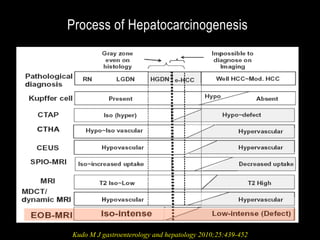
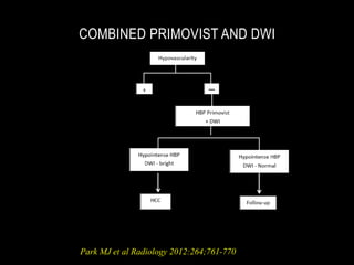
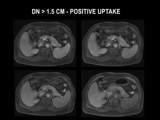
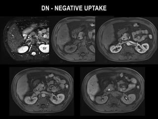
This document discusses the current approach for diagnostic imaging of small hepatocellular carcinoma (HCC) in patients with liver cirrhosis using gadoxetic acid (Primovist) enhanced magnetic resonance imaging (MRI) and diffusion weighted imaging (DWI). It summarizes that the combined use of Primovist MRI and DWI provides high diagnostic accuracy and sensitivity, particularly for detecting HCC lesions smaller than 2cm. This approach can image the step-wise progression of hepatocarcinogenesis by assessing vascularity, hepatocyte function, and cell density. The summary highlights that while Primovist MRI alone has high sensitivity, it has compromised specificity, but combining it with DWI increases specificity by differentiating HCC from
This document summarizes ultrasound findings for cirrhosis of the liver. Key findings include coarse echotexture and nodular surface of the liver. Regenerating nodules appear isoechoic or hypoechoic. Portal hypertension is a complication and is suggested by Doppler findings like dilated portal vein and low venous velocity. Findings of portal hypertension include splenomegaly, ascites, and portosystemic venous collaterals visible with ultrasound.
Metastases are the most common malignant liver lesions and liver imaging is often used to detect them. They can be hypovascular or hypervascular. Hypovascular metastases are best seen on portal venous phase imaging while hypervascular ones are best seen on arterial phase. Differentiating metastases from other lesions such as hemangiomas or cysts requires analyzing features like enhancement pattern and imaging characteristics on multiple sequences/phases. Multiphasic CT or MRI is often used to fully characterize lesions.
This document provides information about a liver ultrasound presentation given by Prof. Dr. Ibrahim Nunow Cabdi, including his medical qualifications and areas of expertise. It outlines the techniques for performing ultrasound of the liver and assessing liver anatomy, vasculature, and segments. Common benign and malignant liver conditions are described, along with how they appear ultrasonographically. Diffuse liver diseases such as hepatitis, cirrhosis, and fatty infiltration are also discussed.
Focal vs diffuse gall bladder wall thickeningairwave12
Focal vs diffuse gallbladder wall thickening can have different causes. [1] Focal thickening may be due to polyps, adenomyomatosis, carcinoma, or other conditions. [2] Diffuse thickening can result from conditions like cholecystitis, adenomyomatosis, or cancer. Imaging findings like wall thickness, presence of stones, diverticula, or masses help differentiate these conditions. Accurately diagnosing the cause of gallbladder wall thickening helps guide patient management.
Presentation1.pptx, ultrasound examination of the liver and gall bladder.Abdellah Nazeer
This document provides an ultrasound protocol and guidelines for examining the liver and gallbladder. It begins with an overview of the role and technique of ultrasound for the liver, including scanning positions and images to capture. Common liver pathologies such as fatty liver, cirrhosis, cysts, hemangiomas, abscesses, and metastases are described. Guidelines are provided for gallbladder ultrasound including patient preparation, technique, and anatomy. Normal findings and pathologies like stones, acute cholecystitis, and emphysematous cholecystitis are outlined. The document concludes with potential vascular disorders of the liver involving blood flow.
This document discusses liver lesions and their appearance on various imaging modalities. It covers benign lesions like hemangioma, focal nodular hyperplasia and hepatic adenoma. Malignant primary lesions discussed are hepatocellular carcinoma and hepatoblastoma. Imaging features of hypervascular and hypovascular lesions on multiphasic CT are summarized. Hepatocellular carcinoma risk factors and clinical presentation are outlined. Imaging appearance of HCC on ultrasound, CT and MRI is described in detail. Hepatic metastases are also discussed along with hypervascular metastatic lesions.
This document provides an overview of renal ultrasound, including normal anatomy, scanning techniques, common findings, and clinical indications. It discusses how to approach scanning both kidneys and describes normal sonographic appearances. Common abnormalities like hydronephrosis, cysts, masses, and medical kidney disease are outlined. Important tips include distinguishing cysts from hydronephrosis and avoiding pitfalls like mistaking prominent pyramids for other findings. The goal of renal ultrasound is to evaluate for obstructive uropathy, renal masses, parenchymal disease, and nephrolithiasis.
Ultrasound of the urinary tract - Renal tumorsSamir Haffar
This document discusses ultrasound imaging of renal tumors. It begins by stating that ultrasound is often the first imaging modality used for the kidneys and plays an important role in diagnosing renal tumors. It then discusses technical advances in ultrasound imaging that have improved detection of renal tumors. The document goes on to describe normal kidney anatomy and various benign and malignant renal tumors that can be identified on ultrasound, including renal cell carcinoma, angiomyolipomas, cysts, and others. It provides ultrasound images and characteristics of different renal pathologies.
This document summarizes ultrasound findings related to the gallbladder. It begins by describing normal gallbladder anatomy and ultrasound appearance. It then discusses various congenital gallbladder abnormalities that can be seen on ultrasound. Finally, it details gallbladder pathologies that can be identified ultrasonographically such as gallstones, sludge, acute and chronic cholecystitis, polyps and carcinoma. For each finding, it provides ultrasound images and descriptions of characteristic ultrasound features.
This document provides an overview of renal isotope studies used to assess renal function and anatomy. It describes the radiopharmaceuticals, patient preparation, indications, and findings for renal scans, renograms, and nuclear cystograms. Key points include that renal scans evaluate renal blood flow, structure, and drainage using agents retained in tubules, filtered by glomeruli, or secreted by tubules. Renograms generate time-activity curves to assess obstruction, transplantation, renovascular hypertension, and hydronephrosis. Nuclear cystograms evaluate vesicoureteral reflux.
This document discusses various gallbladder and biliary tree pathologies that can be detected on sonography. It describes the sonographic appearance and features of gallstones, biliary sludge, acute and chronic cholecystitis, porcelain gallbladder, adenomyomatosis, cholesterol polyps, gallbladder carcinoma, choledochal cysts, Caroli's disease, primary sclerosing cholangitis, Mirizzi syndrome, bacterial cholangitis, and cholangiocarcinoma. Mobility is key to differentiating stones from other entities. Sludge appears as low-level echoes without shadowing. Gallbladder wall thickening and distention indicate acute cholecystitis. Calcification causes hyperechoic
This document provides an overview of MRI in urology, with a focus on MRI of the prostate. It discusses the moderators and professors of the department of urology. It then covers the basic principles of MRI, including magnetic field strength, radiofrequency pulses, T1/T2 weighting, and contrast agents. Applications of MRI for prostate imaging and prostate cancer detection are described, including T2-weighted imaging, diffusion-weighted imaging, and magnetic resonance spectroscopy. The PIRADS scoring system and assessment of extracapsular extension on MRI are also summarized.
Advanced imaging modalities of the liverEnass Khattab
This document discusses advanced imaging modalities for the liver. It begins by classifying liver diseases into focal lesions, diffuse diseases, and biliary diseases. Contrast-enhanced ultrasound and microbubble contrast agents are described for characterizing focal lesions. Hybrid imaging modalities like PET/CT and PET/MRI are summarized for providing functional and anatomical data. Elastography techniques like transient elastography (FibroScan) and MR elastography are presented for assessing liver stiffness and fibrosis noninvasively.
This document discusses Magnetic Resonance Cholangiopancreatography (MRCP), a non-invasive MRI technique used to investigate biliary and pancreatic pathologies. It works by exploiting the inherent differences in T2-weighted contrast between fluid-filled structures and soft tissue. Static or slow moving fluids within the biliary tree and pancreatic duct appear as high signal intensity on MRCP, while surrounding tissue has reduced signal intensity. The document outlines the MRCP technique, imaging parameters, indications including biliary and pancreatic diseases, advantages over ERCP, and some pitfalls.
Congenital anomalies of pancreas and hepatobiliary system radiologyVidya TK
This document discusses congenital anomalies of the pancreas and hepatobiliary system. It describes several pancreatic anomalies including pancreas divisum, annular pancreas, and ectopic pancreas. It also discusses variations in pancreatic duct configuration and number. For the hepatobiliary system, it covers anomalies such as choledochal cysts, Caroli disease, biliary hamartoma, biliary atresia, and variations seen with fibropolycstic liver disease. Imaging plays an important role in diagnosing many of these congenital anomalies.
A presentation about Intravenous Urography (Also known as Intravenous Pyeography).
The presentation contains 41 slides, and is divided into 4 parts :
1 - Introduction.
2 - The procedure.
3 - Examples for abnormal findings.
4 - Studies comparing IVU accuracy with KUB & USG with CT Scan.
This presentation was prepared and presented by me in the tutorials of the Radiology Department of Sebha Medical Center.
This document discusses malignant liver lesions. It describes the different types of primary and secondary malignant tumors that can occur in the liver. The most common are metastatic deposits from other primary cancers, and hepatocellular carcinoma (HCC). HCC is described in detail, including risk factors, pathogenesis, imaging appearance on ultrasound, CT and MRI, staging systems, treatment surveillance, and diagnostic criteria. Other liver cancers such as cholangiocarcinoma are also briefly mentioned.
This document provides a classification and overview of renal tumors. It discusses renal cell carcinoma in depth, including incidence, etiology, pathology, clinical presentation, investigations, staging, treatments for localized and disseminated disease, and follow up care. It also briefly covers other renal tumors such as tumors of the renal parenchyma, pelvis, and Wilms' tumor.
This document discusses paediatric abdominal masses. Ultrasound is generally the initial imaging test which can differentiate cystic from solid masses and indicate organ of origin. CT or MRI may be needed for large or complex masses. Specific masses discussed include renal masses like Wilms tumour, adrenal masses like neuroblastoma, hepatic masses like hepatoblastoma, pancreatic masses, splenic masses, gastrointestinal masses, and pelvic masses like rhabdomyosarcoma. Imaging findings, staging, and treatment options are provided for some of the most common paediatric abdominal masses.
This document discusses the approach to evaluating and diagnosing liver masses. It defines a liver mass and explains how imaging techniques are used in the diagnosis. The differential diagnosis for liver masses can range from benign to malignant lesions. Cystic lesions discussed in detail include pyogenic and amoebic liver abscesses. Solid lesions include inflammatory conditions like abscesses as well as benign and malignant tumors. Treatment options for different lesions are outlined.
Presentation1.pptx, radiological imaging of prostatic diseasesAbdellah Nazeer
This document discusses radiological imaging of prostatic diseases. It begins with an overview of prostate anatomy and zones. It then discusses various imaging modalities used to evaluate the prostate, including MRI, ultrasound, CT, and bone scans. Specific applications are covered such as imaging characteristics of prostate cancer, benign prostatic hypertrophy, prostatitis, and abscesses. Imaging findings of different prostate cancer stages are also reviewed. In summary, the document provides a comprehensive overview of radiological imaging techniques and findings for evaluating diseases of the prostate gland.
The document discusses unilateral small kidneys and the role of imaging in establishing a diagnosis. It describes various causes of unilateral small kidneys including pre-renal issues like renal artery stenosis, intra-renal issues such as renal infarction, and post-renal issues like reflux nephropathy. Imaging modalities like ultrasound, CT, MRI, renal scintigraphy, and angiography are discussed for evaluating the kidneys and determining the underlying etiology. The document emphasizes how different imaging findings can help identify conditions that may cause one kidney to be smaller than the other.
Dr. Navni Garg presented on imaging in benign hepatic masses. Various benign liver lesions were discussed including developmental masses like cysts, inflammatory masses like abscesses, and benign neoplasms. Imaging modalities like ultrasound, CT, MRI, and specialized CT techniques were described for evaluating these lesions. Contrast agents used in MRI like SPIO, USPIO, and hepatobiliary agents were also covered. Specific lesions such as focal nodular hyperplasia, regenerative nodules, and dysplastic nodules were discussed in detail.
This document discusses the imaging modalities used to diagnose abdominal tuberculosis. It begins with an introduction stating that abdominal tuberculosis accounts for 11% of extra-pulmonary tuberculosis cases. It then covers the various routes of infection and clinical presentations. The key imaging investigations discussed are ultrasound, CT and barium studies. Specific findings are highlighted for different types of abdominal tuberculosis including tubercular peritonitis, lymphadenopathy, and involvement of the gastrointestinal tract and viscera. Differential diagnoses such as Crohn's disease and carcinomatosis are also mentioned.
This document discusses benign focal liver lesions of different cellular origins - hepatocellular, cholangiocellular, and mesenchymal. It provides details on common benign liver tumors including cavernous hemangioma, focal nodular hyperplasia (FNH), hepatic adenoma, hepatic cysts, and infantile hemangioendothelioma. Imaging characteristics on ultrasound, CT, and MRI scans are described to help differentiate these benign liver lesions. Common features seen include hypodense lesions on CT, varying signal intensities on MRI, presence of fat, cystic components, enhancement patterns, and visualization of scars.
This document provides a workup algorithm for focal liver lesions. It discusses obtaining a patient history and physical exam findings, as well as blood tests, imaging studies, and biopsy. Common benign and malignant liver lesions are described, including their risk factors, imaging characteristics, and treatment options. For example, hemangiomas are the most common benign tumor, often appearing as well-demarcated lesions on ultrasound and MRI. Hepatocellular carcinoma is the most common primary liver cancer and often appears as a vascular enhancing mass on CT scan. Treatment may involve surgery, chemotherapy, or liver transplantation depending on the type and stage of liver lesion.
Spleen Ultrasound anatomy structure scanning techniques and pathologies Safi. Khan
The spleen is located in the left upper quadrant of the abdomen. It has close anatomical relationships with nearby organs like the stomach, pancreas, kidney and diaphragm. The spleen receives blood supply from the splenic artery and drains into the splenic vein. Accessory spleens and clefts are sometimes seen. Cysts, hemangiomas, lymphangiomas and lymphomas are potential tumors of the spleen. Splenomegaly can result from a variety of causes and is diagnosed based on spleen size exceeding 13cm in length or 6cm in thickness.
This document discusses various malignant liver lesions including primary and secondary tumors. For primary liver cancers, it describes hepatocellular carcinoma (HCC) as the most common type, risk factors such as hepatitis, and imaging features. It also discusses cholangiocarcinoma, hepatoblastoma, and rare tumors such as fibrolamellar carcinoma. Secondary cancers and criteria for staging HCC are also summarized.
This document discusses the appropriate use of ultrasound, CT, and MRI in liver imaging. It provides examples of using each modality to diagnose various common liver conditions like cirrhosis, fatty liver, hepatitis, and liver lesions. Ultrasound is useful as a first-line exam but has limitations. CT is the standard for assessing liver cancer patients but exposes patients to radiation. MRI is now the preferred method for evaluating cirrhosis and differentiating liver lesions as it uses tissue-specific contrast agents without radiation. Biopsy is still often needed where imaging results are doubtful.
J.P. is a 58-year-old man diagnosed with hepatitis B and hepatocellular carcinoma. He underwent various treatments including Y-90 radioembolization, chemoembolization, and sorafenib, but later developed complications including a staph infection and a necrotic liver tumor. V.B. is a 75-year-old woman diagnosed with pancreatic cancer. She received neoadjuvant chemotherapy and radiation, then a Whipple procedure which showed a complete response with no residual tumor. Both patients are being monitored for recurrence.
The document discusses liver shear wave elastography and presents several clinical cases. It provides a table comparing the liver fibrosis classification using liver biopsy and Metavir scores to the corresponding ranges measured by shear wave elastography in kilopascals and meters/second. Several images show shear wave elastography measurements of livers with conditions like steatosis, cirrhosis, and hepatocellular carcinoma. The document demonstrates using shear wave elastography to evaluate liver fibrosis in patients with hepatitis C at different stages of disease.
1) Liver carcinoma, specifically hepatocellular carcinoma (HCC), is a primary tumor of the liver that usually arises in a cirrhotic liver.
2) The main risk factors for HCC are chronic hepatitis B and C infections, which can lead to cirrhosis. Other risk factors include alcoholism and aflatoxin exposure.
3) HCC is often asymptomatic in early stages but can present with abdominal pain or a palpable mass. Diagnosis involves imaging like ultrasound or CT along with blood markers like alpha-fetoprotein.
This document discusses various types of liver lesions including regenerative nodules, dysplastic nodules, hepatocellular adenoma, focal nodular hyperplasia, and hepatocellular carcinoma. It provides details on the histological and immunohistochemical features that can help differentiate these lesions. Key points include that dysplastic nodules are believed to be HCC precursors, hepatocellular adenomas can be single or multifocal and classified based on molecular features, and the distinction between well-differentiated HCC and hepatocellular adenoma can be challenging based on overlapping histological features alone.
O documento apresenta o cronograma de aulas do módulo de Abdome e Neurorradiologia da Liga Acadêmica de Radiologia e Diagnóstico por Imagem da UFSM para 2016/1, com datas e temas como avaliação das adrenais, aneurismas de aorta abdominal, tumores de SNC, esclerose múltipla e AVC isquêmico. As aulas ocorrerão às quartas-feiras no Instituto de Radiologia São Lucas e às quartas-feiras no Serviço de Radiologia do HUSM, ministradas respectivamente
This document discusses liver cirrhosis and its causes, complications, symptoms, investigations, and management. It notes that cirrhosis can be caused by viral infections, alcohol, metabolic disorders, immune disorders, genetic disorders, and drugs. Complications include pruritis, clotting abnormalities, and ascites. Investigations include blood tests of liver enzymes and imaging techniques like ultrasound and biopsy. Management of complications involves treating pruritis with antihistamines and bile acid resins, managing clotting with vitamin K, and mobilizing ascites fluid with sodium restriction and diuretics. Hepatic encephalopathy treatment focuses on reducing ammonia levels.
Liver tumors - A basic guide to diagnose and treat liver tumorsBhavin Vasavada
This document discusses various liver tumors including benign and malignant lesions. The most common primary liver cancer is hepatocellular carcinoma which is often associated with cirrhosis from hepatitis, alcohol, or other causes. Diagnosis involves imaging like ultrasound, CT, or MRI and biopsy if needed. Treatment depends on the size and extent of disease but may include transplantation, resection, ablation, chemoembolization, or other local therapies. Prognosis depends on tumor stage, liver function, and performance status of the patient.
The document discusses non-alcoholic fatty liver disease (NAFLD), which includes a spectrum of conditions from simple steatosis to non-alcoholic steatohepatitis (NASH) and cirrhosis. NAFLD is strongly associated with obesity and metabolic syndrome. The prevalence of NAFLD is increasing globally and varies from 5-30% in different regions. Diagnosis requires imaging and liver biopsy. Treatment focuses on lifestyle modifications and medications to improve insulin resistance.
Presentacion del Residente Jose Chavez Peche en las reuniones de los residentes de Cirugia General y Digestiva del Hospital Nacional Edgardo Rebagliati EsSALUD Lima Perú. Jefe del Departamento Iván Vojvodic
The liver is the largest glandular organ located in the right side of the abdominal cavity. Cirrhosis is a condition where the liver slowly deteriorates and malfunctions due to chronic injury, causing scar tissue to replace healthy liver tissue. Common causes of cirrhosis include heavy alcohol consumption, hepatitis C infection, and obesity. As the disease progresses, complications such as fluid accumulation and infections can occur. Cirrhosis is diagnosed through medical history, exams, imaging tests and sometimes biopsy. Treatment focuses on managing complications, with medications, procedures or liver transplant in severe cases.
Hepatocellular carcinoma is the most common primary liver tumor. Risk factors include hepatitis B and C infections, alcohol use, and exposure to aflatoxins. It typically presents with nonspecific symptoms in patients with underlying liver disease or cirrhosis. Diagnosis involves blood tests like alpha-fetoprotein along with imaging modalities. Treatment options depend on tumor stage and liver function, and may include surgical resection, liver transplantation, ablation, or chemoembolization. Prevention focuses on hepatitis B vaccination and screening high-risk groups to detect cancer early.
3 Things Every Sales Team Needs to Be Thinking About in 2017Drift
Thinking about your sales team's goals for 2017? Drift's VP of Sales shares 3 things you can do to improve conversion rates and drive more revenue.
Read the full story on the Drift blog here: http://blog.drift.com/sales-team-tips
A Guide to SlideShare Analytics - Excerpts from Hubspot's Step by Step Guide ...SlideShare
This document provides a summary of the analytics available through SlideShare for monitoring the performance of presentations. It outlines the key metrics that can be viewed such as total views, actions, and traffic sources over different time periods. The analytics help users identify topics and presentation styles that resonate best with audiences based on view and engagement numbers. They also allow users to calculate important metrics like view-to-contact conversion rates. Regular review of the analytics insights helps users improve future presentations and marketing strategies.
This document summarizes the management of hepatocellular carcinoma (HCC). It discusses the incidence, biological markers, staging evaluations, and treatment options for HCC depending on the stage. For early stage disease (BCLC stages 0 and A), primary curative treatments include surgical resection or liver transplantation. For intermediate stage disease (BCLC stage B), locoregional therapies like radiofrequency ablation, microwave ablation, stereotactic body radiation therapy, and selective internal radiation therapy are options. For more advanced HCC (BCLC stages C and D), palliative treatments like transarterial chemoembolization or systemic therapies like sorafenib are utilized. SBRT is also explored as a bridge to liver transplantation
A 68-year-old man presented with a PSA level of 86 ng/ml but no symptoms. A digital rectal exam found an enlarged prostate that was hard and nodular. This raises concern for prostate cancer. Further tests like biopsy and imaging are needed to confirm the diagnosis and stage the cancer in order to determine the appropriate treatment. Treatment options depend on the risk level and extent of the disease.
RADIATION THERAPY IN BILIARY TRACT CANCERKanhu Charan
This document provides information on biliary tract cancers and the role of chemoradiotherapy in their treatment. It discusses the anatomy and types of biliary cancers, risk factors, presentation, diagnosis, staging, and standard treatment approaches including surgery. It then focuses on the evidence and guidelines for use of radiation therapy, including as adjuvant therapy after surgery for positive margins or nodes, as radical/definitive therapy for unresectable disease, and for palliation of symptoms from local or metastatic disease. Key findings are that chemoradiation improves local control and survival as adjuvant or radical therapy, and brachytherapy and external beam radiation are effective for palliation. Optimal regimens involve fluorouracil or capec
This document discusses 4 case scenarios of renal cell carcinoma (RCC). Case 1 involves an incidental small renal mass found on imaging. Case 2 describes a patient with locally advanced RCC presenting with hematuria. Case 3 involves a patient with metastatic RCC and pathological fracture. Case 4 is about an elderly patient with metastatic RCC presenting with systemic symptoms. For each case, the document discusses relevant imaging findings, biopsy results, staging, prognosis and management options. It also reviews topics like RCC subtypes, grading, risk stratification, nephrometry scores, nephrectomy approaches and adjuvant therapies.
Regional therapy options for tumors include embolization (bland, chemo, radio) and ablation (radiofrequency, cryo). Embolization is useful for colorectal cancer, hepatocellular carcinoma, cholangiocarcinoma, and neuroendocrine metastases. Chemoembolization is palliative for primary and metastatic liver cancer. It provides a survival benefit compared to best supportive care alone in salvage patients. Radioembolization also prolongs survival compared to supportive care. Transarterial chemoembolization improves survival for unresectable cholangiocarcinoma and hepatocellular carcinoma compared to systemic therapy or supportive care alone.
This document discusses neoplasm of the liver (hepatocellular carcinoma or HCC). Some key points:
- HCC is the 5th most common cancer and the 2nd leading cause of cancer death globally. Hepatitis B and C are major risk factors.
- Presentation is usually in men ages 50-60 with right upper quadrant pain, weight loss, and palpable masses. Diagnosis involves imaging like CT/MRI and biomarker alpha-fetoprotein.
- Treatment depends on tumor stage, liver function, and includes surgical resection, transplantation, ablation, embolization, and systemic therapies like sorafenib. Complete resection or transplantation offer the best chance of cure if the
Anselmo A. Cirrosi Epatica e Tumori del Fegato: dalla Resezione al Trapianto....Gianfranco Tammaro
The document discusses cirrhosis and liver tumors, from resection to transplantation. It covers:
- The epidemiology of liver cancer, including risk factors, global incidence, and age-specific incidence.
- The pathogenesis and risk factors involved in the development of hepatocellular carcinoma (HCC).
- The clinical features, diagnosis, and management of HCC according to guidelines from EASL and other sources.
- Treatment options for HCC including resection, transplantation, and prevention.
This document discusses imaging techniques for hepatocellular carcinoma (HCC). It notes that HCC is the 5th most common cancer and 3rd leading cause of cancer death, with risk factors including cirrhosis, hepatitis B/C, alcoholism, and aflatoxin exposure. Ultrasound and serum alpha-fetoprotein levels every 3-6 months allow for early detection of HCC. MRI and CT imaging can detect HCC and characterize features like vascularity, fat content, and iron accumulation that change as HCC progresses from early to advanced stages. Imaging plays a key role in the diagnosis and staging of HCC.
A 60-year-old male presented with a 6-month history of blood in his urine, passing ribbon-like clots on two occasions with right flank pain radiating to his groin. He had weight loss and a previous investigation showed hypercalcemia. Imaging showed a hypoechoic solid mass in the right kidney cortex. The differential diagnosis includes renal cell carcinoma, the most common malignant tumor of the kidney in adults. Renal cell carcinoma originates in the renal cortex and presents with hematuria, flank pain, and weight loss. Staging investigations and surgery or other ablative techniques are used for treatment depending on the stage, with surveillance after for early detection of recurrence.
discusses in detail about approach and management of HCC. Other liver masses and abscesses including cholangiocarcinoma. liver abscess, Hydatid cyst, Hepatic adenoma, hemangioma, Focal Nodular Hyperplasia.
Nuclear medicine in systemic lymphomasGanesh Kumar
Nuclear medicine plays an important role in managing systemic lymphomas through techniques like FDG PET/CT, radioimmunotherapy (RIT), and assessing atypical presentations. PET/CT is useful for staging, prognostication via interim PET, and response evaluation using Deauville criteria. RIT with radiolabeled antibodies like Zevalin and Bexxar is an effective treatment for relapsed lymphomas. Unusual presentations can include neurolymphomatosis where PET/CT helps with diagnosis, and AIDS-related lymphomas where PET/CT aids in differentiation and treatment guidance.
This document provides information on Budd-Chiari syndrome (BCS), including its definition, epidemiology, etiology, clinical features, diagnosis, pathophysiology, management, and role of medical and minimally invasive treatments. BCS is defined as obstruction of the hepatic veins or inferior vena cava, causing hepatic venous outflow obstruction. Common causes include infections, malignancies, and prothrombotic disorders. Clinical features include abdominal pain, ascites, hepatomegaly, and jaundice. Diagnosis involves imaging like ultrasound, CT, MRI and angiography. Management depends on the cause and includes anticoagulation, thrombolysis, angioplasty, stenting, TIPS, and
Hepatocellular carcinoma (HCC) is the 5th most common cancer worldwide and the 3rd leading cause of cancer death. Risk factors include hepatitis B, hepatitis C, and cirrhosis. Definitive diagnosis requires imaging showing a liver mass in the setting of cirrhosis plus elevated AFP or biopsy. The only potentially curative treatments are surgical resection or liver transplantation for small, early-stage tumors. For unresectable HCC, locoregional therapies include ablation or chemoembolization. The targeted drug sorafenib improves survival and is the standard first-line systemic therapy. Combination therapies and sequential targeted agents are under investigation.
This document discusses imaging modalities used in prostate cancer, including transrectal ultrasound (TRUS), multiparametric MRI, CT, and others. TRUS is commonly used to assess prostate size and guide biopsies, but has limitations. Multiparametric MRI, which combines T2-weighted imaging with diffusion-weighted imaging and dynamic contrast enhancement, provides the best imaging for detecting and staging prostate cancer, with a sensitivity of 81% and specificity of 91%. The document recommends a multiparametric MRI protocol using a pelvic phased array coil and optionally an endorectal coil on a 1.5T or 3T magnet for prostate imaging.
This document summarizes information about carcinoma of the gallbladder. It discusses that carcinoma of the gallbladder is rare but more common in females. Risk factors include chronic inflammation from gallstones. It spreads early through lymphatics and blood vessels due to the gallbladder's anatomy. Surgical resection is the main treatment but prognosis is poor due to late stage at presentation. Adjuvant chemotherapy may improve outcomes for high-risk patients but targeted therapies have limited effectiveness for this cancer.
This document provides information on pancreatic cancer including incidence, risk factors, clinical presentation, staging, investigations, surgery, palliation and controversies in management. Some key points are:
- Pancreatic cancer incidence is highest in American Blacks at 11-13 per 100,000 people and 80% of cases occur in those over age 60.
- Only 20% of pancreatic cancers are operable for cure and the 5-year survival is less than 5% due to late presentation and aggressive biology.
- CT scan is the gold standard for staging to assess operability. Surgical resection through pancreaticoduodenectomy or distal pancreatectomy offers the only chance for cure but is only possible in 20-30% of
Liver Transplantation for Hilar Cholangiocarcinoma - Robin D. Kim, MDrick435
The document discusses liver transplantation as a treatment for hilar cholangiocarcinoma (CCA). It notes that liver transplantation (LT), after neoadjuvant chemoradiation therapy, provides superior outcomes for unresectable hilar CCA compared to resection alone. The University of Utah and Huntsman Cancer Institute program began in 2006 and has successfully transplanted 9 of 9 eligible patients, with an 89% overall survival rate. The program follows a strict protocol for patient selection, pre-transplant chemoradiation therapy, and post-transplant surveillance.
This document provides an overview of the management of hepatocellular carcinoma (HCC). It discusses the diagnosis, staging, prognostic factors and various treatment modalities for HCC including surgery, chemotherapy, targeted therapy, radiotherapy, radiofrequency ablation, and transarterial chemoembolization. It provides details on specific surgical procedures, chemotherapy regimens, targeted agents like sorafenib, and radiotherapy techniques including three-dimensional conformal radiotherapy, stereotactic body radiotherapy, and charged particle therapy. It also covers follow-up and potential complications like radiation-induced liver disease.
This document discusses the case of a 63-year-old male patient presenting with jaundice, abdominal pain, weight loss and clay-colored stools. Imaging and biopsy revealed hilar cholangiocarcinoma (Klatskin tumor) involving the left and right hepatic ducts. The patient underwent percutaneous biliary drainage and right hepaticojejunostomy. The document provides details on the diagnosis, staging, risk factors, treatments including surgery and chemotherapy, and prognosis for hilar cholangiocarcinoma.
This document discusses interventional radiology techniques for renal tumors, including biopsy and radiofrequency ablation (RF). It first covers using biopsy for diagnosing small solid renal tumors, noting its accuracy and importance for determining appropriate treatment. RF is then discussed as a treatment option for renal tumors. The technique, evaluation criteria pre-procedure, expected results from studies showing effectiveness rates of 80-90% and predictive factors for success, and post-procedure follow up are summarized. RF ablation is indicated for tumors under 4 cm, especially if exophytic or in high-risk patients.
This document discusses the use of MRI in staging cervical cancer according to the FIGO staging system. It provides details on the MRI protocol, what areas to evaluate for tumor extension, and the accuracy of MRI findings. Key areas MRI can evaluate include parametrial invasion, vaginal involvement, pelvic sidewall extension, hydronephrosis, and lymph node status. Accuracy is high for staging tumor extension locally and detecting lymph node metastases.
Urology gynecology correlative imaging of gynecological diseases t lamJFIM
This document discusses the use of correlative imaging techniques for gynecological diseases. It provides examples of using structural imaging like MRI and CT to detail anatomy and lesion characteristics. It also explores tissue characterization to differentiate tissues and tumors. Cancer staging is discussed, highlighting how imaging can accurately assess tumor extent and involvement of surrounding structures. Functional imaging techniques like PET/CT are also summarized for evaluating tumor response to treatment and detecting metastases in ovarian and other gynecological cancers.
Urology gynecology anapath et imagerie c balleyguierJFIM
This document discusses how to assess benignity in rare ovarian tumors using imaging and pathology. It notes that imaging alone can nearly never determine benignity, except for some functional ovarian lesions, fibrous tumors, and mature teratomas. Pathology is nearly always needed to avoid misdiagnosis, especially for functional lesions. The document provides examples of imaging findings that suggest benignity for certain tumor types like ovarian fibromas and dermoid cysts. It also discusses imaging features that may indicate malignancy and provides examples of rare malignant ovarian tumors.
MDCT plays an expanding role in planning various cardiovascular procedures such as PCI, TAVI, LAA closure, and cardiac resynchronization by identifying high-risk anatomical features. MDCT is useful for planning re-do cardiac surgery and evaluating para-prosthetic valve leaks. Key findings on MDCT can impact surgical approach and reduce risks for re-do procedures. MDCT also helps guide percutaneous treatments for para-prosthetic leaks.
This document discusses various imaging modalities used to image non-small cell lung carcinoma (NSCLC) including anatomical, functional, and molecular imaging. It describes the evolution of imaging from the 1980s using X-ray and CT to visualize anatomy, to current use of functional imaging techniques like dual-energy CT, diffusion-weighted MRI, and PET-CT to provide information on tissue characteristics and metabolism. These advanced imaging methods help evaluate NSCLC for diagnosis, staging, assessing treatment response and detecting recurrence compared to anatomical imaging alone.
Thorax cardio coeur heart evaluation asymptomatic smoker p douekJFIM
The document discusses cardiovascular risk assessment in asymptomatic patients, specifically evaluating a 50-year-old asymptomatic smoker using coronary artery calcium scoring. Coronary artery calcium scoring can help reclassify patients' risk levels compared to traditional risk models and may lead patients to modify risk factors or receive preventative treatments. However, calcium scoring has limitations and its ability to improve outcomes beyond risk factor modification requires further study.
Thorax cardio chest imaging in up to 50 year old non symptomatic smoker g fer...JFIM
Chest imaging in asymptomatic smokers aged 50 and under can identify both normal aging changes in the lungs as well as potentially serious conditions. Normal aging changes include bronchial dilation, enlarged alveolar spaces, and pulmonary cysts in over 25% of those over age 75. Potentially serious conditions identified include emphysema, respiratory bronchiolitis, interstitial lung disease such as NSIP and UIP patterns, and air trapping. Screening studies have found that both the prevalence and progression of interstitial lung abnormalities are higher in current smokers. Low-dose CT is now recommended for lung cancer screening in smokers aged 55-74 with at least 30 pack-year history.
Thorax cardio adult dyspnea imaging g ferrettiJFIM
The document discusses dyspnea (shortness of breath), which is a common symptom in patients presenting to the emergency department with thoracic diseases. High-resolution computed tomography (HRCT) is highlighted as the best non-invasive tool for evaluating dyspnea, as it can identify underlying causes such as emphysema, asthma, pulmonary embolism, and interstitial lung diseases. The document presents several case examples where HRCT provided a diagnosis when other tests were nondiagnostic or revealed atypical features of diseases. HRCT is particularly useful when initial clinical exams and tests fail to identify a cause of dyspnea.
Neurology advanced mr imaging in epilepsy v laiJFIM
This document discusses advanced magnetic resonance (MR) imaging techniques for epilepsy. It provides an overview of various structural and functional imaging findings and concepts. The document outlines several etiologies of epilepsy that can be identified on imaging such as malformations of cortical development, mesial temporal sclerosis, tumors, and vascular or nonvascular insults. Advanced MR techniques discussed include high resolution structural imaging, susceptibility weighted imaging, and functional techniques like radionuclide imaging, T2 relaxometry, MR spectroscopy, diffusion tensor imaging, and arterial spin labeling that can help localize the epileptogenic lesion. The document also presents some of the author's preliminary work utilizing 3T MR imaging and quantitative volumetry to detect subtle lesions, improve detection
This document discusses subarachnoid hemorrhage (SAH), providing information on epidemiology, clinical presentation, causes, imaging techniques, and complications. It can be summarized as follows:
SAH most commonly results from a ruptured intracranial aneurysm (80% of cases), with imaging playing a key role in confirming the presence of SAH, identifying its cause, and detecting complications. CT and CT angiography are the initial imaging modalities, allowing diagnosis of SAH in 95% of cases as well as characterization of aneurysms. MR angiography and cerebral angiography provide alternatives for evaluating SAH of unknown origin or atypical presentations. Managing SAH requires a multidisciplinary approach including emergency
This document provides an overview of multi-modality imaging techniques for dementia. It discusses how dementia is an umbrella term used to describe cognitive disorders caused by specific diseases and conditions. Alzheimer's disease accounts for about 60% of dementia cases. Imaging plays an important role in the diagnosis and evaluation of dementia subtypes. Standard MRI is the primary imaging method and can detect features of various dementias like hippocampal atrophy in Alzheimer's disease or white matter lesions in vascular dementia. Advanced MRI techniques and other modalities like PET are also discussed. The document reviews what constitutes normal age-related brain changes versus abnormalities seen in different neurodegenerative dementias.
1) The document describes cerebrospinal fluid (CSF), including its production in the choroid plexus at a rate of 20 cm3/h, composition, flow through the ventricles and absorption via arachnoid granulations into the dural sinuses.
2) It then discusses two clinical cases of young women presenting with headaches and the differential diagnoses of intracranial hypotension and idiopathic intracranial hypertension.
3) Intracranial hypotension is described as being caused by a CSF leak leading to decreased CSF volume and pressure, with characteristic MRI findings of dural enhancement and venous distension.
This document provides an overview of cerebral vein anatomy and pathology. It describes the external cortical veins that drain into the superior sagittal sinus and internal veins like the vein of Galen. Common pathologies involving cerebral veins include thrombosis of the superior cerebral veins which can cause seizures or bleeding. Malformations of the vein of Galen like aneurysms can cause heart failure in newborns or neurological issues in older children.
1. Arterial spin labeling (ASL) is an MRI technique for assessing cerebral blood flow non-invasively and without the need for contrast agents.
2. ASL uses radiofrequency pulses to magnetically label arterial blood water protons upstream of the imaging region. Images are then acquired with and without labeling to quantify perfusion.
3. ASL acquisitions suffer from low signal-to-noise but various techniques like continuous, pulsed, and pseudo-continuous labeling as well as higher field strengths and multichannel coils have been used to improve quality.
This document discusses imaging of the sacroiliac joint, including radiography and MRI findings. It begins with an overview of sacroiliac joint anatomy. It then describes common radiographic views and classifications of sacroiliac joint damage. The document focuses on using MRI to identify inflammatory lesions in the sacroiliac joints that can indicate early spondyloarthritis. It discusses how stopping NSAIDs before MRI may have little impact on findings. Repeating sacroiliac MRI months later generally does not reveal new cases, except possibly in HLA-B27 positive men. The document recommends sacroiliac MRI over spine MRI alone for axial spondyloarthritis workup given the low yield of isolated spinal findings without sac
This document discusses bone tumors of the pelvis based on a study of 3,398 cases from a bone tumor institute in Italy between 1900-2012. It finds that the most common tumors were primary malignant tumors (40.7%), followed by bone metastases (24.9%), benign tumors (25.4%), and systemic tumors (9.0%). For benign tumors, the most frequent diagnoses were osteochondroma and bone cysts. Examples of specific benign and malignant bone tumors are also presented along with imaging findings.
1. The document discusses the anatomical evolution of the pelvis in primates and humans from quadrupedal to bipedal locomotion.
2. Key anatomical adaptations that enabled human bipedalism include the development of a specialized human foot, a long femoral neck, and a pelvis optimized for weight bearing stability over mobility.
3. The human pelvis and hip joint are uniquely adapted among primates for an upright, bipedal stance and gait under the compressive forces of standing, with implications for conditions like hip osteoarthritis.
The document summarizes a presentation on image-guided musculoskeletal (MSK) interventions. It discusses various core MSK interventions like joint aspiration and injection as well as more advanced procedures. It emphasizes using readily available imaging equipment and low-cost instruments. The goal is to provide high-quality "wrap-up" images to demonstrate expertise to referrers and patients.
- Video recording of this lecture in English language: https://youtu.be/kqbnxVAZs-0
- Video recording of this lecture in Arabic language: https://youtu.be/SINlygW1Mpc
- Link to download the book free: https://nephrotube.blogspot.com/p/nephrotube-nephrology-books.html
- Link to NephroTube website: www.NephroTube.com
- Link to NephroTube social media accounts: https://nephrotube.blogspot.com/p/join-nephrotube-on-social-media.html
Osteoporosis - Definition , Evaluation and Management .pdfJim Jacob Roy
Osteoporosis is an increasing cause of morbidity among the elderly.
In this document , a brief outline of osteoporosis is given , including the risk factors of osteoporosis fractures , the indications for testing bone mineral density and the management of osteoporosis
TEST BANK For Basic and Clinical Pharmacology, 14th Edition by Bertram G. Kat...rightmanforbloodline
TEST BANK For Basic and Clinical Pharmacology, 14th Edition by Bertram G. Katzung, Verified Chapters 1 - 66, Complete Newest Version.
Adhd Medication Shortage Uk - trinexpharmacy.comreignlana06
The UK is currently facing a Adhd Medication Shortage Uk, which has left many patients and their families grappling with uncertainty and frustration. ADHD, or Attention Deficit Hyperactivity Disorder, is a chronic condition that requires consistent medication to manage effectively. This shortage has highlighted the critical role these medications play in the daily lives of those affected by ADHD. Contact : +1 (747) 209 – 3649 E-mail : sales@trinexpharmacy.com
Integrating Ayurveda into Parkinson’s Management: A Holistic ApproachAyurveda ForAll
Explore the benefits of combining Ayurveda with conventional Parkinson's treatments. Learn how a holistic approach can manage symptoms, enhance well-being, and balance body energies. Discover the steps to safely integrate Ayurvedic practices into your Parkinson’s care plan, including expert guidance on diet, herbal remedies, and lifestyle modifications.
share - Lions, tigers, AI and health misinformation, oh my!.pptxTina Purnat
• Pitfalls and pivots needed to use AI effectively in public health
• Evidence-based strategies to address health misinformation effectively
• Building trust with communities online and offline
• Equipping health professionals to address questions, concerns and health misinformation
• Assessing risk and mitigating harm from adverse health narratives in communities, health workforce and health system
8 Surprising Reasons To Meditate 40 Minutes A Day That Can Change Your Life.pptxHolistified Wellness
We’re talking about Vedic Meditation, a form of meditation that has been around for at least 5,000 years. Back then, the people who lived in the Indus Valley, now known as India and Pakistan, practised meditation as a fundamental part of daily life. This knowledge that has given us yoga and Ayurveda, was known as Veda, hence the name Vedic. And though there are some written records, the practice has been passed down verbally from generation to generation.
TEST BANK For Community Health Nursing A Canadian Perspective, 5th Edition by...Donc Test
TEST BANK For Community Health Nursing A Canadian Perspective, 5th Edition by Stamler, Verified Chapters 1 - 33, Complete Newest Version Community Health Nursing A Canadian Perspective, 5th Edition by Stamler, Verified Chapters 1 - 33, Complete Newest Version Community Health Nursing A Canadian Perspective, 5th Edition by Stamler Community Health Nursing A Canadian Perspective, 5th Edition TEST BANK by Stamler Test Bank For Community Health Nursing A Canadian Perspective, 5th Edition Pdf Chapters Download Test Bank For Community Health Nursing A Canadian Perspective, 5th Edition Pdf Download Stuvia Test Bank For Community Health Nursing A Canadian Perspective, 5th Edition Study Guide Test Bank For Community Health Nursing A Canadian Perspective, 5th Edition Ebook Download Stuvia Test Bank For Community Health Nursing A Canadian Perspective, 5th Edition Questions and Answers Quizlet Test Bank For Community Health Nursing A Canadian Perspective, 5th Edition Studocu Test Bank For Community Health Nursing A Canadian Perspective, 5th Edition Quizlet Test Bank For Community Health Nursing A Canadian Perspective, 5th Edition Stuvia Community Health Nursing A Canadian Perspective, 5th Edition Pdf Chapters Download Community Health Nursing A Canadian Perspective, 5th Edition Pdf Download Course Hero Community Health Nursing A Canadian Perspective, 5th Edition Answers Quizlet Community Health Nursing A Canadian Perspective, 5th Edition Ebook Download Course hero Community Health Nursing A Canadian Perspective, 5th Edition Questions and Answers Community Health Nursing A Canadian Perspective, 5th Edition Studocu Community Health Nursing A Canadian Perspective, 5th Edition Quizlet Community Health Nursing A Canadian Perspective, 5th Edition Stuvia Community Health Nursing A Canadian Perspective, 5th Edition Test Bank Pdf Chapters Download Community Health Nursing A Canadian Perspective, 5th Edition Test Bank Pdf Download Stuvia Community Health Nursing A Canadian Perspective, 5th Edition Test Bank Study Guide Questions and Answers Community Health Nursing A Canadian Perspective, 5th Edition Test Bank Ebook Download Stuvia Community Health Nursing A Canadian Perspective, 5th Edition Test Bank Questions Quizlet Community Health Nursing A Canadian Perspective, 5th Edition Test Bank Studocu Community Health Nursing A Canadian Perspective, 5th Edition Test Bank Quizlet Community Health Nursing A Canadian Perspective, 5th Edition Test Bank Stuvia
Rasamanikya is a excellent preparation in the field of Rasashastra, it is used in various Kushtha Roga, Shwasa, Vicharchika, Bhagandara, Vatarakta, and Phiranga Roga. In this article Preparation& Comparative analytical profile for both Formulationon i.e Rasamanikya prepared by Kushmanda swarasa & Churnodhaka Shodita Haratala. The study aims to provide insights into the comparative efficacy and analytical aspects of these formulations for enhanced therapeutic outcomes.
9. Definite HCC
• Wash in (arterial phase hyperenhancement) /
Wash out– AASLD, EASL criteria (>1 cm) and
JSH (any size)
• Dynamic MDCT, MRI (Primovist enhanced MRI),
CEUS
• Only 71% - have ‘wash in’ and ‘wash out’ on
more than one test
Marrero JA et al Liver Transpl 2005;11:281-289
11. Ancillary MRI criteria -
Malignant liver nodules
• T2W Hyperintensity
• Capsular enhancement – LI RADS
• T1 hypointensity
• Lesion size
• Lesional fat
• Lesion growth on follow up
• ‘Nodule in nodule’ pattern
12. PRIMOVIST alone has higher sensitivity
-? Compromised specificity
• Comparing primovist and magnevist,
• Significant increased sensitivity with primovist.
• Hepatocellular phase imaging with Primovist improves HCC detection.
• Primovist MRI has higher diagnostic accuracy (0.88 vs 0.74, p<.
001) and higher sensitivity (0.85 vs0.69, p<.001) than triple phase
MDCT
• Particularly in smaller lesions (<2cm)
• MRI with primovist has equal accuracy as dual-contrast MRI for
HCC detection
• Combined use of extracellular gadolinium and SPIO.
Marin D et al Radiology 2009;251:85-95
Park G et al BJR 2010;83:1010-1016
Kim YK et al Invest Radiol 2010:45:740-746
Martino et al Radiology 2010;256:806
13.
14. Hypervascular nodule
Size does not matter!
Hypervascular nodule without washout
Primovist
Negative uptake
HCC
Positive uptake
Biopsy or Follow up
Kudo M Oncology 2010;78:87-93
15. Hypovascular Nodule- size matters!
Hypovascular nodule
Primovist
Negative uptake
≥1.5cm
HCC – 98%
LGDN – 2%
Positive uptake
<1.5cm
≥1.5cm
<1.5cm
Follow up
Biopsy or
FU
Follow up
17% progress to
HCC in 1 year
HCC
LGDN
17. DIFFUSION WEIGHTED MRI - ↑ Specificity
• SI ratio significantly differentiates malignant and benign
lesions at all b-values.
• Optimal threshold b=600
• SI ratio 1.25.
• For detection of HCC, DWI with b=600 has
• sensitivity of 95.2% compared to 80.6% for
conventional MRI (p=0.023)
• specificity of 82.7% compared to 65.4% (p=0.064%).
• The improved accuracy was most beneficial for differentiating
lesions smaller than 2cm.
Vandecaveye V Eur Radiol 2009;may:1431
19. Small HCC -No lesion seen on T1, T2 or enhanced MRI.
SI ratio=1.5 at b600
20. CURRENT APPROACH
• Combined Primovist enhanced MRI
and Diffusion weighted imaging – able
to image the step wise pathogenesis of
HCC
• Dynamic Primovist MRI - Wash in / Wash out
• Hepatobiliary phase Primovist enhanced MRI - OATP8
expression
• DWI - cell density
• Ancillary features
21. COMBINED PRIMOVIST MRI AND DWI
• Criteria for HCC
• Hypervascular nodules with washout
• Hypervascular nodules without washout, hypointense on HBP
phase (irrespective of DWI)
• Hypervascular nodules without wash out, iso/hyperintense on
HBP phase + Hyperintense DWI
• Hypovascular nodules, hypointense on HBP phase +
Hyperintensity DWI
• Combined Primovist and DWI has better diagnostic
accuracy and sensitivity (93.3%) in detection of HCC <
2cm
• False positive – HGDN
Park MJ et al Radiology 2012:264;761-770
39. FOCAL NODULAR LIVER LESIONS:
1994 INTERNATIONAL CLASSIFICATION
• Regenerative nodule
• Cirrhotic nodule
• Low grade dysplastic nodule (adenomatous
hyperplasia)
• High grade dysplastic nodule (adenomatous
hyperplasia with atypia)
• Dysplastic nodule with subfoci of HCC (early HCC)
• HCC (overt HCC)
International Working Party. Hepatology
1995;22:983-993
40. DEVELOPMENT OF HCC IN
CIRRHOTIC LIVER
• Temporal progression from regenerative nodules
to dysplastic nodules to well differentiated HCC.
• HCC may develop independently of RN and DN.
41. PRIMOVIST MRI MOST SENSITIVE TECHNIQUE IN
DETECTING EARLY HEPATOCARCINOGENESIS
42. CONSENSUS STATEMENTSJAPAN SOCIETY OF HEPATOLOGY
• Typical HCC can be diagnosed by imaging regardless
of the size if a typical vascular pattern is obtained on
dynamic CT, dynamic MRI, CEUS or a combination of
CTHA and CTAP.
• Different from Western guidelines, only one dynamic
study showing the typical pattern is sufficient to
diagnose HCC.
• The typical imaging pattern include hypervascularity in
the arterial phase and washes-out in the portal venous
phase.
43. CONSENSUS STATEMENTSJAPAN SOCIETY OF HEPATOLOGY
• Sonazoid-enhanced ultrasound is more sensitive for
detection of intranodular hypervascularity than MDCT
or dynamic MRI. Therefore to confirm true
hypovascularity, sonazoid-enhanced CEUS is
recommended.
• Nodules with hypovascularity and negative findings on
SPIO-MRI, Kupffer imaging of Sonazoid CEUS,
primovist MRI are likely to be benign. They can be
followed up without treatment.
47. PRIMOVIST CONTRAST ENHANCED
MRI- PROTOCOL OPTIMIZATION AND
EVALUATION OF HEPATIC NODULES IN
LIVER CIRRHOSIS
Dr. Tony Loke
Consultant Radiologist
United Christian Hospital
48. PRIMOVIST - GADOLINIUM-ETHOXYBENZYLDIETHYLENETRIAMINEPENTAACETIC ACID (GDEOB-DTPA)
• Combined extracellular hepatobiliary gadolinium
based contrast agents with liver specific properties
• Multihance and Primovist
• These agents able to assess both vascularity and
hepatocellular function.
49. PROTOCOL OPTIMIZATION - PHARMACOKINETIC
AND PHARMACODYNAMIC PROPERTIES OF
PRIMOVIST IN LIVER CIRRHOSIS.
• Patients with advanced cirrhosis, three important differences are
present.
• Diminished and delayed liver parenchyma enhancement
• diminished parenchymal enhancement in the hepatocyte phase
• time to peak enhancement may be delayed.
• Diminished and delayed biliary excretion.
• In the noncirrhotic liver, primovist produces intense biliary tree
enhancement beginning as early as 5 minutes after contrast injection.
• Enhancement of bile ducts in the cirrhotic liver is delayed and of limited
intensity
50. PROTOCOL OPTIMIZATION - PHARMACOKINETIC
AND PHARMACODYNAMIC PROPERTIES OF
PRIMOVIST IN LIVER CIRRHOSIS.
• Patients with advanced cirrhosis, three important differences are
present.
• Prolonged blood pool enhancement.
• 50% of primovist is cleared by the liver and 50% via the kidneys.
• Patients with advanced cirrhosis, the hepatic elimination pathway is
impaired and the blood vessels appear hyperintense for a longer
duration.
• The relatively low contrast enhancement in portal and hepatic veins
is relevant because it reduces the sensitivity for detecting venous
obstruction and invasion.
51. PROTOCOL OPTIMIZATION – PRIMOVIST
ENHANCED MRI IN CIRRHOTIC LIVER
• Problems with on-label approved dose Gd-EOB-DTPA of
0.025mmol/kg.
• Selecting the appropriate scan delay is difficult from low dose and
small amount of on-label approved dose
• Studies have shown the signal intensity of vessels in the arterial phase
is less with primovist than extracellular gadolinium-based agents using
on-label approved dose.
• Standard dose provide low sensitivity for detection of hypervascular
HCC/lesions despite its higher T1 relaxivitiy
Cruite I et al AJR 2010;195:29-41
52. PROTOCOL OPTIMIZATION-SOLUTION FOR
ACHIEVING OPTIMAL ARTERIAL PHASE
• Optimal arterial phase increases sensitivity for detection of hypervascular
lesions
• Perform consecutive arterial phase data sets.
• Administer the agent at a higher off-label dose (0.0375 – 0.05 mmol/kg).
• This is 50%-100% higher than the approved dose.
• Injecting all 10ml (20ml if patient exceeds the dose rate calculation) -rounded
up to the nearest bottle
• For patients with estimated GFR of less than 60mL/min, a weightadjusted dose is administered without rounding.
• Inject contrast at slower rate of 1cc/second followed by 20ml of saline chaser
at 2cc/second.
• 2cc/sec with higher dose
54. UCH PROTOCOL - PRIMOVIST ENHANCED MRI
LIVER
•
MRCP performed before contrast injection
•
Bolus timing method is used
• Contrast seen at LVOT or ascending aorta, patient asked to take 2 breath holds (8 -10
seconds) and 2 consecutive arterial phases imaging is acquired using 3D VIBE (15
seconds).
• late arterial phase is performed after 2 breath holds.
•
Hepatic phase is performed after another 2 breath holds
•
Equilibrium phase is performed at 120 minutes.
•
Diffusion weighted imaging and 2D axial SPAIR is performed while waiting for delayed 20
mins hepatocyte phase.
•
Hepatocyte phase - 20 minutes delay.
•
Hepatocyte phase - 40 minutes delay
• if hepatic veins and portal veins not cleared
• contrast not visible in biliary tree.
•
Hepatocyte phase - 60 minutes delay may be necessary.
56. WHEN TO USE PRIMOVIST IN PATIENTS WITH
LIVER CIRRHOSIS
• Primovist is routinely use in cirrhosis except for:
• Assessment of ablated lesions for residual or recurrent
disease.
• Reduced vascularity
• Patients whose bilirubin is above 3 mg/dL.
• Sensitivity for lesion detection reduced from diminished liver
enhancement
• Evaluation of vascular patency
• PV and HV remains hyperintense from prolonged blood pool
• Evaluation of hemangiomas
• Appearance same as HCC
57. IS PRIMOVIST BETTER FOR DETECTING HCC?
-COMPARED WITH OTHER AGENTS /IMAGING
MODALITIES
• Combined dynamic and hepatocyte phase of Primovist has greater
diagnoctic accuracy for HCC detection than either dynamic or
MDCT alone
• Comparing primovist and magnevist, significant increase in
sensitivity was achieved with primovist.
• Hepatocellular phase imaging with Primovist improves HCC detection
compared with conventional extracellular gadolinium chelates.
• MRI with primovist has equal accuracy as dual-contrast MRI for HCC
detection (simultaneous use of conventional extracellular gadolinium and
superparamagnetic iron oxide agent).
Marin D et al Radiology 2009;251:85-95
Park G et al BJR 2010;83:1010-1016
Kim YK et al Invest Radiol 2010:45:740-746
58. PRIMOVIST UPTAKE BY HEPATOCYTE BY OATP1
EXCRETION TO BILE JUICE REGULATED BY MRP2
Kudo M J gastroenterology and hepatology 2010;25:439-452
60. HCC (87 lesions)
Hepatobiliary phase
In one study
Hypointense
92%
Isointense
6%
Hyperintense
2%
WDHCC (39 lesions)
Hepatobiliary phase
Another study
Hypointense
35
Isointense
2
Hyperintense
2
DN (8)
Hypointense
3
Isointense
3
Hyperintense
2
Kudo M J gastroenterology and hepatology 2010;25:439-452
61. CRITERIA FOR HCC
• Reading Hepatobiliary phase alone insufficient
• Result in false positives and negatives
• Hepatocyte phase
• Post contrast EOB ratio
• Read the whole exam
• T1 (hypointense)
• T2 (hyperintense)
• Dynamic contrast (hypervascularity +/- washout)
• DWI (restricted diffusion)
62. CRITERIA FOR HCC DIAGNOSIS
• A nodule with increased enhancement on arterial phase and washout on late venous or equilibrium phase
• A nodule with arterial enhancement and hyperintensity on T2WI
(and/or DWI)
• A nodule with isointensity during contrast enhanced arterial phase,
hyperintensity on T2WI (and/or DWI) and no uptake of contrast on
hepatobiliary phase (even < 1.5cm)
• Nodule > 1.5cm with no uptake of contrast agent on hepatobiliary
phase images.
63. HYPOVASCULAR NODULE – SIZE MATTERS
• Hypovascular nodules (on arterial phase) with
negative uptake on hepatobiliary phase are thought to
represent DN or WDHCC
• Lesion > 1.5 will progress to hypervascular nodules in 80%
in 1 year
• Lesion < 1.5 will progress to hypervascular nodules in 17%
in 1 year
Kudo M Oncology 2010;78:87-93
83. FALSE POSITIVE – HEPATOBILIARY PHASE
• Hypointense lesions seen only on hepatobiliary phase (without arterial
enhancement and T2 hyperintensity) can either be WDHCC or Cirrhotic
nodules
• HCC tend to be larger(>1.5cm) than benign nodules(0.5-1.2cm)
• Lesions <1.5cm seen on hepatobiliary phase can still be well-differentiated
HCC and should not be ignored.
• Close monitoring, biopsy or resected in patients with coexisting overt HCC.
• Hypervascular pseudolesions
• 15% shows negative uptake on hepatobiliary phase
• 13% showed T2 hyperintensity
• DWI normal
84. FALSE NEGATIVE - HEPATOBILIARY PHASE
• HCC which are T1 hyperintense may be isointense on
hepatobiliary phase.
• Look for hypervascularity on arterial phase, T2/DWI
hyperintensity
• Hepatic dysfunction or hyperbilirubinemia reduces hepatic
uptake of the contrast agent
• lesion conspicuity on hepatobiliary phase images is
decreased, although false-negative cases can occur in
patients with normal bilirubin level.
• Lesions are less conspicuous in fatty liver
• do 20 min delay without fat sat
• Paradoxical uptake – Green hepatomas
• 2.5 to 8.5% of HCC appear iso/hyperintense
• Altered transporter mechanism
85. SUMMARY- CRITERIA FOR HCC
• Hypervascular nodules with washout – irrespective of delayed
phase
• Hypervascular nodules and hyperintensity on T2WI (and/or DWI) –
irrespective of delayed phase
• Isointense nodule (arterial phase) with hyperintensity on T2WI
(and/or DWI) and negative uptake of contrast on hepatobiliary
phase (even < 1.5cm)
• Nodule > 1.5cm with no uptake of Primovist on hepatobiliary
phase images– irrespective of vascularity
86. SUMMARY
• Negative uptake in hypovascular lesions <1.5 cm can still be
HCC
• FU required as 17% becomes hypervascular in 1 year
• Positive uptake can still be HCC
• DWI (+/- hepatocyte SI ratio) to exclude pseudolesion
• Hypointense rim and/or focal defect on delayed phase
• Nodule in nodule and internal septation helps
• FU(+/-biopsy) necessary