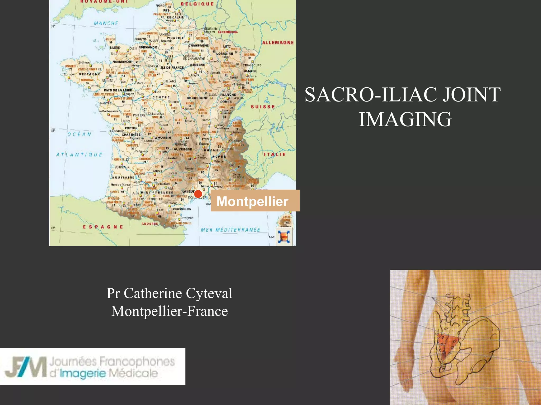
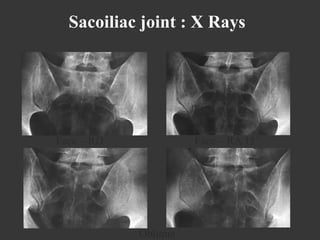
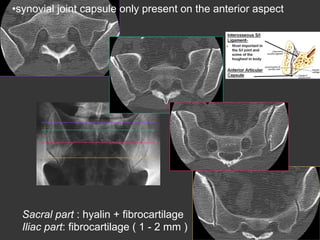
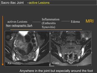
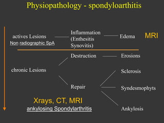
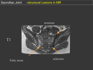
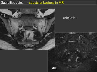
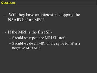
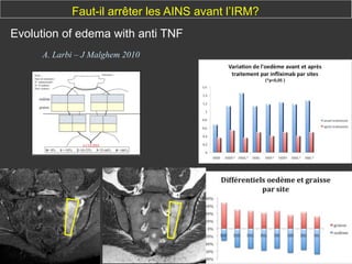
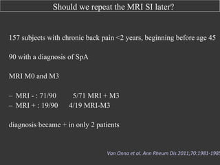
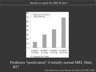
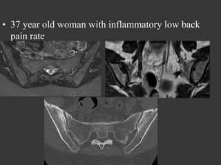
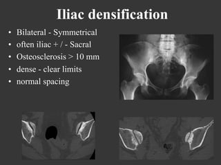
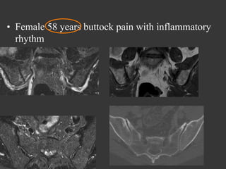
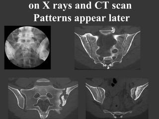
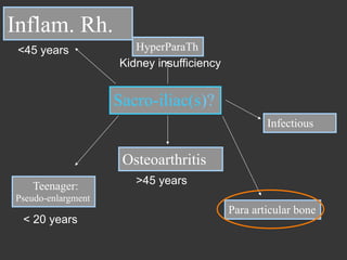
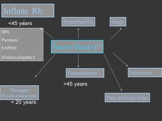
SACRO-ILIAC JOINT
IMAGING

Montpellier

Pr Catherine Cyteval
Montpellier-France
Diarthrodial joint

•  Size and shape of the human ear
•  Articular surface:S1-S3
•  « Key stone in an Arch »
Sacoiliac joint : X Rays

Face – RD

Face – RA 15°

Oblique
• synovial joint capsule only present on the anterior aspect

Sacral part : hyalin + fibrocartilage ( 2 - 5 mm )
Iliac part: fibrocartilage ( 1 - 2 mm )
Mr C
36 year-old
Left Coxalgia for two
months
Inflammatory syndroma
bilateral sacroiliitis
Physiopathology - spondyloarthitis

actives Lesions
Non radiographic SpA

Inflammation
(Enthesitis
Synovitis)

Edema

MRI
Sacro iliac Joint

–active Lesions

Non radiographic SpA

Edema

Destruction

actives Lesions

Inflammation
(Enthesitis
Synovitis)

Erosions

chronic Lesions

Sclerosis
Repair

Ax

MRI

Syndesmophyts

Xrays, CT, MRI

Coro
ankylosing Spondylarthritis

Ankylosis

Anywhere in the joint but especially around the foot
Physiopathology - spondyloarthitis

Non radiographic SpA

Edema

Destruction

actives Lesions

Inflammation
(Enthesitis
Synovitis)

Erosions

chronic Lesions

MRI

Sclerosis
Repair

Syndesmophyts

Xrays, CT, MRI
ankylosing Spondylarthritis

Ankylosis
Sacroiliac Joint

–structural Lesions in MR

erosions

T1

Fatty areas

sclerosis
Sacroiliac Joint

T1

–structural Lesions in MR

ankylosis
Sacroiliac Joint

–structural Lesions

X rays and CT only show chronic lesions

FORESTIER CLASSIFICATION

•  Stade I - Broaden joint
•  Stade II - stamps
•  Stade III - cloody

•  Stade IV - Fusion
Sacroiliac Joint

–structural Lesions

FORESTIER CLASSIFICATION

•  Stade I - Broaden joint
•  Stade II - stamps
•  Stade III - cloody

•  Stade IV - Fusion
Sacroiliac Joint

–structural Lesions

FORESTIER CLASSIFICATION

Stade I - Broaden joint
Stade II - stamps
Stade III - cloody
Stade IV - Fusion
Sacroiliac Joint

– Structural Lesions

FORESTIER CLASSIFICATION

•  Grade I - Pseudo – élargissement
•  Grade II - Timbre – poste
•  Grade III - Aspect marécageux

•  Grade IV - Fusion
SpA Diagnostic

Several diagnostic criteria based on
clinical and radiological signs

AMOR

ESSG
European
Spondylarthropathy
Study Group

New York
modifié

Existence of an X-ray, at least, sacroiliitis grade
2 bilaterally or grade 3 unilateral

ASAS
Assessment of
SpondyloArthritis
international
Society

Sacro-iliite IRM
ou radiographique
SPA Diagnostic

AMOR

ESSG
European
Spondylarthropathy
Study Group

New York
modifié

Existence of an X-ray, at least, sacroiliitis grade
2 bilaterally or grade 3 unilateral

ASAS
Assessment of
SpondyloArthritis
international
Society

MRI
(Or Xray) Sacroiliitis
2009 ASAS Criteria

axial spondyloarthritis
chronic Lombalgia and < 45 year-old
And
MRI or Xray
Sacroiliitis + 1 élément

arthritis
enthesitis
uveitis
dactylitis

or

HLA B27 +
+ 2 éléments

Psoriasis

AINS good Reponse

Enthérocolopathy

High CRP

family history

HLA B27
Our patient

axial spondyloarthritis
chronic Lombalgia and < 45 year-old
And
MRI or Xray
Sacroiliitis + 1 élément

arthritis
enthesitis
uveitis
dactylitis

or

HLA B27 +
+ 2 éléments

Psoriasis

AINS good Reponse

Enthérocolopathy

High CRP

family history

HLA B27
inflammatory lesions / ASAS criterias
sub chondral Œdema on 2 concecutive slices or 2
localisations on the same slice (STIR)

(Rudwaleit M et al. Ann. Rheum.Dis.2008)
ASAS 2009
ASAS 2009

enthesitis

Not in account

chronic Lesions

T1

sacro-tuberal Ligament
sacro-iliac interosseous Ligament
•  Aseptic Osteitis (SAPHO):
Unilateral
Massive, sacral and iliac sclerosis
Diagnostic value of MRI
Sensitivity ?
SI MR+ in 75% SpA patients
Weber	
  U	
  et	
  al	
  Eular	
  2013	
  OP	
  273	
  

Specificity ?
inflammatory Signal in 11% of the control patients
Aydin et al. Ann Rheum Dis 2012

Edema and fatty infiltration in 27% of the control patients
U Weber et al; Arthritis & Rheum; octobre 2010

retrospective study : 110

p. (28 SPA)

Abnormality in 21% of the control patients
C Cyteval Skeletal Radiology 2013
Questions

• 

Will they have an interest in stopping the
NSAID before MRI?

•  If the MRI is the first SI –  Should we repeat the MRI SI later?
–  Should we do an MRI of the spine (or after a
negative MRI SI)?
Faut-il arrêter les AINS avant l’IRM?
Evolution of edema with anti TNF
A. Larbi – J Malghem 2010
Should we stop NSAIDs before the MRI?

20 patients with SpA treated by NSAIDs (etoricoxib)
•  MRI lumbar spine and SI to S0 and S6 (20 patients)
•  15/20 patients (71%) had lesions on MRI S0 (63 lesions in total)
•  12/20 (60%) responders
• SI : S0 : 11 MRI + (25 lesions) à S6 : 9 MRI + (22 lesions)

• Spine: A S0 : 11 MRI + (38 lesions) à S6 : 9 MRI + (36 lesions)

Low impact of etoricoxib on inflammatory
lesions in MRI, even if clinical improvement
Jarre4	
  SJ	
  et	
  al.	
  Ann	
  Rheum	
  Dis	
  2009	
  
Should we stop NSAIDs before the MRI?

INFAST Study Design
Part I: Treatment Phase
Screening
/Washout

•
•


2:1 randomization
IV infusions: weeks 0, 2, 6, 12, 18,


  and 24

	
  Anti TNF+ AINS


  Placebo + AINS

M0


  MRI

105 p
51 p

M6


  MRI

ET=early termination; IFX=infliximab (anti TNF); IV=intravenous; NPX=Naproxen (AINS); PBO=placebo.
Should we stop NSAIDs before the MRI?

Characteristic

Gender,male

Anti TNF+
AINS

Anti TNF+
AINS

a

Placebo+
AINS

Placebo+
AINS

b

(N=105)
M0

M6

69%

(N=51)
M0

M6

78%

Age,years,mean(SD)

31.7(8.51)

30.7(7.34)

Years since symptom
onset,mean(SD)

1.76(0.896)

1.91(1.439)

Spine
SI joint
Spine or SI joint

59%
88%
91%

40%
72.4%
21.9%

Patients with readable MRIs and
active
Lesions ats creening

BASFI=Bath Ankylosing Spondylitis Functional Index; HLA=human leukocyte antigen.
aFor disease characteristics, N=106. bFor disease characteristics, N=52.

59%
90%
94%

54.9%
93.9%
100%
Should we stop NSAIDs before the MRI?

Seems that the response is NO
Should we repeat the MRI SI later?
–  68 patients with recent inflammatory back pain (38% men, age
34.9 ± 10.3 years)

–  MRI SI M0, M24
–  44 with negative MRI at baseline
◊ 15% became +
–  24 with positive MRI at baseline
◊ 30% became-

Van	
  den	
  Berg	
  ACR2012	
  (SPACE)	
  
Should we repeat the MRI SI later?

157 subjects with chronic back pain <2 years, beginning before age 45
90 with a diagnosis of SpA
MRI M0 and M3
–  MRI - : 71/90
–  MRI + : 19/90

5/71 MRI + M3
4/19 MRI-M3

diagnosis became + in only 2 patients

Van	
  Onna	
  et	
  al.	
  Ann	
  Rheum	
  Dis	
  2011;70:1981-­‐1985
Should we repeat the MRI SI later?

Downloaded from ard.bmj.com on April 15, 2013 - Published by group.bmj.com

Extended report

ccording to the ASAS/OMERACT MRI
tients with early inflammatory low back
SpAC

1 Year

2 Years

HLA-B27 positive

+
+
–
+
–
NA
NA
NA
–
–
–
+
+
+
NA

+
NA
+
–
–
+
–
NA
–
+
NA
+
–
NA
NA

8
4
2
0
0
1
0
1
6
0
3
1
0
2
3

s Society; ESpAC, Early Spondyloarthritis Cohort;
7; NA, not available; OMERACT, Outcome
cal Trials.

d either one or two follow-up MRI.
5%), the MRI became positive at 1
HLA-B27-positive patient of these
ed positive at 2 years follow-up, in
nt the MRI became negative again
wo HLA-B27-negative patients the
ars follow-up and in two HLA-B27-

Figure 1 Likelihood of a positive MRI at any time point in patients with
short-standing inflammatory back pain assessed at baseline, 1 year and
2 years of follow-up in function of HLA-B27 status and gender. HLA-B27,
human leucocyte antigen B27.

Predictors "positivation" if initially normal MRI: Male,
B27

The likelihood of a positiveVan	
  Onna	
  et	
  al.	
  Ain the case
MRI is negligible (<5%) nn	
  Rheum	
  Dis	
  2011;70:1981-­‐1985	
  	
  
Should we repeat the MRI SI later?

Seems that the response is NO for female
Possible for male
Should make sacroiliac and / or spine MRI?
Patients avec atteinte axiale
symptomatique(n=362)

SA axiale nonradiologique (n = 160)

SA axiale radiologique =
SA (n = 202)

43,8 %

63,5 %

36,3 (10,3)

39,1 (11,3)

Duration of the ilness (ans) [DS]

5,7 (6,8)

11,2 (10,0)

BASDAI (0-10) [DS]

4,1 (2,0)

4,3 (2,0)

78,0 %

86,9 %

Males
Âge (ans) [DS]

HLA B27+

%	
  
90	
  
80	
  
70	
  
60	
  
50	
  
40	
  
30	
  
20	
  
10	
  
0	
  

p	
  =	
  0,978	
  
77,8	
  %	
  
76,7	
  %	
  
(112/144)	
   (132/172)	
  

No	
  X	
  Rays	
  sign	
  of	
  sacroilii's	
  
p	
  =	
  0,004	
  
56,3	
  %	
  
(54/96)	
  
28,8	
  %	
  
(17/59)	
  

MRI	
  ac've	
  sacroilii's	
  

Active MRI inflammatory
lesions spinal

Sacroilii's	
  seen	
  on	
  X	
  Rays	
  
p	
  =	
  0,569	
  
6,8	
  %	
  
(3/44)	
  

10,0	
  %	
  
(6/60)	
  

Active MRI inflammatory lesions
spinal (without sacroiliitis)

Whatever the clinical, sacroiliac MRI seems to be the most profitable review. Only
6.8% of non-radiological axial SA have isolated spinal inflammatory lesions
without sacroiliitis
Rudwaleit-­‐	
  Song	
  -­‐ACR	
  2010	
  -­‐	
  (519)	
  
Should make sacroiliac and / or spine MRI?

•  Reading MRI of the spine in addition to the SI increases the number of nonradiographic forms that are ultimately recognized positive MRI compared to
reading only SI
Sog IH et al. Arthritis Rheum 2008

•  It increases quite significantly the number of false positives (mechanical or
healthy)
•  In addition it allows to diagnose other causes of lumbar pain
Protocole
•  Sacro iliac joint :
–  coronal T1- STIR
–  Axial STIR

•  Spine from T7 to L5:
–  Sagittal STIR (or Fat sat T2)
•  37 year old woman with inflammatory low back
pain rate
Iliac densification
• 
• 
• 
• 
• 

Bilateral - Symmetrical
often iliac + / - Sacral
Osteosclerosis > 10 mm
dense - clear limits
normal spacing
•  Female 58 years buttock pain with inflammatory
rhythm
Osteoarthritis
• 
• 
• 
• 

Multiple pregnancies.. Elderly… Weight
Location middle 1/3 , anterior
Common - Bilateral asymmetric - Spurs
Check the pubic symphysis
upper1/3

Front

Middle

Middle 1/3

Back
Inf 1/3
Hyperostosis
Inflam. Rh.
SPA
Psoriasis
SAPHO

Sacro-iliac(s)?

(Entérocolopathy)

Osteoarthritis
17 years old male with low back pain
Inflam. Rh.
<45 y ears

HyperParaTh

drugs

Sacro-iliac(s)?
Osteoarthritis
Teenager:

>45 years

Pseudo-enlargment

< 20 years
When growth has not finished

Infectious
•  67 years old woman with Kidney
insufficiency for 17 years
2rd hyperparathyroidism
Inflam. Rh.
HyperParaTh
SPA
Psoriasis
SAPHO

Sacro-iliac(s)?

(Entérocolopathy)

Osteoarthritis
Teenager:
Pseudo-enlargment
• 
• 
• 
• 
• 

C Noémie
24 year-old
A week after delivery
Inflammatory left buttock pain
infl Sd ++++
C.Noémie

sept 2011

oct 2011
infectious Sacroiliitis staph
Effusion
Unilateral

infectious Sacroiliitis
Infectious sacro-iliitis
•  Unilateral
•  Soft tissus +++
•  Effusion
on X rays and CT scan
Patterns appear later

1
Inflam. Rh.
<45 years

HyperParaTh
Kidney insufficiency

Sacro-iliac(s)?
Infectious

Osteoarthritis
Teenager:
Pseudo-enlargment

< 20 years

>45 years
26 year-old female

fracture
Inflam. Rh.
<45 years

HyperParaTh
Kidney insufficiency

Sacro-iliac(s)?
Infectious

Osteoarthritis
Teenager:
Pseudo-enlargment

< 20 years

>45 years
Para articular bone
Para articular Decalcification

Hemochromatosis
Inflam. Rh.
<45 years

HyperParaTh
Kidney insufficiency

Sacro-iliac(s)?
Infectious

Osteoarthritis
Teenager:
Pseudo-enlargment

< 20 years

>45 years
Para articular bone
Mr B., 20 years old
Inflammatory Low back pain
?
Inflam. Rh.
HyperParaTh
SPA
Psoriasis
SAPHO

Sacro-iliac(s)?

(Entérocolopathy)

Osteoarthritis

Infectious

Teenager:
Pseudo-enlargment

Para articular bone
Inflam. Rh.
HyperParaTh
SPA
Psoriasis
SAPHO

Sacro-iliac(s)?

(Entérocolopathy)

Osteoarthritis

Infectious

Teenager:
Pseudo-enlargment

Para articular bone
SPA

?

Mr B
Inflam. Rh.
HyperParaTh
SPA
Psoriasis
SAPHO

Sacro-iliac(s)?

(Entérocolopathy)

Osteoarthritis

Infectious

Teenager:
Pseudo-enlargment

Para articular bone
Roaccutane (isotrétinoïne):
8 months in 2007 and 4 months in
2010 !!!
Isotrétinoïne and bones
•  Sacroiliitis : seldom
*E. Eksioglu et al., Sacroiliitis and polyneuropathy during isotrtinoin treatment 2007
*Elias et al.,Acne fulminans and bilateral seronegative sacroiliitis triggered by isotretinoin 1991
*Bachmeyer et al., Isotretinoin induced bilateral sacroiliitis 2003
•  Diffuse Idiopathic Skeletal Hyperostosis like
Hyperostosis and calcification of tendons and ligaments
Bone bridges along the anterior longitudinal ligament (6
vertebrae at least)
*J. DiGiovanna et al., Isotretinoin effects on bone
Mr S. (26 ans)
Roaccutane for 7 years
Inflam. Rh.
<45 years

HyperParaTh

drugs

SPA
Psoriasis
SAPHO

Sacro-iliac(s)?

(Entérocolopathy)

Osteoarthritis

Infectious

>45 years
Teenager:
Pseudo-enlargment

< 20 years

Para articular bone
Traps
•  Accessory Sacroiliac Joint

Thank you

Msk imaging sacro iliac c cyteval