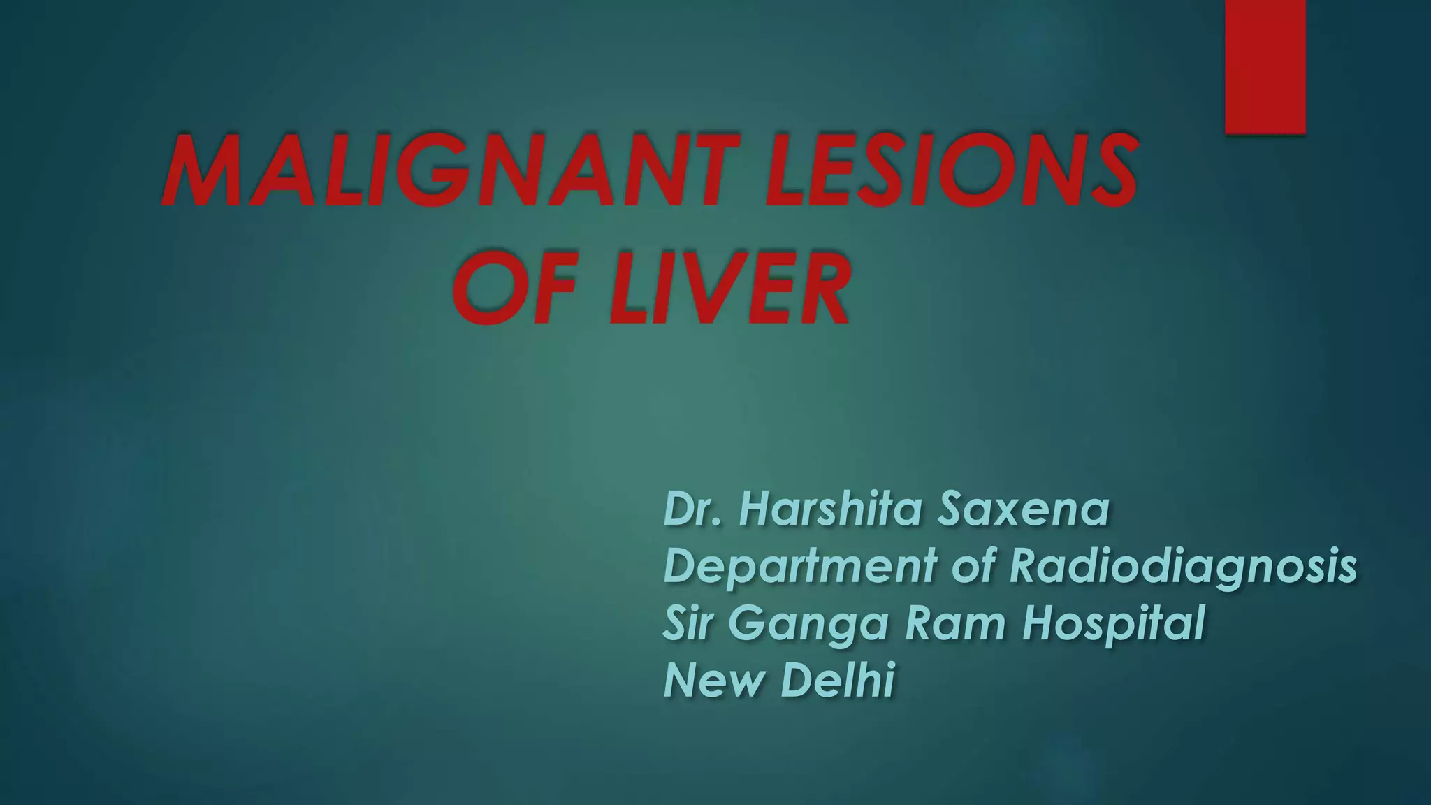
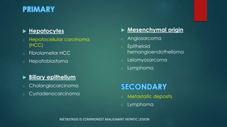
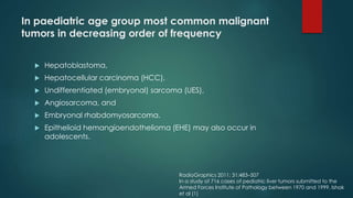
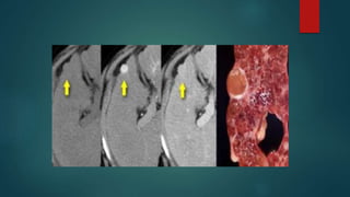
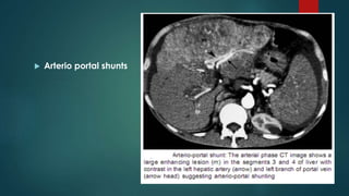
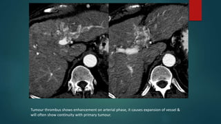
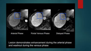
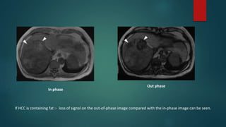
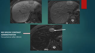
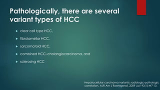
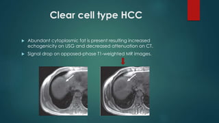
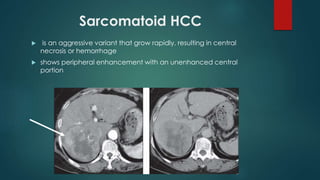
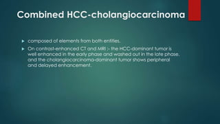
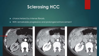
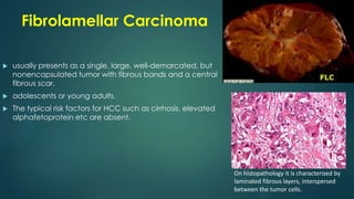
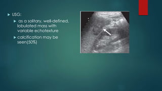
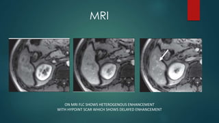
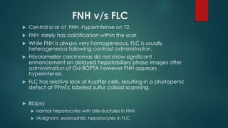
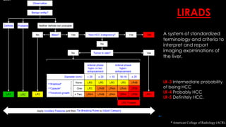
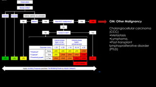
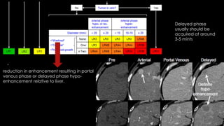
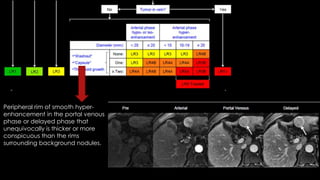
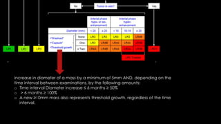
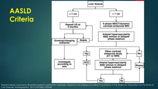
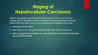
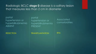
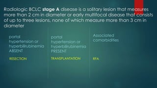
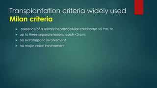
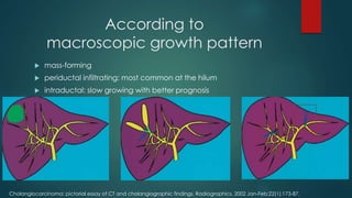
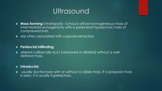
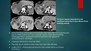
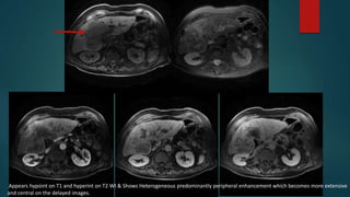
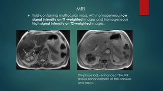
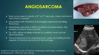
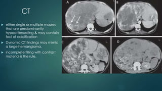
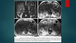
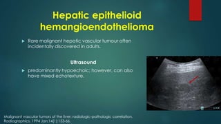
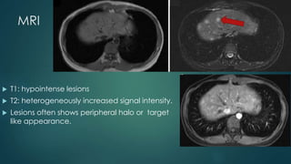
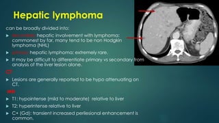
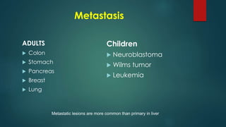
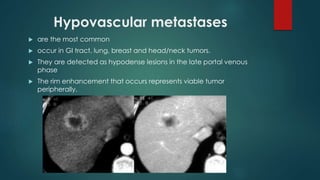
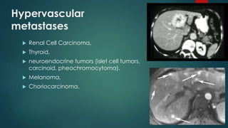
SlideShare a Scribd company logo
MALIGNANT LESIONS
OF LIVER
Dr. Harshita Saxena
Department of Radiodiagnosis
Sir Ganga Ram Hospital
New Delhi
PRIMARY
 Hepatocytes
o Hepatocellular carcinoma
(HCC)
o Fibrolamellar HCC
o Hepatoblastoma
 Biliary epithelium
o Cholangiocarcinoma
o Cystadenocarcinoma
 Mesenchymal origin
o Angiosarcoma
o Epitheloid
hemangioendothelioma
o Leiomyosarcoma
o Lymphoma
SECONDARY
o Metastatic deposits
o Lymphoma
METASTASIS IS COMMONEST MALIGNANT HEPATIC LESION
In paediatric age group most common malignant
tumors in decreasing order of frequency
 Hepatoblastoma,
 Hepatocellular carcinoma (HCC),
 Undifferentiated (embryonal) sarcoma (UES),
 Angiosarcoma, and
 Embryonal rhabdomyosarcoma.
 Epithelioid hemangioendothelioma (EHE) may also occur in
adolescents.
RadioGraphics 2011; 31:483–507
In a study of 716 cases of pediatric liver tumors submitted to the
Armed Forces Institute of Pathology between 1970 and 1999, Ishak
et al (1)
Hepatocellular Carcinoma
 Most common primary malignancy of the liver
 Male : female > 4 : 1
 Rising incidence, attributed to a rise in hepatitis B and C infection
Risk factors
 hepatitis B (HBV) infection
 hepatitis C (HCV) infection
 alcoholism
 biliary cirrhosis
 food toxins e.g. aflatoxins
 congenital biliary atresia
 inborn errors of metabolism
haemochromatosis
alpha-1 antitrypsin deficiency
type 1 glycogen storage disease
Wilson disease
Clinical presentation
 HCC are typically diagnosed in adults in late middle age or elderly
 Patients with cirrhosis usually present earlier.
 less aggressive - pyrexia of unknown origin, abdominal pain,
malaise, weight loss, and hepatomegaly. Jaundice is rare.
 Aggressive - hemoperitoneum.
Investigation
 alpha-fetoprotein (AFP) levels are elevated in 50-75 % of cases
 Radiological investigation including ultrasound, CT and MRI
 Biopsy
USG
• Variable appearance
• Small (<3cm) usually
hypoechoic
• Larger tumors often are
heterogeneous
• May invade the portal vein
• Most tumors will show central
vascularity on Doppler study.
CT
 Focal calcification -7.5%
 Majority – hyperdense on
arterial phase, as HCCs take
supply from the hepatic artery
rather than portal vein
 Heterogenous enhancement
due to central necrosis
 Arterio portal shunts
Tumour thrombus shows enhancement on arterial phase, it causes expansion of vessel &
will often show continuity with primary tumour.
In phase
Out phase
If HCC is containing fat :- loss of signal on the out-of-phase image compared with the in-phase image can be seen.
T1 T2
SPIO
RES SPECIFIC CONTRAST
ADMINISTRATION
Ferucarbotran (after 20min)
Pathologically, there are several
variant types of HCC
 clear cell type HCC,
 fibrolamellar HCC,
 sarcomatoid HCC,
 combined HCC–cholangiocarcinoma, and
 sclerosing HCC
Hepatocellular carcinoma variants: radiologic-pathologic
correlation. AJR Am J Roentgenol. 2009 Jul;193(1):W7-13.
Clear cell type HCC
 Abundant cytoplasmic fat is present resulting increased
echogenicity on USG and decreased attenuation on CT.
 Signal drop on opposed-phase T1-weighted MR images.
Sarcomatoid HCC
 is an aggressive variant that grow rapidly, resulting in central
necrosis or hemorrhage
 shows peripheral enhancement with an unenhanced central
portion
Combined HCC-cholangiocarcinoma
 composed of elements from both entities.
 On contrast-enhanced CT and MRI :- the HCC-dominant tumor is
well enhanced in the early phase and washed out in the late phase,
and the cholangiocarcinoma-dominant tumor shows peripheral
and delayed enhancement.
Sclerosing HCC
 characterized by intense fibrosis.
 With remarkable progressive and prolonged enhancement
Fibrolamellar Carcinoma
 usually presents as a single, large, well-demarcated, but
nonencapsulated tumor with fibrous bands and a central
fibrous scar.
 adolescents or young adults.
 The typical risk factors for HCC such as cirrhosis, elevated
alphafetoprotein etc are absent.
On histopathology it is characterized by
laminated fibrous layers, interspersed
between the tumor cells.
 USG:
 as a solitary, well-defined,
lobulated mass with
variable echotexture
 calcification may be
seen(50%)
CT:
 Hypodense mass
 Central scar – Calcification
 Predominant heterogenous enhancement
MRI
ON MRI FLC SHOWS HETEROGENOUS ENHANCEMENT
WITH HYPOINT SCAR WHICH SHOWS DELAYED ENHANCEMENT
FNH v/s FLC
 Central scar of FNH -hyperintense on T2.
 FNH rarely has calcification within the scar.
 While FNH is always very homogeneous, FLC is usually
heterogeneous following contrast administration.
 Fibrolamellar carcinomas do not show significant
enhancement on delayed hepatobiliary phase images after
administration of Gd-BOPTA however FNH appears
hyperintense.
 FLC has relative lack of Kupffer cells, resulting in a photopenic
defect at 99mTc labeled sulfur colloid scanning
 Biopsy
 normal hepatocytes with bile ductules in FNH
 Malignant, eosinophilic hepatocytes in FLC
* American College of Radiology (ACR).
LIRADS
A system of standardized
terminology and criteria to
interpret and report
imaging examinations of
the liver.
LR-3 Intermediate probability
of being HCC
LR-4 Probably HCC
LR-5 Definitely HCC.
OM: Other Malignancy
Cholangiocellular carcinoma
(CCC)
•Metastasis
•Lymphoma,
•Post-transplant
lymphoproliferative disorder
(PTLD)
LR5V: Definitely HCC with
Tumor in vein
The term tumor in vein is
preferred over the term tumor
thrombus
reduction in enhancement resulting in portal
venous phase or delayed phase hypo-
enhancement relative to liver.
Delayed phase
usually should be
acquired at around
3-5 mints
Peripheral rim of smooth hyper-
enhancement in the portal venous
phase or delayed phase that
unequivocally is thicker or more
conspicuous than the rims
surrounding background nodules.
increase in diameter of a mass by a minimum of 5mm AND, depending on the
time interval between examinations, by the following amounts:
o Time interval Diameter increase ≤ 6 months ≥ 50%
o > 6 months ≥ 100%
o A new ≥10mm mass also represents threshold growth, regardless of the time
interval.
AASLD
Criteria
Hepatocellular carcinoma: illustrated guide to systematic radiologic diagnosis and staging according to guidelines of the American Association for the Study of
Liver Diseases. Radiographics. 2013 Oct;33(6):1653-68.
Staging of
Hepatocellular Carcinoma
AASLD advocates use of the BCLC (Barcelona Clinic Liver Cancer )
staging system because it is the only system that encompasses the three
factors that have been shown to be independent predictors of survival :-
 radiologic tumor extent,
 liver function { by using the Child-Turcotte-Pugh (CTP) score} and
 patient’s performance status { by using the Eastern Cooperative Oncology
Group (ECOG) scale}
Radiologic BCLC stage 0 disease is a solitary lesion
that measures less than 2 cm in diameter
portal
hypertension or
hyperbilirubinemia
ABSENT
RESECTION
portal
hypertension or
hyperbilirubinemia
PRESENT
TRANSPLANTATION
Associated
comorbidities
RFA
Radiologic BCLC stage A disease is a solitary lesion that measures
more than 2 cm in diameter or early multifocal disease that consists
of up to three lesions, none of which measure more than 3 cm in
diameter
portal
hypertension or
hyperbilirubinemia
ABSENT
RESECTION
portal
hypertension or
hyperbilirubinemia
PRESENT
TRANSPLANTATION
Associated
comorbidities
RFA
Radiologic BCLC stage B disease is advanced
multifocal disease that consists of more than one
lesion, with at least one that is larger than 3 cm, or
of more than three lesions regardless of size
TACE
Radiologic BCLC stage C disease is
hepatocellular carcinoma with either vascular
invasion or nodal or metastatic disease.
SORAFENIB
BCLC stage D disease is not a radiologic stage. It
is determined only on the basis of poor liver
function and poor patient performance (CTP = C,
ECOG > 2).
Management consist of only supportive therapies
Transplantation criteria widely used
Milan criteria
 presence of a solitary hepatocellular carcinoma <5 cm, or
 up to three separate lesions, each <3 cm.
 no extrahepatic involvement
 no major vessel involvement
HEPATOBLASTOMA
 Most common primary liver tumor in childhood.
 Usually occurs in first 3 yrs of life.
 Present as painless abdominal mass, anorexia, wt loss, pain &
jaundice.
 S.AFP is usually elevated.
 Usually unifocal but multiple nodules or diffuse liver
involvement may be seen. calcification in about 33%.
 Epithelial hepatoblastomas demonstrate a more
homogeneous appearance, while mixed tumors are more
heterogeneous in attenuation.
USG
 large, inhomogeneous echogenic mass sometime
with calcification.
CT
 well defined heterogeneous
hypodense mass. Frequently
with areas of necrosis,
haemorrhage and
calcification.
 Like HCC, hepatoblastoma
can invade the portal or
hepatic veins
Arterial neovascularity can be seen with washout
on delayed scans.
MRI:
 T1: generally hypointense
 C+ (Gd): can show heterogeneous enhancement
 T2 : generally hyperintense compared to liver
 areas of necrosis and haemorrhage are common
Cholangiocarcinoma
 second most common primary hepatic tumour
 usually in the elderly (7th decade).
 There is slight male predilection.
 Elevated tumor markers: CA19-9, CEA
Risk factors • primary sclerosing cholangitis (PSC)
• recurrent pyogenic cholangitis
• liver flukes
• Opisthorchis viverrini
• Clonorchis sinensis (clonorchiasis)
• Caroli's disease/choledochal cysts
• toxins
• thorotrast
• dioxin
• polyvinyl chloride
• heavy alcohol use
• viral infection(s)
• HIV
• hepatitis B and hepatitis C
• EBV
According to
macroscopic growth pattern
 mass-forming
 periductal infiltrating: most common at the hilum
 intraductal: slow growing with better prognosis
Cholangiocarcinoma: pictorial essay of CT and cholangiographic findings. Radiographics. 2002 Jan-Feb;22(1):173-87.
Ultrasound
 Mass-forming intrahepatic: tumours will be homogeneous mass of
intermediate echogenicity with a peripheral hypoechoic halo of
compressed liver.
 are often associated with capsular retraction
 Periductal infiltrating:
 altered calibre bile duct (narrowed or dilated) without a well-
defined mass.
 Intraductal:
 usually ductectasia with or without a visible mass. If a polypoid mass
is seen, it is usually hyperechoic.
 Mass-forming cholangiocarcinomas: are typically homogeneously
low in attenuation on non-contrast scans, and demonstrate
heterogeneous peripheral enhancement with gradual
enhancement centrally.
 capsular retraction may be seen.
 The bile ducts distal to the mass are typically dilated.
 unlike HCC, cholangiocarcinoma only rarely forms a tumour
thrombus .
The lesion appears hyperdense in the
equilibrium phase due to dens fibrous tissue
retaining contrast
Appears hypoint on T1 and hyperint on T2 WI & Shows Heterogeneous predominantly peripheral enhancement which becomes more extensive
and central on the delayed images.
Cystadenocarcinoma
 rare, usually slow growing, multilocular cystic tumors
 in middle-aged women
 Symptoms are related to the mass effect of the
lesion and consist of intermittent pain or biliary
obstruction.
Imaging features
 USG & CT: it appears as a solitary cystic mass with a
well-defined thick fibrous capsule, mural nodules,
internal septa, and rarely capsular calcification.
 Polypoid, pedunculated excrescences
MRI
 fluid-containing multilocular mass, with homogeneous low
signal intensity on T1-weighted images and homogeneous
high signal intensity on T2-weighted images.
PV-phase Gd - enhanced T1w MR
shows enhancement of the capsule
and septa.
ANGIOSARCOMA
 Rare tumor seen in adults of 5th to 7th decade, more common in
men than in women.
 associated with chemical carcinogens exposure including
Thorotrast.
 Metastases are seen in 60% of patients at presentation, the
median survival is only 6 months.
 On USG, either multiple nodules or a solitary mass can be
demonstrated.
 The echotexture of an angiosarcoma varies according to the
degree of internal necrosis and hemorrhage.
Malignant vascular tumors of the liver: radiologic-pathologic correlation.
Radiographics. 1994 Jan;14(1):153-66.
CT
 either single or multiple masses
that are predominantly
hypoattenuating & may contain
foci of calcification
 Dynamic CT findings may mimic
a large hemangioma,
 incomplete filling with contrast
material is the rule.
Hepatic epithelioid
hemangioendothelioma
 Rare malignant hepatic vascular tumour often
incidentally discovered in adults.
Ultrasound
 predominantly hypoechoic; however, can also
have mixed echotexture.
Malignant vascular tumors of the liver: radiologic-pathologic correlation.
Radiographics. 1994 Jan;14(1):153-66.
CT
 Typically seen as multiple hypo-attenuating lesions that
coalesce to form larger confluent hypo-attenuating regions
in a peripheral or subcapsular distribution with peripheral and
progressive centripetal enhancement in larger lesions with
incomplete fill-in on delayed phase images.
 Subcapsular lesion often present with capsular retraction.
MRI
 T1: hypointense lesions
 T2: heterogeneously increased signal intensity.
 Lesions often shows peripheral halo or target
like appearance.
Hepatic lymphoma
can be broadly divided into:
 secondary hepatic involvement with lymphoma:
commonest by far, many tend to be non Hodgkin
lymphoma (NHL)
 primary hepatic lymphoma: extremely rare.
 It may be difficult to differentiate primary vs secondary from
analysis of the liver lesion alone.
CT
 Lesions are generally reported to be hypo attenuating on
CT.
MRI
 T1: hypointense (mild to moderate) relative to liver
 T2: hyperintense relative to liver
 C+ (Gd): transient increased perilesional enhancement is
common.
Metastasis
ADULTS
 Colon
 Stomach
 Pancreas
 Breast
 Lung
Children
 Neuroblastoma
 Wilms tumor
 Leukemia
Metastatic lesions are more common than primary in liver
Hypovascular metastases
 are the most common
 occur in GI tract, lung, breast and head/neck tumors.
 They are detected as hypodense lesions in the late portal venous
phase
 The rim enhancement that occurs represents viable tumor
peripherally.
Hypervascular
metastases
 Renal Cell Carcinoma,
 Thyroid,
 neuroendocrine tumors (islet cell tumors,
carcinoid, pheochromocytoma).
 Melanoma,
 Choriocarcinoma.
Hemorrhagic Liver Metastases
 Colon
 Thyroid
 Breast
 Choriocarcinoma
 Melanoma
 RCC
Metastatic tumor with internal fluid-fluid level (arrow)
suggesting intratumoral hemorrhage.
Published by the University of Washington in the public interest. ISSN
1930-0433
Calcified metastases
 Mucinous CA of GI tract (colon, stomach,
rectum),
 Melanoma
 Ovarian ca.
Cystic metastases
 Mucinous ovarian ca,
 Colonic ca
Main References
 Buetow PC, Buck JL, Ros PR, Goodman ZD. Malignant vascular tumors of the
liver: radiologic-pathologic correlation. Radiographics. 1994 Jan;14(1):153-66.
 McEvoy SH, McCarthy CJ, Lavelle LP, Moran DE, Cantwell CP, Skehan SJ et al.
Hepatocellular carcinoma: illustrated guide to systematic radiologic diagnosis
and staging according to guidelines of the American Association for the
Study of Liver Diseases. Radiographics. 2013 Oct;33(6):1653-68.
 Chung EM, Lattin GE Jr, Cube R, Lewis RB, Marichal-Hernández C, Shawhan R,
Conran RM. From the archives of the AFIP: Pediatric liver masses: radiologic-
pathologic correlation. Part 2. Malignant tumors. Radiographics. 2011 Mar-
Apr;31(2):483-507.
 Chung YE, Park MS, Park YN, Lee HJ, Seok JY, Yu JS, Kim MJ. Hepatocellular
carcinoma variants: radiologic-pathologic correlation. AJR Am J Roentgenol.
2009 Jul;193(1):W7-13.
 McEvoy SH, McCarthy CJ, Lavelle LP, Moran DE, Cantwell CP, Skehan SJ et al.
Hepatocellular carcinoma: illustrated guide to systematic radiologic diagnosis
and staging according to guidelines of the American Association for the
Study of Liver Diseases. Radiographics. 2013 Oct;33(6):1653-68.
 Han JK, Choi BI, Kim AY, An SK, Lee JW, Kim TK et al. Cholangiocarcinoma:
pictorial essay of CT and cholangiographic findings. Radiographics. 2002 Jan-
Feb;22(1):173-87.
 Liver Masses - Common Tumors. Richard Baron. Radiology Assistant.

More Related Content

What's hot

Presentation1.pptx, radiological imaging of cholangiocarcinoma.
Presentation1.pptx, radiological imaging of cholangiocarcinoma.Presentation1.pptx, radiological imaging of cholangiocarcinoma.
Presentation1.pptx, radiological imaging of cholangiocarcinoma.Abdellah Nazeer
 
CT and MRI Imaging of Hepatic metastases
CT and MRI Imaging of Hepatic metastasesCT and MRI Imaging of Hepatic metastases
CT and MRI Imaging of Hepatic metastases
Dr Priyanka Vishwakarma
 
Liver lesions
Liver lesionsLiver lesions
Liver lesionsairwave12
 
Presentation1, radiological imaging of gastro intestinal stromal tumour(gist).
Presentation1, radiological imaging of gastro intestinal stromal tumour(gist).Presentation1, radiological imaging of gastro intestinal stromal tumour(gist).
Presentation1, radiological imaging of gastro intestinal stromal tumour(gist).
Abdellah Nazeer
 
Gastric carcinoma radiology ppt
Gastric carcinoma radiology  ppt Gastric carcinoma radiology  ppt
Gastric carcinoma radiology ppt
Sidharthan Selvaraju
 
Focal liver lesions- in the eye of a radiologist
Focal liver lesions- in the eye of a radiologistFocal liver lesions- in the eye of a radiologist
Focal liver lesions- in the eye of a radiologist
Dr.Santosh Atreya
 
Liver lesions SYMPOSIUM RADIOLOGY
Liver lesions SYMPOSIUM RADIOLOGYLiver lesions SYMPOSIUM RADIOLOGY
Liver lesions SYMPOSIUM RADIOLOGY
Satyendra Raghuwanshi
 
Imaging of Large Bowel Polyp
Imaging of Large Bowel PolypImaging of Large Bowel Polyp
Imaging of Large Bowel Polyp
Dr. Yash Kumar Achantani
 
Radiology spotters
Radiology spottersRadiology spotters
Radiology spotters
priyanka rana
 
Imaging in small bowel tumors Dr. Muhammad Bin Zulfiqar
Imaging in small bowel tumors Dr. Muhammad Bin ZulfiqarImaging in small bowel tumors Dr. Muhammad Bin Zulfiqar
Imaging in small bowel tumors Dr. Muhammad Bin Zulfiqar
Dr. Muhammad Bin Zulfiqar
 
Cystic hepatic lesions
Cystic hepatic lesionsCystic hepatic lesions
Cystic hepatic lesions
Dr Varun Bansal
 
Imaging of portal hypertension
Imaging of portal hypertensionImaging of portal hypertension
Imaging of portal hypertension
Dev Lakhera
 
Radioanatomy of mediastinum and approach to mediastinal masses
Radioanatomy of mediastinum and approach to mediastinal massesRadioanatomy of mediastinum and approach to mediastinal masses
Radioanatomy of mediastinum and approach to mediastinal masses
AkankshaMalviya3
 
INFLAMMATORY BOWEL DISEASE IMAGING(RADIOLOGY)
INFLAMMATORY BOWEL DISEASE IMAGING(RADIOLOGY)INFLAMMATORY BOWEL DISEASE IMAGING(RADIOLOGY)
INFLAMMATORY BOWEL DISEASE IMAGING(RADIOLOGY)
Khyati Vadera
 
Thyroid us
Thyroid usThyroid us
Thyroid us
Wafik Ebrahim
 
Prostate carcinoma raiology
Prostate carcinoma raiologyProstate carcinoma raiology
Prostate carcinoma raiology
Dr. Mohit Goel
 
Imaging in Pediatric Retroperitoneal Masses
Imaging in Pediatric Retroperitoneal MassesImaging in Pediatric Retroperitoneal Masses
Imaging in Pediatric Retroperitoneal Masses
Dr.Suhas Basavaiah
 
CT Imaging of CA Esophagus
CT Imaging of CA EsophagusCT Imaging of CA Esophagus
CT Imaging of CA Esophagus
Dr. Yash Kumar Achantani
 

What's hot (20)

Presentation1.pptx, radiological imaging of cholangiocarcinoma.
Presentation1.pptx, radiological imaging of cholangiocarcinoma.Presentation1.pptx, radiological imaging of cholangiocarcinoma.
Presentation1.pptx, radiological imaging of cholangiocarcinoma.
 
CT and MRI Imaging of Hepatic metastases
CT and MRI Imaging of Hepatic metastasesCT and MRI Imaging of Hepatic metastases
CT and MRI Imaging of Hepatic metastases
 
Liver lesions
Liver lesionsLiver lesions
Liver lesions
 
Presentation1, radiological imaging of gastro intestinal stromal tumour(gist).
Presentation1, radiological imaging of gastro intestinal stromal tumour(gist).Presentation1, radiological imaging of gastro intestinal stromal tumour(gist).
Presentation1, radiological imaging of gastro intestinal stromal tumour(gist).
 
Gastric carcinoma radiology ppt
Gastric carcinoma radiology  ppt Gastric carcinoma radiology  ppt
Gastric carcinoma radiology ppt
 
Focal liver lesions- in the eye of a radiologist
Focal liver lesions- in the eye of a radiologistFocal liver lesions- in the eye of a radiologist
Focal liver lesions- in the eye of a radiologist
 
Liver lesions SYMPOSIUM RADIOLOGY
Liver lesions SYMPOSIUM RADIOLOGYLiver lesions SYMPOSIUM RADIOLOGY
Liver lesions SYMPOSIUM RADIOLOGY
 
Imaging of Large Bowel Polyp
Imaging of Large Bowel PolypImaging of Large Bowel Polyp
Imaging of Large Bowel Polyp
 
Radiology spotters
Radiology spottersRadiology spotters
Radiology spotters
 
Imaging in small bowel tumors Dr. Muhammad Bin Zulfiqar
Imaging in small bowel tumors Dr. Muhammad Bin ZulfiqarImaging in small bowel tumors Dr. Muhammad Bin Zulfiqar
Imaging in small bowel tumors Dr. Muhammad Bin Zulfiqar
 
Focal liver lesion
Focal liver lesionFocal liver lesion
Focal liver lesion
 
Hepatic imaging
Hepatic imagingHepatic imaging
Hepatic imaging
 
Cystic hepatic lesions
Cystic hepatic lesionsCystic hepatic lesions
Cystic hepatic lesions
 
Imaging of portal hypertension
Imaging of portal hypertensionImaging of portal hypertension
Imaging of portal hypertension
 
Radioanatomy of mediastinum and approach to mediastinal masses
Radioanatomy of mediastinum and approach to mediastinal massesRadioanatomy of mediastinum and approach to mediastinal masses
Radioanatomy of mediastinum and approach to mediastinal masses
 
INFLAMMATORY BOWEL DISEASE IMAGING(RADIOLOGY)
INFLAMMATORY BOWEL DISEASE IMAGING(RADIOLOGY)INFLAMMATORY BOWEL DISEASE IMAGING(RADIOLOGY)
INFLAMMATORY BOWEL DISEASE IMAGING(RADIOLOGY)
 
Thyroid us
Thyroid usThyroid us
Thyroid us
 
Prostate carcinoma raiology
Prostate carcinoma raiologyProstate carcinoma raiology
Prostate carcinoma raiology
 
Imaging in Pediatric Retroperitoneal Masses
Imaging in Pediatric Retroperitoneal MassesImaging in Pediatric Retroperitoneal Masses
Imaging in Pediatric Retroperitoneal Masses
 
CT Imaging of CA Esophagus
CT Imaging of CA EsophagusCT Imaging of CA Esophagus
CT Imaging of CA Esophagus
 

Viewers also liked

liver mass - how to investigate?
liver mass - how to investigate?liver mass - how to investigate?
liver mass - how to investigate?
hr77
 
Abdominal imaging hcc t loke
Abdominal imaging hcc t lokeAbdominal imaging hcc t loke
Abdominal imaging hcc t lokeJFIM
 
MR imaging of liver
MR imaging of liverMR imaging of liver
MR imaging of liver
Parvathy Nair
 
Hepatocellular carcinoma
Hepatocellular carcinomaHepatocellular carcinoma
Hepatocellular carcinoma
yinnshang
 
4.11.15 GI Symposium
4.11.15 GI Symposium4.11.15 GI Symposium
4.11.15 GI SymposiumBeth Lynn
 
Magnetic resonance imaging of focal hepatic lesions
Magnetic resonance imaging of focal hepatic lesionsMagnetic resonance imaging of focal hepatic lesions
Magnetic resonance imaging of focal hepatic lesions
Mozammil Rabbani
 
Dr. masciotra liver shear wave elastography clinical cases
Dr. masciotra liver shear wave elastography clinical casesDr. masciotra liver shear wave elastography clinical cases
Dr. masciotra liver shear wave elastography clinical cases
antonio pio masciotra
 
Focal hepatic lesions
Focal hepatic lesionsFocal hepatic lesions
Focal hepatic lesions
Yasser Asiri
 
An approach to cystic hepatic lesions jk 05-aprl-2016
An approach to cystic hepatic lesions jk 05-aprl-2016An approach to cystic hepatic lesions jk 05-aprl-2016
An approach to cystic hepatic lesions jk 05-aprl-2016
Parth Bhut
 
Cronograma Aulas Li-RADs 2016/1
Cronograma Aulas Li-RADs 2016/1Cronograma Aulas Li-RADs 2016/1
Cronograma Aulas Li-RADs 2016/1
Lucas Samuel
 
Cystic diseases of liver includes pyogenic . amoebic and the hydatid diseases
Cystic diseases of liver includes pyogenic . amoebic and the hydatid diseases Cystic diseases of liver includes pyogenic . amoebic and the hydatid diseases
Cystic diseases of liver includes pyogenic . amoebic and the hydatid diseases
nikhilameerchetty
 
Imaging of knee by mr and usg
Imaging of knee by mr and usgImaging of knee by mr and usg
Imaging of knee by mr and usg
Dr. Muhammad Bin Zulfiqar
 
Transjugular intrahepatic porto systemic shunt
Transjugular intrahepatic porto systemic shuntTransjugular intrahepatic porto systemic shunt
Transjugular intrahepatic porto systemic shuntairwave12
 
Ct anatomy of paranasal sinus
Ct  anatomy of paranasal sinusCt  anatomy of paranasal sinus
Ct anatomy of paranasal sinus
Shweta Thakur
 
Lect 4-gastric tumors
Lect 4-gastric tumorsLect 4-gastric tumors
Lect 4-gastric tumors
Mohanad Mohanad
 
Budd chiari syndrome
Budd chiari syndromeBudd chiari syndrome
Budd chiari syndrome
Jino Justin
 
Radiology in portal hypertension
Radiology in portal hypertensionRadiology in portal hypertension
Radiology in portal hypertension
Sunil Kumar
 

Viewers also liked (19)

liver mass - how to investigate?
liver mass - how to investigate?liver mass - how to investigate?
liver mass - how to investigate?
 
Abdominal imaging hcc t loke
Abdominal imaging hcc t lokeAbdominal imaging hcc t loke
Abdominal imaging hcc t loke
 
MR imaging of liver
MR imaging of liverMR imaging of liver
MR imaging of liver
 
Hepatocellular carcinoma
Hepatocellular carcinomaHepatocellular carcinoma
Hepatocellular carcinoma
 
4.11.15 GI Symposium
4.11.15 GI Symposium4.11.15 GI Symposium
4.11.15 GI Symposium
 
Magnetic resonance imaging of focal hepatic lesions
Magnetic resonance imaging of focal hepatic lesionsMagnetic resonance imaging of focal hepatic lesions
Magnetic resonance imaging of focal hepatic lesions
 
Dr. masciotra liver shear wave elastography clinical cases
Dr. masciotra liver shear wave elastography clinical casesDr. masciotra liver shear wave elastography clinical cases
Dr. masciotra liver shear wave elastography clinical cases
 
30
3030
30
 
Focal hepatic lesions
Focal hepatic lesionsFocal hepatic lesions
Focal hepatic lesions
 
An approach to cystic hepatic lesions jk 05-aprl-2016
An approach to cystic hepatic lesions jk 05-aprl-2016An approach to cystic hepatic lesions jk 05-aprl-2016
An approach to cystic hepatic lesions jk 05-aprl-2016
 
Cronograma Aulas Li-RADs 2016/1
Cronograma Aulas Li-RADs 2016/1Cronograma Aulas Li-RADs 2016/1
Cronograma Aulas Li-RADs 2016/1
 
Treatment of portal hypertension
Treatment of portal hypertensionTreatment of portal hypertension
Treatment of portal hypertension
 
Cystic diseases of liver includes pyogenic . amoebic and the hydatid diseases
Cystic diseases of liver includes pyogenic . amoebic and the hydatid diseases Cystic diseases of liver includes pyogenic . amoebic and the hydatid diseases
Cystic diseases of liver includes pyogenic . amoebic and the hydatid diseases
 
Imaging of knee by mr and usg
Imaging of knee by mr and usgImaging of knee by mr and usg
Imaging of knee by mr and usg
 
Transjugular intrahepatic porto systemic shunt
Transjugular intrahepatic porto systemic shuntTransjugular intrahepatic porto systemic shunt
Transjugular intrahepatic porto systemic shunt
 
Ct anatomy of paranasal sinus
Ct  anatomy of paranasal sinusCt  anatomy of paranasal sinus
Ct anatomy of paranasal sinus
 
Lect 4-gastric tumors
Lect 4-gastric tumorsLect 4-gastric tumors
Lect 4-gastric tumors
 
Budd chiari syndrome
Budd chiari syndromeBudd chiari syndrome
Budd chiari syndrome
 
Radiology in portal hypertension
Radiology in portal hypertensionRadiology in portal hypertension
Radiology in portal hypertension
 

Similar to Malignant liver lesions

Liver Neoplasms
Liver   NeoplasmsLiver   Neoplasms
Liver NeoplasmsDeep Deep
 
Ultrasonography of hepatic mass lesion
Ultrasonography of hepatic mass lesionUltrasonography of hepatic mass lesion
Ultrasonography of hepatic mass lesion
Abhilasha Singh
 
Hepatocellular carcinoma (HCC)
Hepatocellular carcinoma (HCC)Hepatocellular carcinoma (HCC)
Hepatocellular carcinoma (HCC)
Saood Malik
 
Hepatocellular carcinoma
Hepatocellular  carcinomaHepatocellular  carcinoma
Hepatocellular carcinoma
Arphan Azaad
 
Veeru ca pancreas
Veeru ca pancreasVeeru ca pancreas
Veeru ca pancreas
Veeru Reddy
 
Ampullary carcinoma
Ampullary carcinomaAmpullary carcinoma
Ampullary carcinoma
Dr Tauqeer A Siddiqui MD FACP
 
Carcinoma stomach seminar
Carcinoma stomach seminarCarcinoma stomach seminar
Carcinoma stomach seminar
Rushabh Shah
 
1080823-常見的大腸疾病
1080823-常見的大腸疾病1080823-常見的大腸疾病
1080823-常見的大腸疾病
Ks doctor
 
Nodular hyperplasia of the liver
Nodular hyperplasia of the liverNodular hyperplasia of the liver
Nodular hyperplasia of the liver
Dr./ Ihab Samy
 
Primary%20and%20Secondary%20malignant%20conditions%20of%20Liver,.pptx
Primary%20and%20Secondary%20malignant%20conditions%20of%20Liver,.pptxPrimary%20and%20Secondary%20malignant%20conditions%20of%20Liver,.pptx
Primary%20and%20Secondary%20malignant%20conditions%20of%20Liver,.pptx
AmandeepSingh952
 
Role of Radiotherapy in Primary and Metastatic Liver Tumors
Role of Radiotherapy in Primary and Metastatic Liver Tumors Role of Radiotherapy in Primary and Metastatic Liver Tumors
Role of Radiotherapy in Primary and Metastatic Liver Tumors
Anil Gupta
 
Ca bladder diagnosis
Ca bladder diagnosisCa bladder diagnosis
Ca bladder diagnosis
Deepesh Kalra
 
liver mass by atiq popal
liver mass by atiq popalliver mass by atiq popal
liver mass by atiq popal
Atiq Popal
 
renal cell carcinoma radiology
renal cell carcinoma radiologyrenal cell carcinoma radiology
renal cell carcinoma radiology
docaashishgupt
 
Hcc
HccHcc
Hepatic & pancreatic tumors
Hepatic & pancreatic tumorsHepatic & pancreatic tumors
Hepatic & pancreatic tumors
ABDUL QADEER MEMON
 
BENIGN TUMORS OF KIDNEY URETER & BLADDER
BENIGN TUMORS OF KIDNEY URETER & BLADDERBENIGN TUMORS OF KIDNEY URETER & BLADDER
BENIGN TUMORS OF KIDNEY URETER & BLADDER
Arif S
 

Similar to Malignant liver lesions (20)

Liver Neoplasms
Liver   NeoplasmsLiver   Neoplasms
Liver Neoplasms
 
Overview Of Hepatocellular Cas
Overview Of Hepatocellular CasOverview Of Hepatocellular Cas
Overview Of Hepatocellular Cas
 
Ultrasonography of hepatic mass lesion
Ultrasonography of hepatic mass lesionUltrasonography of hepatic mass lesion
Ultrasonography of hepatic mass lesion
 
Hepatocellular carcinoma (HCC)
Hepatocellular carcinoma (HCC)Hepatocellular carcinoma (HCC)
Hepatocellular carcinoma (HCC)
 
13 liver cancer
13 liver cancer13 liver cancer
13 liver cancer
 
Hepatocellular carcinoma
Hepatocellular  carcinomaHepatocellular  carcinoma
Hepatocellular carcinoma
 
Veeru ca pancreas
Veeru ca pancreasVeeru ca pancreas
Veeru ca pancreas
 
Ampullary carcinoma
Ampullary carcinomaAmpullary carcinoma
Ampullary carcinoma
 
Carcinoma stomach seminar
Carcinoma stomach seminarCarcinoma stomach seminar
Carcinoma stomach seminar
 
1080823-常見的大腸疾病
1080823-常見的大腸疾病1080823-常見的大腸疾病
1080823-常見的大腸疾病
 
Renal malignancies
Renal  malignanciesRenal  malignancies
Renal malignancies
 
Nodular hyperplasia of the liver
Nodular hyperplasia of the liverNodular hyperplasia of the liver
Nodular hyperplasia of the liver
 
Primary%20and%20Secondary%20malignant%20conditions%20of%20Liver,.pptx
Primary%20and%20Secondary%20malignant%20conditions%20of%20Liver,.pptxPrimary%20and%20Secondary%20malignant%20conditions%20of%20Liver,.pptx
Primary%20and%20Secondary%20malignant%20conditions%20of%20Liver,.pptx
 
Role of Radiotherapy in Primary and Metastatic Liver Tumors
Role of Radiotherapy in Primary and Metastatic Liver Tumors Role of Radiotherapy in Primary and Metastatic Liver Tumors
Role of Radiotherapy in Primary and Metastatic Liver Tumors
 
Ca bladder diagnosis
Ca bladder diagnosisCa bladder diagnosis
Ca bladder diagnosis
 
liver mass by atiq popal
liver mass by atiq popalliver mass by atiq popal
liver mass by atiq popal
 
renal cell carcinoma radiology
renal cell carcinoma radiologyrenal cell carcinoma radiology
renal cell carcinoma radiology
 
Hcc
HccHcc
Hcc
 
Hepatic & pancreatic tumors
Hepatic & pancreatic tumorsHepatic & pancreatic tumors
Hepatic & pancreatic tumors
 
BENIGN TUMORS OF KIDNEY URETER & BLADDER
BENIGN TUMORS OF KIDNEY URETER & BLADDERBENIGN TUMORS OF KIDNEY URETER & BLADDER
BENIGN TUMORS OF KIDNEY URETER & BLADDER
 

Recently uploaded

The Normal Electrocardiogram - Part I of II
The Normal Electrocardiogram - Part I of IIThe Normal Electrocardiogram - Part I of II
The Normal Electrocardiogram - Part I of II
MedicoseAcademics
 
HOT NEW PRODUCT! BIG SALES FAST SHIPPING NOW FROM CHINA!! EU KU DB BK substit...
HOT NEW PRODUCT! BIG SALES FAST SHIPPING NOW FROM CHINA!! EU KU DB BK substit...HOT NEW PRODUCT! BIG SALES FAST SHIPPING NOW FROM CHINA!! EU KU DB BK substit...
HOT NEW PRODUCT! BIG SALES FAST SHIPPING NOW FROM CHINA!! EU KU DB BK substit...
GL Anaacs
 
Physiology of Chemical Sensation of smell.pdf
Physiology of Chemical Sensation of smell.pdfPhysiology of Chemical Sensation of smell.pdf
Physiology of Chemical Sensation of smell.pdf
MedicoseAcademics
 
Surat @ℂall @Girls ꧁❤8527049040❤꧂@ℂall @Girls Service Vip Top Model Safe
Surat @ℂall @Girls ꧁❤8527049040❤꧂@ℂall @Girls Service Vip Top Model SafeSurat @ℂall @Girls ꧁❤8527049040❤꧂@ℂall @Girls Service Vip Top Model Safe
Surat @ℂall @Girls ꧁❤8527049040❤꧂@ℂall @Girls Service Vip Top Model Safe
Savita Shen $i11
 
New Directions in Targeted Therapeutic Approaches for Older Adults With Mantl...
New Directions in Targeted Therapeutic Approaches for Older Adults With Mantl...New Directions in Targeted Therapeutic Approaches for Older Adults With Mantl...
New Directions in Targeted Therapeutic Approaches for Older Adults With Mantl...
i3 Health
 
Factory Supply Best Quality Pmk Oil CAS 28578–16–7 PMK Powder in Stock
Factory Supply Best Quality Pmk Oil CAS 28578–16–7 PMK Powder in StockFactory Supply Best Quality Pmk Oil CAS 28578–16–7 PMK Powder in Stock
Factory Supply Best Quality Pmk Oil CAS 28578–16–7 PMK Powder in Stock
rebeccabio
 
Couples presenting to the infertility clinic- Do they really have infertility...
Couples presenting to the infertility clinic- Do they really have infertility...Couples presenting to the infertility clinic- Do they really have infertility...
Couples presenting to the infertility clinic- Do they really have infertility...
Sujoy Dasgupta
 
BRACHYTHERAPY OVERVIEW AND APPLICATORS
BRACHYTHERAPY OVERVIEW  AND  APPLICATORSBRACHYTHERAPY OVERVIEW  AND  APPLICATORS
BRACHYTHERAPY OVERVIEW AND APPLICATORS
Krishan Murari
 
Flu Vaccine Alert in Bangalore Karnataka
Flu Vaccine Alert in Bangalore KarnatakaFlu Vaccine Alert in Bangalore Karnataka
Flu Vaccine Alert in Bangalore Karnataka
addon Scans
 
KDIGO 2024 guidelines for diabetologists
KDIGO 2024 guidelines for diabetologistsKDIGO 2024 guidelines for diabetologists
KDIGO 2024 guidelines for diabetologists
د.محمود نجيب
 
Are There Any Natural Remedies To Treat Syphilis.pdf
Are There Any Natural Remedies To Treat Syphilis.pdfAre There Any Natural Remedies To Treat Syphilis.pdf
Are There Any Natural Remedies To Treat Syphilis.pdf
Little Cross Family Clinic
 
Maxilla, Mandible & Hyoid Bone & Clinical Correlations by Dr. RIG.pptx
Maxilla, Mandible & Hyoid Bone & Clinical Correlations by Dr. RIG.pptxMaxilla, Mandible & Hyoid Bone & Clinical Correlations by Dr. RIG.pptx
Maxilla, Mandible & Hyoid Bone & Clinical Correlations by Dr. RIG.pptx
Dr. Rabia Inam Gandapore
 
TEST BANK for Operations Management, 14th Edition by William J. Stevenson, Ve...
TEST BANK for Operations Management, 14th Edition by William J. Stevenson, Ve...TEST BANK for Operations Management, 14th Edition by William J. Stevenson, Ve...
TEST BANK for Operations Management, 14th Edition by William J. Stevenson, Ve...
kevinkariuki227
 
Hemodialysis: Chapter 3, Dialysis Water Unit - Dr.Gawad
Hemodialysis: Chapter 3, Dialysis Water Unit - Dr.GawadHemodialysis: Chapter 3, Dialysis Water Unit - Dr.Gawad
Hemodialysis: Chapter 3, Dialysis Water Unit - Dr.Gawad
NephroTube - Dr.Gawad
 
Prix Galien International 2024 Forum Program
Prix Galien International 2024 Forum ProgramPrix Galien International 2024 Forum Program
Prix Galien International 2024 Forum Program
Levi Shapiro
 
Charaka Samhita Sutra sthana Chapter 15 Upakalpaniyaadhyaya
Charaka Samhita Sutra sthana Chapter 15 UpakalpaniyaadhyayaCharaka Samhita Sutra sthana Chapter 15 Upakalpaniyaadhyaya
Charaka Samhita Sutra sthana Chapter 15 Upakalpaniyaadhyaya
Dr KHALID B.M
 
ARTIFICIAL INTELLIGENCE IN HEALTHCARE.pdf
ARTIFICIAL INTELLIGENCE IN  HEALTHCARE.pdfARTIFICIAL INTELLIGENCE IN  HEALTHCARE.pdf
ARTIFICIAL INTELLIGENCE IN HEALTHCARE.pdf
Anujkumaranit
 
ARTHROLOGY PPT NCISM SYLLABUS AYURVEDA STUDENTS
ARTHROLOGY PPT NCISM SYLLABUS AYURVEDA STUDENTSARTHROLOGY PPT NCISM SYLLABUS AYURVEDA STUDENTS
ARTHROLOGY PPT NCISM SYLLABUS AYURVEDA STUDENTS
Dr. Vinay Pareek
 
MANAGEMENT OF ATRIOVENTRICULAR CONDUCTION BLOCK.pdf
MANAGEMENT OF ATRIOVENTRICULAR CONDUCTION BLOCK.pdfMANAGEMENT OF ATRIOVENTRICULAR CONDUCTION BLOCK.pdf
MANAGEMENT OF ATRIOVENTRICULAR CONDUCTION BLOCK.pdf
Jim Jacob Roy
 
Alcohol_Dr. Jeenal Mistry MD Pharmacology.pdf
Alcohol_Dr. Jeenal Mistry MD Pharmacology.pdfAlcohol_Dr. Jeenal Mistry MD Pharmacology.pdf
Alcohol_Dr. Jeenal Mistry MD Pharmacology.pdf
Dr Jeenal Mistry
 

Recently uploaded (20)

The Normal Electrocardiogram - Part I of II
The Normal Electrocardiogram - Part I of IIThe Normal Electrocardiogram - Part I of II
The Normal Electrocardiogram - Part I of II
 
HOT NEW PRODUCT! BIG SALES FAST SHIPPING NOW FROM CHINA!! EU KU DB BK substit...
HOT NEW PRODUCT! BIG SALES FAST SHIPPING NOW FROM CHINA!! EU KU DB BK substit...HOT NEW PRODUCT! BIG SALES FAST SHIPPING NOW FROM CHINA!! EU KU DB BK substit...
HOT NEW PRODUCT! BIG SALES FAST SHIPPING NOW FROM CHINA!! EU KU DB BK substit...
 
Physiology of Chemical Sensation of smell.pdf
Physiology of Chemical Sensation of smell.pdfPhysiology of Chemical Sensation of smell.pdf
Physiology of Chemical Sensation of smell.pdf
 
Surat @ℂall @Girls ꧁❤8527049040❤꧂@ℂall @Girls Service Vip Top Model Safe
Surat @ℂall @Girls ꧁❤8527049040❤꧂@ℂall @Girls Service Vip Top Model SafeSurat @ℂall @Girls ꧁❤8527049040❤꧂@ℂall @Girls Service Vip Top Model Safe
Surat @ℂall @Girls ꧁❤8527049040❤꧂@ℂall @Girls Service Vip Top Model Safe
 
New Directions in Targeted Therapeutic Approaches for Older Adults With Mantl...
New Directions in Targeted Therapeutic Approaches for Older Adults With Mantl...New Directions in Targeted Therapeutic Approaches for Older Adults With Mantl...
New Directions in Targeted Therapeutic Approaches for Older Adults With Mantl...
 
Factory Supply Best Quality Pmk Oil CAS 28578–16–7 PMK Powder in Stock
Factory Supply Best Quality Pmk Oil CAS 28578–16–7 PMK Powder in StockFactory Supply Best Quality Pmk Oil CAS 28578–16–7 PMK Powder in Stock
Factory Supply Best Quality Pmk Oil CAS 28578–16–7 PMK Powder in Stock
 
Couples presenting to the infertility clinic- Do they really have infertility...
Couples presenting to the infertility clinic- Do they really have infertility...Couples presenting to the infertility clinic- Do they really have infertility...
Couples presenting to the infertility clinic- Do they really have infertility...
 
BRACHYTHERAPY OVERVIEW AND APPLICATORS
BRACHYTHERAPY OVERVIEW  AND  APPLICATORSBRACHYTHERAPY OVERVIEW  AND  APPLICATORS
BRACHYTHERAPY OVERVIEW AND APPLICATORS
 
Flu Vaccine Alert in Bangalore Karnataka
Flu Vaccine Alert in Bangalore KarnatakaFlu Vaccine Alert in Bangalore Karnataka
Flu Vaccine Alert in Bangalore Karnataka
 
KDIGO 2024 guidelines for diabetologists
KDIGO 2024 guidelines for diabetologistsKDIGO 2024 guidelines for diabetologists
KDIGO 2024 guidelines for diabetologists
 
Are There Any Natural Remedies To Treat Syphilis.pdf
Are There Any Natural Remedies To Treat Syphilis.pdfAre There Any Natural Remedies To Treat Syphilis.pdf
Are There Any Natural Remedies To Treat Syphilis.pdf
 
Maxilla, Mandible & Hyoid Bone & Clinical Correlations by Dr. RIG.pptx
Maxilla, Mandible & Hyoid Bone & Clinical Correlations by Dr. RIG.pptxMaxilla, Mandible & Hyoid Bone & Clinical Correlations by Dr. RIG.pptx
Maxilla, Mandible & Hyoid Bone & Clinical Correlations by Dr. RIG.pptx
 
TEST BANK for Operations Management, 14th Edition by William J. Stevenson, Ve...
TEST BANK for Operations Management, 14th Edition by William J. Stevenson, Ve...TEST BANK for Operations Management, 14th Edition by William J. Stevenson, Ve...
TEST BANK for Operations Management, 14th Edition by William J. Stevenson, Ve...
 
Hemodialysis: Chapter 3, Dialysis Water Unit - Dr.Gawad
Hemodialysis: Chapter 3, Dialysis Water Unit - Dr.GawadHemodialysis: Chapter 3, Dialysis Water Unit - Dr.Gawad
Hemodialysis: Chapter 3, Dialysis Water Unit - Dr.Gawad
 
Prix Galien International 2024 Forum Program
Prix Galien International 2024 Forum ProgramPrix Galien International 2024 Forum Program
Prix Galien International 2024 Forum Program
 
Charaka Samhita Sutra sthana Chapter 15 Upakalpaniyaadhyaya
Charaka Samhita Sutra sthana Chapter 15 UpakalpaniyaadhyayaCharaka Samhita Sutra sthana Chapter 15 Upakalpaniyaadhyaya
Charaka Samhita Sutra sthana Chapter 15 Upakalpaniyaadhyaya
 
ARTIFICIAL INTELLIGENCE IN HEALTHCARE.pdf
ARTIFICIAL INTELLIGENCE IN  HEALTHCARE.pdfARTIFICIAL INTELLIGENCE IN  HEALTHCARE.pdf
ARTIFICIAL INTELLIGENCE IN HEALTHCARE.pdf
 
ARTHROLOGY PPT NCISM SYLLABUS AYURVEDA STUDENTS
ARTHROLOGY PPT NCISM SYLLABUS AYURVEDA STUDENTSARTHROLOGY PPT NCISM SYLLABUS AYURVEDA STUDENTS
ARTHROLOGY PPT NCISM SYLLABUS AYURVEDA STUDENTS
 
MANAGEMENT OF ATRIOVENTRICULAR CONDUCTION BLOCK.pdf
MANAGEMENT OF ATRIOVENTRICULAR CONDUCTION BLOCK.pdfMANAGEMENT OF ATRIOVENTRICULAR CONDUCTION BLOCK.pdf
MANAGEMENT OF ATRIOVENTRICULAR CONDUCTION BLOCK.pdf
 
Alcohol_Dr. Jeenal Mistry MD Pharmacology.pdf
Alcohol_Dr. Jeenal Mistry MD Pharmacology.pdfAlcohol_Dr. Jeenal Mistry MD Pharmacology.pdf
Alcohol_Dr. Jeenal Mistry MD Pharmacology.pdf
 

Malignant liver lesions

  • 1. MALIGNANT LESIONS OF LIVER Dr. Harshita Saxena Department of Radiodiagnosis Sir Ganga Ram Hospital New Delhi
  • 2. PRIMARY  Hepatocytes o Hepatocellular carcinoma (HCC) o Fibrolamellar HCC o Hepatoblastoma  Biliary epithelium o Cholangiocarcinoma o Cystadenocarcinoma  Mesenchymal origin o Angiosarcoma o Epitheloid hemangioendothelioma o Leiomyosarcoma o Lymphoma SECONDARY o Metastatic deposits o Lymphoma METASTASIS IS COMMONEST MALIGNANT HEPATIC LESION
  • 3. In paediatric age group most common malignant tumors in decreasing order of frequency  Hepatoblastoma,  Hepatocellular carcinoma (HCC),  Undifferentiated (embryonal) sarcoma (UES),  Angiosarcoma, and  Embryonal rhabdomyosarcoma.  Epithelioid hemangioendothelioma (EHE) may also occur in adolescents. RadioGraphics 2011; 31:483–507 In a study of 716 cases of pediatric liver tumors submitted to the Armed Forces Institute of Pathology between 1970 and 1999, Ishak et al (1)
  • 4. Hepatocellular Carcinoma  Most common primary malignancy of the liver  Male : female > 4 : 1  Rising incidence, attributed to a rise in hepatitis B and C infection
  • 5. Risk factors  hepatitis B (HBV) infection  hepatitis C (HCV) infection  alcoholism  biliary cirrhosis  food toxins e.g. aflatoxins  congenital biliary atresia  inborn errors of metabolism haemochromatosis alpha-1 antitrypsin deficiency type 1 glycogen storage disease Wilson disease
  • 6. Clinical presentation  HCC are typically diagnosed in adults in late middle age or elderly  Patients with cirrhosis usually present earlier.  less aggressive - pyrexia of unknown origin, abdominal pain, malaise, weight loss, and hepatomegaly. Jaundice is rare.  Aggressive - hemoperitoneum.
  • 7. Investigation  alpha-fetoprotein (AFP) levels are elevated in 50-75 % of cases  Radiological investigation including ultrasound, CT and MRI  Biopsy
  • 8. USG • Variable appearance • Small (<3cm) usually hypoechoic • Larger tumors often are heterogeneous • May invade the portal vein • Most tumors will show central vascularity on Doppler study.
  • 9. CT  Focal calcification -7.5%  Majority – hyperdense on arterial phase, as HCCs take supply from the hepatic artery rather than portal vein  Heterogenous enhancement due to central necrosis
  • 10.
  • 12. Tumour thrombus shows enhancement on arterial phase, it causes expansion of vessel & will often show continuity with primary tumour.
  • 13.
  • 14. In phase Out phase If HCC is containing fat :- loss of signal on the out-of-phase image compared with the in-phase image can be seen.
  • 15. T1 T2 SPIO RES SPECIFIC CONTRAST ADMINISTRATION Ferucarbotran (after 20min)
  • 16. Pathologically, there are several variant types of HCC  clear cell type HCC,  fibrolamellar HCC,  sarcomatoid HCC,  combined HCC–cholangiocarcinoma, and  sclerosing HCC Hepatocellular carcinoma variants: radiologic-pathologic correlation. AJR Am J Roentgenol. 2009 Jul;193(1):W7-13.
  • 17. Clear cell type HCC  Abundant cytoplasmic fat is present resulting increased echogenicity on USG and decreased attenuation on CT.  Signal drop on opposed-phase T1-weighted MR images.
  • 18. Sarcomatoid HCC  is an aggressive variant that grow rapidly, resulting in central necrosis or hemorrhage  shows peripheral enhancement with an unenhanced central portion
  • 19. Combined HCC-cholangiocarcinoma  composed of elements from both entities.  On contrast-enhanced CT and MRI :- the HCC-dominant tumor is well enhanced in the early phase and washed out in the late phase, and the cholangiocarcinoma-dominant tumor shows peripheral and delayed enhancement.
  • 20. Sclerosing HCC  characterized by intense fibrosis.  With remarkable progressive and prolonged enhancement
  • 21. Fibrolamellar Carcinoma  usually presents as a single, large, well-demarcated, but nonencapsulated tumor with fibrous bands and a central fibrous scar.  adolescents or young adults.  The typical risk factors for HCC such as cirrhosis, elevated alphafetoprotein etc are absent. On histopathology it is characterized by laminated fibrous layers, interspersed between the tumor cells.
  • 22.  USG:  as a solitary, well-defined, lobulated mass with variable echotexture  calcification may be seen(50%)
  • 23. CT:  Hypodense mass  Central scar – Calcification  Predominant heterogenous enhancement
  • 24. MRI ON MRI FLC SHOWS HETEROGENOUS ENHANCEMENT WITH HYPOINT SCAR WHICH SHOWS DELAYED ENHANCEMENT
  • 25. FNH v/s FLC  Central scar of FNH -hyperintense on T2.  FNH rarely has calcification within the scar.  While FNH is always very homogeneous, FLC is usually heterogeneous following contrast administration.  Fibrolamellar carcinomas do not show significant enhancement on delayed hepatobiliary phase images after administration of Gd-BOPTA however FNH appears hyperintense.  FLC has relative lack of Kupffer cells, resulting in a photopenic defect at 99mTc labeled sulfur colloid scanning  Biopsy  normal hepatocytes with bile ductules in FNH  Malignant, eosinophilic hepatocytes in FLC
  • 26. * American College of Radiology (ACR). LIRADS A system of standardized terminology and criteria to interpret and report imaging examinations of the liver. LR-3 Intermediate probability of being HCC LR-4 Probably HCC LR-5 Definitely HCC.
  • 27. OM: Other Malignancy Cholangiocellular carcinoma (CCC) •Metastasis •Lymphoma, •Post-transplant lymphoproliferative disorder (PTLD)
  • 28. LR5V: Definitely HCC with Tumor in vein The term tumor in vein is preferred over the term tumor thrombus
  • 29. reduction in enhancement resulting in portal venous phase or delayed phase hypo- enhancement relative to liver. Delayed phase usually should be acquired at around 3-5 mints
  • 30. Peripheral rim of smooth hyper- enhancement in the portal venous phase or delayed phase that unequivocally is thicker or more conspicuous than the rims surrounding background nodules.
  • 31. increase in diameter of a mass by a minimum of 5mm AND, depending on the time interval between examinations, by the following amounts: o Time interval Diameter increase ≤ 6 months ≥ 50% o > 6 months ≥ 100% o A new ≥10mm mass also represents threshold growth, regardless of the time interval.
  • 32. AASLD Criteria Hepatocellular carcinoma: illustrated guide to systematic radiologic diagnosis and staging according to guidelines of the American Association for the Study of Liver Diseases. Radiographics. 2013 Oct;33(6):1653-68.
  • 33. Staging of Hepatocellular Carcinoma AASLD advocates use of the BCLC (Barcelona Clinic Liver Cancer ) staging system because it is the only system that encompasses the three factors that have been shown to be independent predictors of survival :-  radiologic tumor extent,  liver function { by using the Child-Turcotte-Pugh (CTP) score} and  patient’s performance status { by using the Eastern Cooperative Oncology Group (ECOG) scale}
  • 34. Radiologic BCLC stage 0 disease is a solitary lesion that measures less than 2 cm in diameter portal hypertension or hyperbilirubinemia ABSENT RESECTION portal hypertension or hyperbilirubinemia PRESENT TRANSPLANTATION Associated comorbidities RFA
  • 35. Radiologic BCLC stage A disease is a solitary lesion that measures more than 2 cm in diameter or early multifocal disease that consists of up to three lesions, none of which measure more than 3 cm in diameter portal hypertension or hyperbilirubinemia ABSENT RESECTION portal hypertension or hyperbilirubinemia PRESENT TRANSPLANTATION Associated comorbidities RFA
  • 36. Radiologic BCLC stage B disease is advanced multifocal disease that consists of more than one lesion, with at least one that is larger than 3 cm, or of more than three lesions regardless of size TACE
  • 37. Radiologic BCLC stage C disease is hepatocellular carcinoma with either vascular invasion or nodal or metastatic disease. SORAFENIB
  • 38. BCLC stage D disease is not a radiologic stage. It is determined only on the basis of poor liver function and poor patient performance (CTP = C, ECOG > 2). Management consist of only supportive therapies
  • 39. Transplantation criteria widely used Milan criteria  presence of a solitary hepatocellular carcinoma <5 cm, or  up to three separate lesions, each <3 cm.  no extrahepatic involvement  no major vessel involvement
  • 40. HEPATOBLASTOMA  Most common primary liver tumor in childhood.  Usually occurs in first 3 yrs of life.  Present as painless abdominal mass, anorexia, wt loss, pain & jaundice.  S.AFP is usually elevated.  Usually unifocal but multiple nodules or diffuse liver involvement may be seen. calcification in about 33%.  Epithelial hepatoblastomas demonstrate a more homogeneous appearance, while mixed tumors are more heterogeneous in attenuation.
  • 41. USG  large, inhomogeneous echogenic mass sometime with calcification.
  • 42. CT  well defined heterogeneous hypodense mass. Frequently with areas of necrosis, haemorrhage and calcification.  Like HCC, hepatoblastoma can invade the portal or hepatic veins Arterial neovascularity can be seen with washout on delayed scans.
  • 43. MRI:  T1: generally hypointense  C+ (Gd): can show heterogeneous enhancement  T2 : generally hyperintense compared to liver  areas of necrosis and haemorrhage are common
  • 44. Cholangiocarcinoma  second most common primary hepatic tumour  usually in the elderly (7th decade).  There is slight male predilection.  Elevated tumor markers: CA19-9, CEA
  • 45. Risk factors • primary sclerosing cholangitis (PSC) • recurrent pyogenic cholangitis • liver flukes • Opisthorchis viverrini • Clonorchis sinensis (clonorchiasis) • Caroli's disease/choledochal cysts • toxins • thorotrast • dioxin • polyvinyl chloride • heavy alcohol use • viral infection(s) • HIV • hepatitis B and hepatitis C • EBV
  • 46. According to macroscopic growth pattern  mass-forming  periductal infiltrating: most common at the hilum  intraductal: slow growing with better prognosis Cholangiocarcinoma: pictorial essay of CT and cholangiographic findings. Radiographics. 2002 Jan-Feb;22(1):173-87.
  • 47. Ultrasound  Mass-forming intrahepatic: tumours will be homogeneous mass of intermediate echogenicity with a peripheral hypoechoic halo of compressed liver.  are often associated with capsular retraction  Periductal infiltrating:  altered calibre bile duct (narrowed or dilated) without a well- defined mass.  Intraductal:  usually ductectasia with or without a visible mass. If a polypoid mass is seen, it is usually hyperechoic.
  • 48.  Mass-forming cholangiocarcinomas: are typically homogeneously low in attenuation on non-contrast scans, and demonstrate heterogeneous peripheral enhancement with gradual enhancement centrally.  capsular retraction may be seen.  The bile ducts distal to the mass are typically dilated.  unlike HCC, cholangiocarcinoma only rarely forms a tumour thrombus . The lesion appears hyperdense in the equilibrium phase due to dens fibrous tissue retaining contrast
  • 49. Appears hypoint on T1 and hyperint on T2 WI & Shows Heterogeneous predominantly peripheral enhancement which becomes more extensive and central on the delayed images.
  • 50. Cystadenocarcinoma  rare, usually slow growing, multilocular cystic tumors  in middle-aged women  Symptoms are related to the mass effect of the lesion and consist of intermittent pain or biliary obstruction.
  • 51. Imaging features  USG & CT: it appears as a solitary cystic mass with a well-defined thick fibrous capsule, mural nodules, internal septa, and rarely capsular calcification.  Polypoid, pedunculated excrescences
  • 52. MRI  fluid-containing multilocular mass, with homogeneous low signal intensity on T1-weighted images and homogeneous high signal intensity on T2-weighted images. PV-phase Gd - enhanced T1w MR shows enhancement of the capsule and septa.
  • 53. ANGIOSARCOMA  Rare tumor seen in adults of 5th to 7th decade, more common in men than in women.  associated with chemical carcinogens exposure including Thorotrast.  Metastases are seen in 60% of patients at presentation, the median survival is only 6 months.  On USG, either multiple nodules or a solitary mass can be demonstrated.  The echotexture of an angiosarcoma varies according to the degree of internal necrosis and hemorrhage. Malignant vascular tumors of the liver: radiologic-pathologic correlation. Radiographics. 1994 Jan;14(1):153-66.
  • 54. CT  either single or multiple masses that are predominantly hypoattenuating & may contain foci of calcification  Dynamic CT findings may mimic a large hemangioma,  incomplete filling with contrast material is the rule.
  • 55.
  • 56. Hepatic epithelioid hemangioendothelioma  Rare malignant hepatic vascular tumour often incidentally discovered in adults. Ultrasound  predominantly hypoechoic; however, can also have mixed echotexture. Malignant vascular tumors of the liver: radiologic-pathologic correlation. Radiographics. 1994 Jan;14(1):153-66.
  • 57. CT  Typically seen as multiple hypo-attenuating lesions that coalesce to form larger confluent hypo-attenuating regions in a peripheral or subcapsular distribution with peripheral and progressive centripetal enhancement in larger lesions with incomplete fill-in on delayed phase images.  Subcapsular lesion often present with capsular retraction.
  • 58. MRI  T1: hypointense lesions  T2: heterogeneously increased signal intensity.  Lesions often shows peripheral halo or target like appearance.
  • 59. Hepatic lymphoma can be broadly divided into:  secondary hepatic involvement with lymphoma: commonest by far, many tend to be non Hodgkin lymphoma (NHL)  primary hepatic lymphoma: extremely rare.  It may be difficult to differentiate primary vs secondary from analysis of the liver lesion alone. CT  Lesions are generally reported to be hypo attenuating on CT. MRI  T1: hypointense (mild to moderate) relative to liver  T2: hyperintense relative to liver  C+ (Gd): transient increased perilesional enhancement is common.
  • 60. Metastasis ADULTS  Colon  Stomach  Pancreas  Breast  Lung Children  Neuroblastoma  Wilms tumor  Leukemia Metastatic lesions are more common than primary in liver
  • 61. Hypovascular metastases  are the most common  occur in GI tract, lung, breast and head/neck tumors.  They are detected as hypodense lesions in the late portal venous phase  The rim enhancement that occurs represents viable tumor peripherally.
  • 62. Hypervascular metastases  Renal Cell Carcinoma,  Thyroid,  neuroendocrine tumors (islet cell tumors, carcinoid, pheochromocytoma).  Melanoma,  Choriocarcinoma.
  • 63. Hemorrhagic Liver Metastases  Colon  Thyroid  Breast  Choriocarcinoma  Melanoma  RCC Metastatic tumor with internal fluid-fluid level (arrow) suggesting intratumoral hemorrhage. Published by the University of Washington in the public interest. ISSN 1930-0433
  • 64. Calcified metastases  Mucinous CA of GI tract (colon, stomach, rectum),  Melanoma  Ovarian ca.
  • 65. Cystic metastases  Mucinous ovarian ca,  Colonic ca
  • 66. Main References  Buetow PC, Buck JL, Ros PR, Goodman ZD. Malignant vascular tumors of the liver: radiologic-pathologic correlation. Radiographics. 1994 Jan;14(1):153-66.  McEvoy SH, McCarthy CJ, Lavelle LP, Moran DE, Cantwell CP, Skehan SJ et al. Hepatocellular carcinoma: illustrated guide to systematic radiologic diagnosis and staging according to guidelines of the American Association for the Study of Liver Diseases. Radiographics. 2013 Oct;33(6):1653-68.  Chung EM, Lattin GE Jr, Cube R, Lewis RB, Marichal-Hernández C, Shawhan R, Conran RM. From the archives of the AFIP: Pediatric liver masses: radiologic- pathologic correlation. Part 2. Malignant tumors. Radiographics. 2011 Mar- Apr;31(2):483-507.  Chung YE, Park MS, Park YN, Lee HJ, Seok JY, Yu JS, Kim MJ. Hepatocellular carcinoma variants: radiologic-pathologic correlation. AJR Am J Roentgenol. 2009 Jul;193(1):W7-13.  McEvoy SH, McCarthy CJ, Lavelle LP, Moran DE, Cantwell CP, Skehan SJ et al. Hepatocellular carcinoma: illustrated guide to systematic radiologic diagnosis and staging according to guidelines of the American Association for the Study of Liver Diseases. Radiographics. 2013 Oct;33(6):1653-68.  Han JK, Choi BI, Kim AY, An SK, Lee JW, Kim TK et al. Cholangiocarcinoma: pictorial essay of CT and cholangiographic findings. Radiographics. 2002 Jan- Feb;22(1):173-87.  Liver Masses - Common Tumors. Richard Baron. Radiology Assistant.