Download as PDF, PPTX


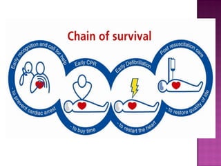
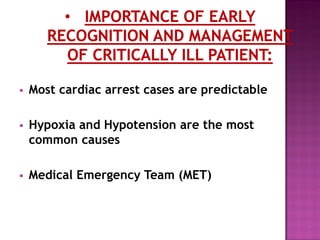
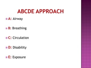
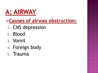
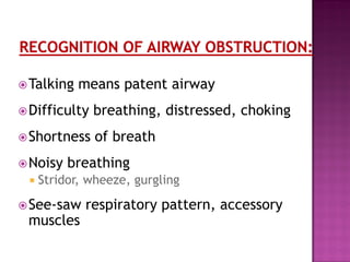
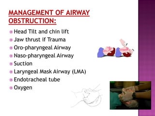
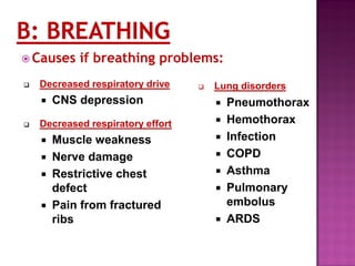

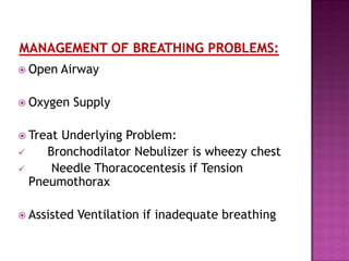

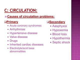

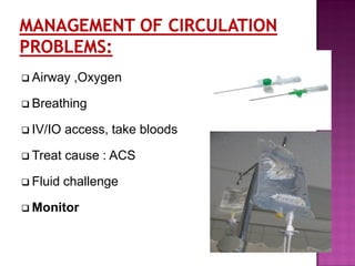
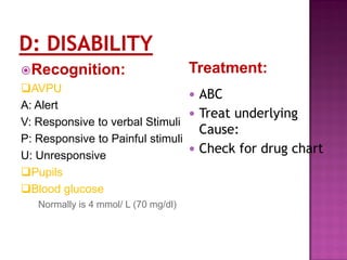


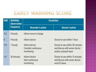


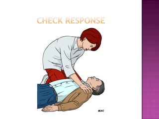

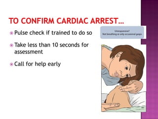

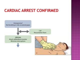
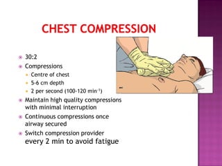


This document provides information on assessing and managing critically ill patients using the ABCDE approach. It discusses: - Common early signs of critical illness including hypoxia and hypotension. - The ABCDE approach which prioritizes establishing a patient's airway, breathing, circulation, disability level and exposure for examination. - Techniques for assessing and intervening on airway, breathing and circulation issues including providing oxygen, treating underlying causes, and starting IV fluids. - The importance of continuous reassessment, calling for help early, and following basic life support protocols when indicated to stabilize critically ill patients.


![ONFH[AVN HIP] -TRIPLE REGIME -A NOVAL SURGICAL CONCEPT .pptx](https://cdn.slidesharecdn.com/ss_thumbnails/onfhavnhip2026koaconcalicutdrgokuldevdrmashraf-260210064517-213ec005-thumbnail.jpg?width=640&height=640&fit=bounds)


