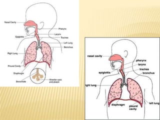
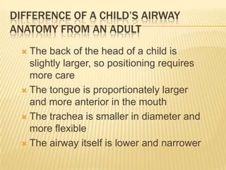
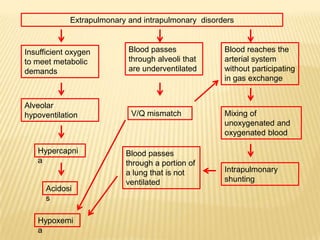
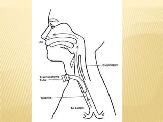
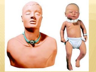
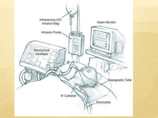
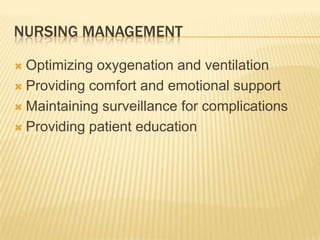
Respiratory disorders
Difference of a child’s airway anatomy from an adultThe back of the head of a child is slightly larger, so positioning requires more careThe tongue is proportionately larger and more anterior in the mouthThe trachea is smaller in diameter and more flexibleThe airway itself is lower and narrower
Acute respiratory failure
Acute respiratory FailureA clinical condition in which the pulmonary system fails to maintain adequate gas exchangeMost common organ failure in the ICUMortality rate: 22% to 75%Results from a deficiency in the performance of the pulmonary systemUsually occurs secondary to another disorder that has altered the normal function of the pulmonary system in such a way as to decrease the ventilatory drive, decrease muscle strength, decrease chest wall elasticity, decrease lung’s capacity for gas exchange, increase airway resistance, or increase metabolic O2 requirements
Etiology - extrapulmonaryBrain – drug overdose, brain trauma or lesion, post-op anesthesia depressionSpinal cord – Guillain-Barre syndrome, poliomyelitis, spinal cord trauma or lesionNeuromuscular system – Myasthenia gravis, Multiple sclerosis, neuromuscular-blocking agents, organophosphate poisoningThorax – massive obesity, chest traumaPleura – pleural effusion, pneumothoraxUpper airways – sleep apnea, tracheal obstruction, epiglottitis
Etiology - intrapulmonaryLower airways and alveoli – COPD, asthma, bronchiolitis,pneumoniaPulmonary circulation – pulmomary emboliAlveolar-capillary membrane – acute lung injury, inhalation of toxic gases, near-drowning
Extrapulmonary and intrapulmonary  disordersBlood passes through alveoli that are underventilatedBlood reaches the arterial system without participating in gas exchangeInsufficient oxygen to meet metabolic demandsAlveolar hypoventilationV/Q mismatchMixing of unoxygenated and oxygenated bloodHypercapniaBlood passes through a portion of a lung that is not ventilatedIntrapulmonary shuntingAcidosis  Hypoxemia
Assessment and diagnosisClinical manifestations are related to the development of hypoxemia, hypercapnia, and acidosisClinical manifestations are so varied that they considered unreliable in predicting the degree of hypoxemia or hypercapnia or the severityABG: PaO2 less than 60 mm Hg and the PaCO2 is greater than 45 mm HgBronchoscopy, chest X-ray, thoracic CT
Nursing diagnosis prioritiesImpaired gas exchange related to alveolar hypoventilationImpaired gas exchange related to ventilation/perfusion mismatching or intrapulmonary shuntingIneffective breathing pattern related to musculoskeletal fatigue or neuromuscular impairment
Medical managementAimed at treating the underlying cause, promoting adequate gas exchange, correcting acidosis, initiating nutrition support, and preventing complicationsMedical interventions to promote gas exchange are aimed at improving oxygenation and ventilation
1. OxygenationPurpose is to correct hypoxemia – aim is to keep the arterial hemoglobin oxygen saturation greater than 90%Goal is to keep the tissues’ needs satisfied but not produce hypercapnia or oxygen toxicitySupplemental oxygenation administration is effective in treating hypoxemia related to alveolar hypoventilation and V/Q mismatchingPositive pressure is necessary when there is intrapulmonary shunting (to open collapsed alveoli) can be delivered via nasal or oronasal mask (to avoid intubation)
2. ventilationDepending on the underlying cause and severity, the patient may be initially treated with noninvasive ventilationMechanical ventilationPEEP – positive end expiratory pressure
PEEPOpens collapsed alveoliStabilizes flooded alveoliIncreases FRCHowever,Decreases cardiac output, decreasing venous return secondary to increased intrathoracic pressureBarotrauma, as a result of gas escaping into the surrounding spaces secondary to alveolar rupture
3. pharmacologyBronchodilators – Beta-agonists and antocholinergic agentsSteroidsSedation can be used to comfort the patient and decrease the work of breathing, particularly if the patient is fighting the ventilatorAnalgesics for pain controlMethylxanthines and mucolytics are no longer used because of their negative side effects
4. Acidosis - treatmentOnce the patient is adequately oxygenated and ventilated, the acidosis should correct itselfUse of sodium bicarbonate has been shown to be of minimal benefit and is no longer recommended, even in the presence of severe acidosis
5. Nutrition supportGoals are to meet the overall nutritional needs of the patient, while avoiding overfeeding, to prevent nutrition delivery-related complications and to improve patient outcomesThe enteral route is the preferred method of nutrition administrationParenteral nutrition for those who cannot tolerate enteral feedings or cannot receive enough nutrients enterally
6. Complications - treatmentMaintaining oxygenation, normalizing electrolytes, and monitoring drug levels will facilitate the prevention and treatment of encephalopathy and dysrhythmiasVenous thromboembolism can be prevented by using compression stockings and low-dose unfractionated heparin or low-molecular weight heparinGIT bleeding can be prevented through the use of histamine-2 antagonists, cytoprotective agents, or gastric proton pump inhibitorsPatient is at risk of developing complications associated with artificial airway, mechanical ventilation, enteral and parenteral nutrition, and peripheral cannulation
Guideline Values for estimating Fio2 with low flow o2 devices
Nursing management Optimizing oxygenation and ventilationProviding comfort and emotional supportMaintaining surveillance for complications Providing patient education
1. Optimizing oxygenation and ventilationPositioning – the goal is to place the least affected area of the affected lung in the most dependent position – gravity normally facilitates preferential ventilation and perfusion to the dependent areas of the lungs- the best gas exchange would take place in the dependent areas of the lungs
1. Optimizing oxygenation and ventilationPositioningPatients with diffuse lung disease may benefit from being positioned with the right lung down, because it is larger and more vascular than the left lungFor those with alveolar hypoventilation, a nonrecumbent position (sitting or semierect) may be beneficialSemirecumbency position can help prevent aspiration and inhibit the development of hospital-associated pneumoniaFrequent positioning (at least every 2 hours) is beneficial in optimizing the patient’s ventilatory pattern and V/Q matching
1. Optimizing oxygenation and ventilationPreventing desaturation – performing procedures as needed: oxygenating before suctioning, providing adequate rest and recovery time between various procedures, and minimizing oxygen consumption Promoting secretion clearance – providing adequate systemic hydration, humidifying supplemental oxygen, coughing, and suctioningNote: postural drainage and chest percussion and vibration have been found to be of little benefit in the critically ill patient; to facilitate breathing, the thorax should be maintained in alignment and the head of the bed elevated 30 to 45 degrees
2. Patient educationPathophysiology of the diseaseSpecific etiologyPrecipitating factor modificationImportance of taking medicationsBreathing techniques (e.g., pursed-lip breathing diaphragmatic breathing)Energy conservation techniques
2. Patient educationMeasures to prevent pulmonary infections (e.g., proper nutrition, hand washing, immunization against S. pneumoniae and influenza viruses)Signs and symptoms of pulmonary infections (e.g., sputum color change, shortness of breath, fever)Cough enhancement techniques
Collaborative managementIdentify and treat underlying causeAdminister oxygen therapyIntubate patientAdminister medicationsPosition patient to optimize ventilation/perfusion matchingSuction as needed
Collaborative managementProvide adequate rest and recovery time between various proceduresCorrect acidosisInitiate nutritional supportMaintain surveillance for complications: encephalopathy, dysrhythmias, venous thromboembolism, GI bleedingProvide comfort and emotional support
Pneumonia
pneumoniaAn acute inflammation of the lung parenchyma that is caused by an infectious agent that can lead to alveolar consolidationCAP – community acquired pneumoniaHAP – hospital acquired pneumoniaVAP – ventilator-associated pneumonia
Precipitating conditions of pneumonia
Microorganisms/noninfectious agentsInhalation/aspirationLower airwaysLoss of cough reflex, damage to cilia of the respiratory tract, impaired host defensesColonization of the lower respiratory tractLung tissue attempts to undergo healingHepatizationRelease of histamine and other vasoactive chemical mediators Stage of fibrosisLung tissue reacts to accumulating exudates and  microorganismsVasodilationPulmonary function impairedMore and more exudates accumulateStage of congestionClinical manifestations
EtiologySevere CAP – S. pneumoniae, Legionella species, H. influenzae, S. aureus, M. pneumoniae, respiratory viruses, Chlamydia pneumoniae, and P. aerugionosaHAP – S. aureus, S. pneumoniae, P. aeruginosa, Acinetobacterbaumannii, Klebsiella species, Proteus species, Serratia species, fungi, and respiratory viruses
Assessment and DiagnosisIneffective airway clearance related to excessive secretions or abnormal viscosity of mucusImpaired gas exchange related to ventilatory/perfusion mismatching or intrapulmonary shuntingRisk for infection, risk factor: invasive monitoring devicesPowerlessness related to lack of control over current situation or disease progression
Assessment and diagnosisChest radiographSputum Gram stain and cultureDiagnostic bronchoscopyCBC with differential countChemistry panelBlood culturesABG
Medical managementAntibiotic therapyOxygen therapyFluid managementNutritional supportTreatment of associated medical problems and complicationsTherapeutic bronchoscopy may be necessary in patients who have difficulty mobilizing secretions
Nursing managementOptimizing oxygenation and ventilationPreventing the spread of infectionProviding comfort and emotional supportMaintaining surveillance for complications
Collaborative managementAdminister oxygen therapyInitiate mechanical ventilation as requiredAdminister medications: antibiotics, bronchodilatorsPosition patient to optimize ventilation/perfusion matchingSuction as neededProvide adequate rest and recovery time between various proceduresMaintain surveillance for complications: acute respiratory failureProvide comfort and emotional support
Pulmonary embolism
Pulmonary embolism (PE)Occurs when a clot (thrombotic emboli) or other matter (nonthrombotic emboli) lodges in the pulmonary arterial system, disrupting the blood flow to a region of the lungsMajority come form the deep leg veins, particularly the iliac, femoral, and popliteal veinsOther sources: RV, the upper extremities, and the pelvic veinsNonthrombotic emboli: fat, tumors, amniotic fluid, air, and foreign bodies
etiologyThree predisposing factors: hypercoagulability, injury to the vascular endothelium and venous stasis (Virchow’s triad)Venous stasis: AF, decreased CO, immobilityInjury to the vascular endothelium: local vessel injury, infection, incision, atherosclerosisHypercoagulability: polycythemia
Abc 2011 2012 respiratory disorders

Abc 2011 2012 respiratory disorders

  • 1.
  • 3.
    Difference of achild’s airway anatomy from an adultThe back of the head of a child is slightly larger, so positioning requires more careThe tongue is proportionately larger and more anterior in the mouthThe trachea is smaller in diameter and more flexibleThe airway itself is lower and narrower
  • 4.
  • 5.
    Acute respiratory FailureAclinical condition in which the pulmonary system fails to maintain adequate gas exchangeMost common organ failure in the ICUMortality rate: 22% to 75%Results from a deficiency in the performance of the pulmonary systemUsually occurs secondary to another disorder that has altered the normal function of the pulmonary system in such a way as to decrease the ventilatory drive, decrease muscle strength, decrease chest wall elasticity, decrease lung’s capacity for gas exchange, increase airway resistance, or increase metabolic O2 requirements
  • 6.
    Etiology - extrapulmonaryBrain– drug overdose, brain trauma or lesion, post-op anesthesia depressionSpinal cord – Guillain-Barre syndrome, poliomyelitis, spinal cord trauma or lesionNeuromuscular system – Myasthenia gravis, Multiple sclerosis, neuromuscular-blocking agents, organophosphate poisoningThorax – massive obesity, chest traumaPleura – pleural effusion, pneumothoraxUpper airways – sleep apnea, tracheal obstruction, epiglottitis
  • 7.
    Etiology - intrapulmonaryLowerairways and alveoli – COPD, asthma, bronchiolitis,pneumoniaPulmonary circulation – pulmomary emboliAlveolar-capillary membrane – acute lung injury, inhalation of toxic gases, near-drowning
  • 8.
    Extrapulmonary and intrapulmonary disordersBlood passes through alveoli that are underventilatedBlood reaches the arterial system without participating in gas exchangeInsufficient oxygen to meet metabolic demandsAlveolar hypoventilationV/Q mismatchMixing of unoxygenated and oxygenated bloodHypercapniaBlood passes through a portion of a lung that is not ventilatedIntrapulmonary shuntingAcidosis Hypoxemia
  • 9.
    Assessment and diagnosisClinicalmanifestations are related to the development of hypoxemia, hypercapnia, and acidosisClinical manifestations are so varied that they considered unreliable in predicting the degree of hypoxemia or hypercapnia or the severityABG: PaO2 less than 60 mm Hg and the PaCO2 is greater than 45 mm HgBronchoscopy, chest X-ray, thoracic CT
  • 10.
    Nursing diagnosis prioritiesImpairedgas exchange related to alveolar hypoventilationImpaired gas exchange related to ventilation/perfusion mismatching or intrapulmonary shuntingIneffective breathing pattern related to musculoskeletal fatigue or neuromuscular impairment
  • 11.
    Medical managementAimed attreating the underlying cause, promoting adequate gas exchange, correcting acidosis, initiating nutrition support, and preventing complicationsMedical interventions to promote gas exchange are aimed at improving oxygenation and ventilation
  • 12.
    1. OxygenationPurpose isto correct hypoxemia – aim is to keep the arterial hemoglobin oxygen saturation greater than 90%Goal is to keep the tissues’ needs satisfied but not produce hypercapnia or oxygen toxicitySupplemental oxygenation administration is effective in treating hypoxemia related to alveolar hypoventilation and V/Q mismatchingPositive pressure is necessary when there is intrapulmonary shunting (to open collapsed alveoli) can be delivered via nasal or oronasal mask (to avoid intubation)
  • 13.
    2. ventilationDepending onthe underlying cause and severity, the patient may be initially treated with noninvasive ventilationMechanical ventilationPEEP – positive end expiratory pressure
  • 14.
    PEEPOpens collapsed alveoliStabilizesflooded alveoliIncreases FRCHowever,Decreases cardiac output, decreasing venous return secondary to increased intrathoracic pressureBarotrauma, as a result of gas escaping into the surrounding spaces secondary to alveolar rupture
  • 15.
    3. pharmacologyBronchodilators –Beta-agonists and antocholinergic agentsSteroidsSedation can be used to comfort the patient and decrease the work of breathing, particularly if the patient is fighting the ventilatorAnalgesics for pain controlMethylxanthines and mucolytics are no longer used because of their negative side effects
  • 16.
    4. Acidosis -treatmentOnce the patient is adequately oxygenated and ventilated, the acidosis should correct itselfUse of sodium bicarbonate has been shown to be of minimal benefit and is no longer recommended, even in the presence of severe acidosis
  • 17.
    5. Nutrition supportGoalsare to meet the overall nutritional needs of the patient, while avoiding overfeeding, to prevent nutrition delivery-related complications and to improve patient outcomesThe enteral route is the preferred method of nutrition administrationParenteral nutrition for those who cannot tolerate enteral feedings or cannot receive enough nutrients enterally
  • 18.
    6. Complications -treatmentMaintaining oxygenation, normalizing electrolytes, and monitoring drug levels will facilitate the prevention and treatment of encephalopathy and dysrhythmiasVenous thromboembolism can be prevented by using compression stockings and low-dose unfractionated heparin or low-molecular weight heparinGIT bleeding can be prevented through the use of histamine-2 antagonists, cytoprotective agents, or gastric proton pump inhibitorsPatient is at risk of developing complications associated with artificial airway, mechanical ventilation, enteral and parenteral nutrition, and peripheral cannulation
  • 19.
    Guideline Values forestimating Fio2 with low flow o2 devices
  • 34.
    Nursing management Optimizingoxygenation and ventilationProviding comfort and emotional supportMaintaining surveillance for complications Providing patient education
  • 36.
    1. Optimizing oxygenationand ventilationPositioning – the goal is to place the least affected area of the affected lung in the most dependent position – gravity normally facilitates preferential ventilation and perfusion to the dependent areas of the lungs- the best gas exchange would take place in the dependent areas of the lungs
  • 37.
    1. Optimizing oxygenationand ventilationPositioningPatients with diffuse lung disease may benefit from being positioned with the right lung down, because it is larger and more vascular than the left lungFor those with alveolar hypoventilation, a nonrecumbent position (sitting or semierect) may be beneficialSemirecumbency position can help prevent aspiration and inhibit the development of hospital-associated pneumoniaFrequent positioning (at least every 2 hours) is beneficial in optimizing the patient’s ventilatory pattern and V/Q matching
  • 38.
    1. Optimizing oxygenationand ventilationPreventing desaturation – performing procedures as needed: oxygenating before suctioning, providing adequate rest and recovery time between various procedures, and minimizing oxygen consumption Promoting secretion clearance – providing adequate systemic hydration, humidifying supplemental oxygen, coughing, and suctioningNote: postural drainage and chest percussion and vibration have been found to be of little benefit in the critically ill patient; to facilitate breathing, the thorax should be maintained in alignment and the head of the bed elevated 30 to 45 degrees
  • 39.
    2. Patient educationPathophysiologyof the diseaseSpecific etiologyPrecipitating factor modificationImportance of taking medicationsBreathing techniques (e.g., pursed-lip breathing diaphragmatic breathing)Energy conservation techniques
  • 40.
    2. Patient educationMeasuresto prevent pulmonary infections (e.g., proper nutrition, hand washing, immunization against S. pneumoniae and influenza viruses)Signs and symptoms of pulmonary infections (e.g., sputum color change, shortness of breath, fever)Cough enhancement techniques
  • 41.
    Collaborative managementIdentify andtreat underlying causeAdminister oxygen therapyIntubate patientAdminister medicationsPosition patient to optimize ventilation/perfusion matchingSuction as needed
  • 42.
    Collaborative managementProvide adequaterest and recovery time between various proceduresCorrect acidosisInitiate nutritional supportMaintain surveillance for complications: encephalopathy, dysrhythmias, venous thromboembolism, GI bleedingProvide comfort and emotional support
  • 44.
  • 45.
    pneumoniaAn acute inflammationof the lung parenchyma that is caused by an infectious agent that can lead to alveolar consolidationCAP – community acquired pneumoniaHAP – hospital acquired pneumoniaVAP – ventilator-associated pneumonia
  • 46.
  • 47.
  • 48.
    Microorganisms/noninfectious agentsInhalation/aspirationLower airwaysLossof cough reflex, damage to cilia of the respiratory tract, impaired host defensesColonization of the lower respiratory tractLung tissue attempts to undergo healingHepatizationRelease of histamine and other vasoactive chemical mediators Stage of fibrosisLung tissue reacts to accumulating exudates and microorganismsVasodilationPulmonary function impairedMore and more exudates accumulateStage of congestionClinical manifestations
  • 49.
    EtiologySevere CAP –S. pneumoniae, Legionella species, H. influenzae, S. aureus, M. pneumoniae, respiratory viruses, Chlamydia pneumoniae, and P. aerugionosaHAP – S. aureus, S. pneumoniae, P. aeruginosa, Acinetobacterbaumannii, Klebsiella species, Proteus species, Serratia species, fungi, and respiratory viruses
  • 51.
    Assessment and DiagnosisIneffectiveairway clearance related to excessive secretions or abnormal viscosity of mucusImpaired gas exchange related to ventilatory/perfusion mismatching or intrapulmonary shuntingRisk for infection, risk factor: invasive monitoring devicesPowerlessness related to lack of control over current situation or disease progression
  • 52.
    Assessment and diagnosisChestradiographSputum Gram stain and cultureDiagnostic bronchoscopyCBC with differential countChemistry panelBlood culturesABG
  • 59.
    Medical managementAntibiotic therapyOxygentherapyFluid managementNutritional supportTreatment of associated medical problems and complicationsTherapeutic bronchoscopy may be necessary in patients who have difficulty mobilizing secretions
  • 60.
    Nursing managementOptimizing oxygenationand ventilationPreventing the spread of infectionProviding comfort and emotional supportMaintaining surveillance for complications
  • 61.
    Collaborative managementAdminister oxygentherapyInitiate mechanical ventilation as requiredAdminister medications: antibiotics, bronchodilatorsPosition patient to optimize ventilation/perfusion matchingSuction as neededProvide adequate rest and recovery time between various proceduresMaintain surveillance for complications: acute respiratory failureProvide comfort and emotional support
  • 67.
  • 68.
    Pulmonary embolism (PE)Occurswhen a clot (thrombotic emboli) or other matter (nonthrombotic emboli) lodges in the pulmonary arterial system, disrupting the blood flow to a region of the lungsMajority come form the deep leg veins, particularly the iliac, femoral, and popliteal veinsOther sources: RV, the upper extremities, and the pelvic veinsNonthrombotic emboli: fat, tumors, amniotic fluid, air, and foreign bodies
  • 69.
    etiologyThree predisposing factors:hypercoagulability, injury to the vascular endothelium and venous stasis (Virchow’s triad)Venous stasis: AF, decreased CO, immobilityInjury to the vascular endothelium: local vessel injury, infection, incision, atherosclerosisHypercoagulability: polycythemia