STATUS ASTHMATICUS
BY
ANCY JAYAKUMARI BHUVANENDRAN
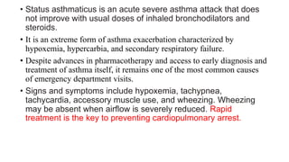
• Status asthmaticus is an acute severe asthma attack that does
not improve with usual doses of inhaled bronchodilators and
steroids.
• It is an extreme form of asthma exacerbation characterized by
hypoxemia, hypercarbia, and secondary respiratory failure.
• Despite advances in pharmacotherapy and access to early diagnosis and
treatment of asthma itself, it remains one of the most common causes
of emergency department visits.
• Signs and symptoms include hypoxemia, tachypnea,
tachycardia, accessory muscle use, and wheezing. Wheezing
may be absent when airflow is severely reduced. Rapid
treatment is the key to preventing cardiopulmonary arrest.
• Other symptoms of an asthma attack associated with status
asthmaticus include:
• difficulty breathing
• heavy sweating
• trouble speaking
• fatigue and weakness
• abdominal, back, or neck muscle pain
• panic or confusion
• blue-tinted lips or skin
• loss of consciousnes
What causes it ???
Experts aren’t sure why some people with asthma develop severe asthma, or
why it doesn’t respond to typical asthma treatments.
But it’s usually caused by the same triggers that contribute to traditional
asthma attacks, which include:
•respiratory infections
•severe stress
•cold weather
•severe allergic reactions
•air pollution
•exposure to chemicals and other irritants
•smoking
It might also be related to poorly controlled asthma, often due to not sticking
with a treatment plan prescribed by a doctor.
PATHOPHYSIOLOGY
At a physiological level, premature airway closure during
exhalation causes an increase in functional residual capacity
and air trapping. Heterogeneous distribution of air trapping
results in ventilation-perfusion mismatch and hypoxemia-
triggering anaerobic metabolism and lactic acidosis. It is
offset initially by respiratory alkalosis and is compounded
once respiratory fatigue and respiratory acidosis ensue.
DIAGNOSIS
Status asthmaticus is typically diagnosed by symptoms and supported by
various tests that measure respiration rate and blood oxygen levels.
Measurement of airflow obstruction can be challenging to perform but is best achieved at the bedside
with an assessment of PEFR than FEV1.The reduction of both values by 50% of the patient's
personal best is an indicator of status.
The absolute value of PEFR less than 120 L per minute and FEV1 less than 1 L corresponds with the
proportional reduction.
Initial blood gas results indicate respiratory alkalosis with hypoxemia. Therefore, developing
respiratory acidosis or elevated PCO2 are indicators of status asthmaticus that indicate the need for
ventilatory support.
However, it should not be the lone decision-maker and should be coupled with a serial physical
examination, evidence of worsening mentation, and fatigability or hemodynamic alterations.
ECG may also show transient and reversible signs of right heart strain, including peaked p wave or
right axis deviation.
MANAGEMENT
The serial measurement of PEFR is a practical and reliable predictor of severity and the
need for hospitalization
A favorable response to initial treatment of status asthmaticus should be a visible
improvement in symptoms that sustains 30 minutes or beyond the last bronchodilator dose
and a PEFR greater than 70% of predicted.
On the other hand, patients with evidence of continuing clinical decline or less than 10%
improvement in PEFR or less than 40% of predicted, should be considered for
admission to the intensive care unit.
PHARMACOLOGICAL MANAGEMENT
• Short-acting inhaled Beta-agonists are the drug of the first choice in
acute asthma. Albuterol is preferred because of its higher beta 2
selectivities and longer duration of action.
• Initial treatment consists of 2.5 mg of albuterol by nebulization every
20 minutes for 60 minutes (three doses) followed by treatments hourly
during the first several hours of therapy.
• CORTICOSTEROIDS reduce airway inflammation and mucus
production and potentiate beta-agonist activity in smooth muscles and
reduce beta-agonists tachyphylaxis in patients with severe asthma.
• IV magnesium sulfate is indicated in the management of acute, very
severe asthma (FEV1 <25% predicted).
• The magnesium dose is 1 to 2 grams IV over 30 minutes.
• Nebulized magnesium is effective and may also improve pulmonary
function in severe asthma when it follows aggressive β-agonist and steroid
therapy.
• When using magnesium in any form, monitor blood pressure and deep
tendon reflexes during administration because hypotension or
neuromuscular blockade may occur.
• NONINVASIVE POSITIVE-PRESSURE VENTILATION
• Noninvasive positive-pressure ventilation improves airflow and
respirations compared with usual care.
• it is commonly used in clinical practice for acute life-threatening
asthma.
• Noninvasive positive-pressure ventilation decreases the need
for intubation, results in clinical improvement, and decreases
the need for hospitalization.
Mechanical Ventilation and Sedation
• The decision to intubate a patient presenting with status asthmaticus is a clinical one
and does not unequivocally require a blood gas assessment.
• Immediate indications for intubation include:
• Acute cardiopulmonary arrest
• Severe obtundation or coma
• Frank evidence of respiratory fatigue with gasping or inability to speak at all
• “Intubation and Mechanical Ventilation”) are necessary to prevent respiratory
arrest.
• Mechanical ventilation does not relieve the airflow obstruction—it
merely eliminates the work of breathing and enables the patient to rest
while the airflow obstruction is resolved.
Ketamine
Ketamine has sedative, analgesic, anesthetic, and bronchodilatory properties and has been
increasingly recommended for emergency intubation in status asthmaticus .The usual dose is 1
to 2 mg/kg given intravenously at a rate of 0.5 mg/kg per minute to provide 10 to 15 minutes
of general anesthesia without significant respiratory depression.
Potential risks to consider before deciding in favor of ketamine include:
•Ability to cause hypertension and tachycardia with sympathetic stimulation. Thus it is to be
avoided in patients with uncontrolled hypertension, preeclampsia, or raised intracranial
pressure.
•Lowering of seizure threshold
•Increase in laryngeal secretion
•Metabolism through the liver, thus causing some accumulation with the continuous infusion in
liver failure.
DISPOSITION AND FOLLOW-UP
• Disposition decisions should take into account a combination of subjective
parameters, such as resolution of wheezing and improvement in air
exchange, as assessed by auscultation and patient opinion.
• Objective measures, such as normalization of FEV1 or PEFR.
• Advise discharged patients to use a short-acting β-agonist on a scheduled
basis for several days and to complete any oral corticosteroids regimens.
• Add inhaled corticosteroids in patients with a history of persistent asthma
who are not already using this regimen.
• A good response to treatment resolves symptoms and results in a PEFR or
FEV1 of >70% predicted; these patients can be safely discharged home
Cont….
• Patients with a poor response to treatment have persistent
symptoms and FEV1 or PEFR of <40% predicted; these patients are
usually best observed or admitted.
• An incomplete response to treatment the middle ground, is defined as
some persistence of symptoms and a PEF or FEV1 between 40% and
69% predicted.
Most asthmatics treated in the ED fall into this category and may be
discharged home safely, although some benefit from prolonged observation
or admission.
• Patients who fail to improve adequately over a period of several hours
because they are in the late phase of their exacerbation and those with
significant risk factors for death from asthma are best placed in an
observation unit or hospital bed.
• Arrange follow-up care to ensure resolution and to review the
long-term medication plan for the chronic management of
asthma. High previous relapse rates suggest the need for
follow-up within 1 to 4 weeks of the ED visit.
• Deliver an appropriate written discharge plan of action that
addresses routine care and care of worsening symptoms
• Educate patients on asthma triggers, and review all discharge
medications and the correct use of the inhaler and a peak flow
meter (for daily tracking).
REFERENCE
• Tintinallis emergency medicine guide
• https://www.ncbi.nlm.nih.gov/books/NBK526070/
• https://www.healthline.com/health/status-asthmaticus#symptoms
• https://www.verywellhealth.com/status-asthmaticus-overview-
3866901#:~:text=Status%20asthmaticus%20is%20typically%20diagno
sed,able%20to%20speak%20at%20all
Status asthmaticus ancy ppt

Status asthmaticus ancy ppt

  • 1.
  • 2.
    • Status asthmaticusis an acute severe asthma attack that does not improve with usual doses of inhaled bronchodilators and steroids. • It is an extreme form of asthma exacerbation characterized by hypoxemia, hypercarbia, and secondary respiratory failure. • Despite advances in pharmacotherapy and access to early diagnosis and treatment of asthma itself, it remains one of the most common causes of emergency department visits. • Signs and symptoms include hypoxemia, tachypnea, tachycardia, accessory muscle use, and wheezing. Wheezing may be absent when airflow is severely reduced. Rapid treatment is the key to preventing cardiopulmonary arrest.
  • 3.
    • Other symptomsof an asthma attack associated with status asthmaticus include: • difficulty breathing • heavy sweating • trouble speaking • fatigue and weakness • abdominal, back, or neck muscle pain • panic or confusion • blue-tinted lips or skin • loss of consciousnes
  • 4.
    What causes it??? Experts aren’t sure why some people with asthma develop severe asthma, or why it doesn’t respond to typical asthma treatments. But it’s usually caused by the same triggers that contribute to traditional asthma attacks, which include: •respiratory infections •severe stress •cold weather •severe allergic reactions •air pollution •exposure to chemicals and other irritants •smoking It might also be related to poorly controlled asthma, often due to not sticking with a treatment plan prescribed by a doctor.
  • 5.
    PATHOPHYSIOLOGY At a physiologicallevel, premature airway closure during exhalation causes an increase in functional residual capacity and air trapping. Heterogeneous distribution of air trapping results in ventilation-perfusion mismatch and hypoxemia- triggering anaerobic metabolism and lactic acidosis. It is offset initially by respiratory alkalosis and is compounded once respiratory fatigue and respiratory acidosis ensue.
  • 7.
    DIAGNOSIS Status asthmaticus istypically diagnosed by symptoms and supported by various tests that measure respiration rate and blood oxygen levels. Measurement of airflow obstruction can be challenging to perform but is best achieved at the bedside with an assessment of PEFR than FEV1.The reduction of both values by 50% of the patient's personal best is an indicator of status. The absolute value of PEFR less than 120 L per minute and FEV1 less than 1 L corresponds with the proportional reduction. Initial blood gas results indicate respiratory alkalosis with hypoxemia. Therefore, developing respiratory acidosis or elevated PCO2 are indicators of status asthmaticus that indicate the need for ventilatory support. However, it should not be the lone decision-maker and should be coupled with a serial physical examination, evidence of worsening mentation, and fatigability or hemodynamic alterations. ECG may also show transient and reversible signs of right heart strain, including peaked p wave or right axis deviation.
  • 8.
    MANAGEMENT The serial measurementof PEFR is a practical and reliable predictor of severity and the need for hospitalization A favorable response to initial treatment of status asthmaticus should be a visible improvement in symptoms that sustains 30 minutes or beyond the last bronchodilator dose and a PEFR greater than 70% of predicted. On the other hand, patients with evidence of continuing clinical decline or less than 10% improvement in PEFR or less than 40% of predicted, should be considered for admission to the intensive care unit.
  • 9.
    PHARMACOLOGICAL MANAGEMENT • Short-actinginhaled Beta-agonists are the drug of the first choice in acute asthma. Albuterol is preferred because of its higher beta 2 selectivities and longer duration of action. • Initial treatment consists of 2.5 mg of albuterol by nebulization every 20 minutes for 60 minutes (three doses) followed by treatments hourly during the first several hours of therapy. • CORTICOSTEROIDS reduce airway inflammation and mucus production and potentiate beta-agonist activity in smooth muscles and reduce beta-agonists tachyphylaxis in patients with severe asthma.
  • 10.
    • IV magnesiumsulfate is indicated in the management of acute, very severe asthma (FEV1 <25% predicted). • The magnesium dose is 1 to 2 grams IV over 30 minutes. • Nebulized magnesium is effective and may also improve pulmonary function in severe asthma when it follows aggressive β-agonist and steroid therapy. • When using magnesium in any form, monitor blood pressure and deep tendon reflexes during administration because hypotension or neuromuscular blockade may occur.
  • 11.
    • NONINVASIVE POSITIVE-PRESSUREVENTILATION • Noninvasive positive-pressure ventilation improves airflow and respirations compared with usual care. • it is commonly used in clinical practice for acute life-threatening asthma. • Noninvasive positive-pressure ventilation decreases the need for intubation, results in clinical improvement, and decreases the need for hospitalization.
  • 12.
    Mechanical Ventilation andSedation • The decision to intubate a patient presenting with status asthmaticus is a clinical one and does not unequivocally require a blood gas assessment. • Immediate indications for intubation include: • Acute cardiopulmonary arrest • Severe obtundation or coma • Frank evidence of respiratory fatigue with gasping or inability to speak at all • “Intubation and Mechanical Ventilation”) are necessary to prevent respiratory arrest. • Mechanical ventilation does not relieve the airflow obstruction—it merely eliminates the work of breathing and enables the patient to rest while the airflow obstruction is resolved.
  • 13.
    Ketamine Ketamine has sedative,analgesic, anesthetic, and bronchodilatory properties and has been increasingly recommended for emergency intubation in status asthmaticus .The usual dose is 1 to 2 mg/kg given intravenously at a rate of 0.5 mg/kg per minute to provide 10 to 15 minutes of general anesthesia without significant respiratory depression. Potential risks to consider before deciding in favor of ketamine include: •Ability to cause hypertension and tachycardia with sympathetic stimulation. Thus it is to be avoided in patients with uncontrolled hypertension, preeclampsia, or raised intracranial pressure. •Lowering of seizure threshold •Increase in laryngeal secretion •Metabolism through the liver, thus causing some accumulation with the continuous infusion in liver failure.
  • 14.
    DISPOSITION AND FOLLOW-UP •Disposition decisions should take into account a combination of subjective parameters, such as resolution of wheezing and improvement in air exchange, as assessed by auscultation and patient opinion. • Objective measures, such as normalization of FEV1 or PEFR. • Advise discharged patients to use a short-acting β-agonist on a scheduled basis for several days and to complete any oral corticosteroids regimens. • Add inhaled corticosteroids in patients with a history of persistent asthma who are not already using this regimen. • A good response to treatment resolves symptoms and results in a PEFR or FEV1 of >70% predicted; these patients can be safely discharged home
  • 15.
    Cont…. • Patients witha poor response to treatment have persistent symptoms and FEV1 or PEFR of <40% predicted; these patients are usually best observed or admitted. • An incomplete response to treatment the middle ground, is defined as some persistence of symptoms and a PEF or FEV1 between 40% and 69% predicted. Most asthmatics treated in the ED fall into this category and may be discharged home safely, although some benefit from prolonged observation or admission. • Patients who fail to improve adequately over a period of several hours because they are in the late phase of their exacerbation and those with significant risk factors for death from asthma are best placed in an observation unit or hospital bed.
  • 16.
    • Arrange follow-upcare to ensure resolution and to review the long-term medication plan for the chronic management of asthma. High previous relapse rates suggest the need for follow-up within 1 to 4 weeks of the ED visit. • Deliver an appropriate written discharge plan of action that addresses routine care and care of worsening symptoms • Educate patients on asthma triggers, and review all discharge medications and the correct use of the inhaler and a peak flow meter (for daily tracking).
  • 17.
    REFERENCE • Tintinallis emergencymedicine guide • https://www.ncbi.nlm.nih.gov/books/NBK526070/ • https://www.healthline.com/health/status-asthmaticus#symptoms • https://www.verywellhealth.com/status-asthmaticus-overview- 3866901#:~:text=Status%20asthmaticus%20is%20typically%20diagno sed,able%20to%20speak%20at%20all