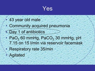
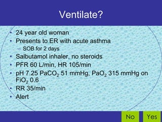
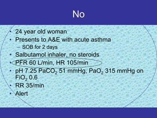
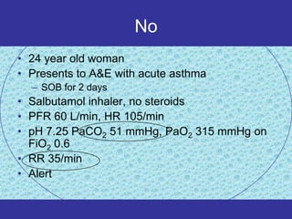
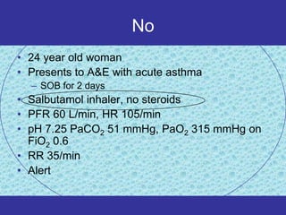
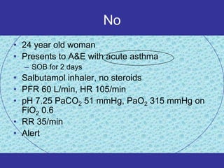
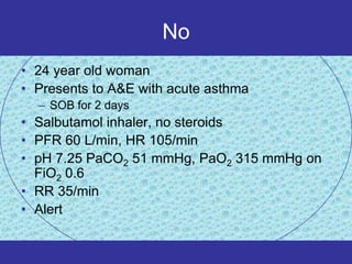
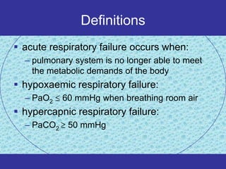
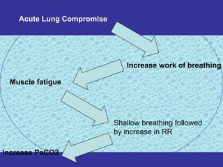
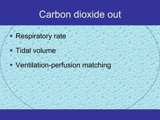
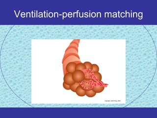
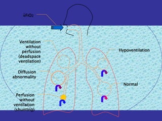
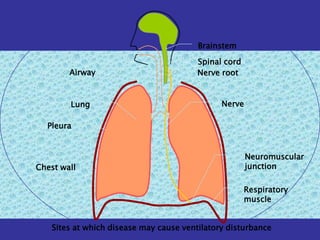
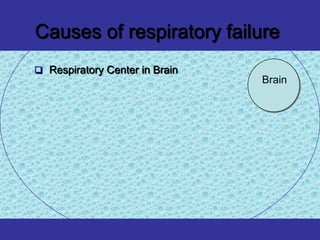
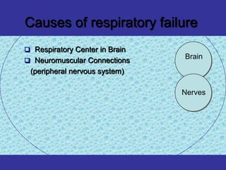
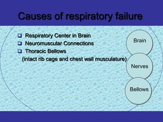
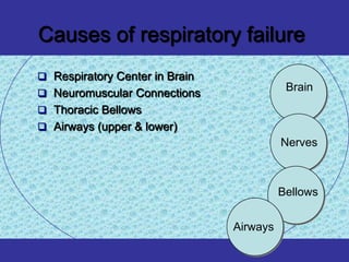
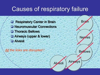
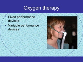
Acute respiratory failure
Topics
   Definition of acute respiratory failure
   Basic respiratory physiology
   Pathophysiology
   Respiratory monitoring
   Treatment
Definitions
 acute respiratory failure occurs when:
  – pulmonary system is no longer able to meet
    the metabolic demands of the body
 hypoxaemic respiratory failure:
  – PaO2    60 mmHg when breathing room air
 hypercapnic respiratory failure:
  – PaCO2    50 mmHg
Basic respiratory physiology
CO2   O2
Pulmonary Ventilation and pressures
Oxygen in
• Depends on
  – PAO2
  – Diffusing capacity
  – Perfusion
  – Ventilation-perfusion matching
Oxygen


                                        Carbon
                                        dioxide


                                          Water
                                          vapour

                               Nitrogen


Alveolar pressure PAO2 PACO2 PAH2O PAN2
Alveolar Oxygen tension


PAO2= (PB-PH2O)FIO2 – CO2/0.8

PAO2= (760-47)0.21 -40/0.8=100
How much oxygen is in the
        blood

 PaO2

 SaO2

 Oxygen content (CaO2)
How much oxygen is in the blood

PaO2
• The amount of dissolved oxygen in the plasma phase --
  and hence the PaO2 -- is determined by alveolar PO2
  and lung architecture only

SaO2
• The percentage of hemoglobin molecule bounded with
  oxygen.

Oxygen Content CaO2
• CaO2 = Hb (gm/dl) x 1.34 ml O2/gm Hb x SaO2 + PaO2 x
  (.003 ml O2/mm Hg/dl)
Oxygen dessociation Curve
Oxygen in
• Depends on
  – PAO2
    •   FIO2
    •   PACO2
    •   Ventilation
    •   Alveolar pressure
  – Ventilation-perfusion matching
  – Perfusion
  – Diffusing capacity
Carbon dioxide out

 Largely dependent on alveolar ventilation


   Alveolar ventilatio n RR x (V - V )T       D


 Anatomical dead space constant but
  physiological dead space depends on
  ventilation-perfusion matching
Carbon dioxide out
• Patient    Vt    f         Ve          Description
  –A        (400) (20) = 8.0 L/min      slow and deep
  –B        (200) (40) = 8.o L/min       fast/shallow


• Patient     Va-Vd     f     Va        Description
  –A        (400-150)(20) = 5.0 L/min   slow and deep
  –B        (200-150)(40) = 2.0 L/min    fast/shallow
Acute Lung Compromise


                               Increase work of breathing

 Muscle fatigue


                            Shallow breathing followed
                            by increase in RR

Increase PaCO2
Carbon dioxide out

 Respiratory rate

 Tidal volume

 Ventilation-perfusion matching
Ventilation-perfusion matching
Dead space
Shunt
Pathophysiology
Pathophysiology
• Low inspired Po2
• Hypoventilation
• Ventilation-perfusion mismatch
  – Shunting
  – Dead space ventilation
• Diffusion abnormality
PAO2=105 mmHg
      PACO2=37 mmHg


75%                   100%
Pathophysiology
•   Low inspired oxygen concentration
•   Hypoventilation
•   Shunting
•   Dead space ventilation
•   Diffusion abnormality
F I O2


   Ventilation
      without
    perfusion    Hypoventilation
  (deadspace
  ventilation)

  Diffusion
abnormality
                   Normal

 Perfusion
   without
ventilation
(shunting)
Brainstem

                                         Spinal cord
        Airway                           Nerve root


        Lung                                   Nerve

  Pleura


                                                       Neuromuscular
Chest wall                                             junction

                                                       Respiratory
                                                       muscle


    Sites at which disease may cause ventilatory disturbance
Causes of respiratory failure
 Respiratory Center in Brain
                                Brain
Causes of respiratory failure
 Respiratory Center in Brain
                                Brain
 Neuromuscular Connections
 (peripheral nervous system)

                                Nerves
Causes of respiratory failure
 Respiratory Center in Brain
                                                 Brain
 Neuromuscular Connections
 Thoracic Bellows
 (intact rib cage and chest wall musculature)
                                                Nerves


                                                Bellows
Causes of respiratory failure
 Respiratory Center in Brain
                                           Brain
 Neuromuscular Connections
 Thoracic Bellows
 Airways (upper & lower)
                                          Nerves


                                          Bellows


                                Airways
Causes of respiratory failure
     Respiratory Center in Brain                   Brain
     Neuromuscular Connections
     Thoracic Bellows
     Airways (upper & lower)                   Nerves
     Alveoli


All the links are disrupted !                   Bellows


                                          Airways
                                Alveoli
Shunting
• Intra-pulmonary
  –   Pneumonia
  –   Pulmonary oedema
  –   Atelectasis
  –   Collapse
  –   Pulmonary haemorrhage or contusion
• Intra-cardiac
  – Any cause of right to left shunt
       • eg Fallot’s, Eisenmenger,
       • Pulmonary hypertension with patent foramen ovale
Respiratory monitoring
Clinical
•   Respiratory compensation
•   Sympathetic stimulation
•   Tissue hypoxia
•   Haemoglobin desaturation
Clinical
• Respiratory compensation
  – Tachypnoea
  – Accessory muscles
  – Recesssion
  – Nasal flaring
• Sympathetic stimulation
• Tissue hypoxia
• Haemoglobin desaturation
Clinical
• Respiratory compensation
• Sympathetic stimulation
  – HR
  – BP (early)
  – sweating
• Tissue hypoxia
• Haemoglobin desaturation
Clinical
• Respiratory compensation
• Sympathetic stimulation
• Tissue hypoxia
  – Altered mental state
  – HR and BP (late)
• Haemoglobin desaturation
Summary

• worry if
     •   RR > 30/min (or < 8/min)
     •   unable to speak 1/2 sentence without pausing
     •   agitated, confused or comatose
     •   cyanosed or SpO2 < 90%
     •   deteriorating despite therapy
• remember
     • normal SpO2 does not mean severe
       ventilatory problems are not present
Treatment
Treatment

• Treat the cause
• Supportive treatment
  – Oxygen therapy
  – CPAP
  – Mechanical ventilation
Oxygen therapy
• Fixed performance
  devices
• Variable performance
  devices
Variable performance device

       30


                        100% O2
Flow
                                  6 l/min O2


        6


        0
                 Time
Variable performance device

       30                              24 l/min air


                          37% O2
Flow
                                   6 l/min O2


        6


        0
                 Time
Fixed performance device
            Venturi mask

       30


                                  60% O2   60% O2 30 l/min
Flow
                                                 15 l/min air

                                           100% O2 15 l/min


        0
                           Time
Other devices
      Reservoir face mask •
     Bag valve resuscitator •
CPAP
• reduces shunt by recruiting partially
  collapsed alveoli
Mechanical ventilation
• Decision to ventilate
  – Complex
  – Multifactorial
  – No simple rules
Ventilate?
• Severity of respiratory failure
• Cardiopulmonary reserve
• Adequacy of compensation
  – Ventilatory requirement
• Expected speed of response
  – Underlying disease
  – Treatment already given
• Risks of mechanical ventilation
Ventilate?
• 43 year old male
• Community acquired pneumonia
• Day 1 of antibiotics
• PaO2 60 mmHg, PaCO2 30 mmHg, pH
  7.15 on 15 l/min via reservoir facemask
• Respiratory rate 35/min
• Agitated


                                    No      Yes
Yes
• 43 year old male
• Community acquired pneumonia
• Day 1 of antibiotics
• PaO2 60 mmHg, PaCO2 30 mmHg, pH
  7.15 on 15 l/min via reservoir facemask
• Respiratory rate 35/min
• Agitated
Yes
• 43 year old male
• Community acquired pneumonia
• Day 1 of antibiotics
• PaO2 60 mmHg, PaCO2 30 mmHg, pH
  7.15 on 15 l/min via reservoir facemask
• Respiratory rate 35/min
• Agitated
Yes
• 43 year old male
• Community acquired pneumonia
• Day 1 of antibiotics
• PaO2 60 mmHg, PaCO2 30 mmHg, pH
  7.15 on 15 l/min via reservoir facemask
• Respiratory rate 35/min
• Agitated
Yes
• 43 year old male
• Community acquired pneumonia
• Day 1 of antibiotics
• PaO2 60 mmHg, PaCO2 30 mmHg, pH 7.15
  on 15 l/min via reservoir facemask
• Respiratory rate 35/min
• Agitated
Yes
• 43 year old male
• Community acquired pneumonia
• Day 1 of antibiotics
• PaO2 60 mmHg, PaCO2 30 mmHg, pH
  7.15 on 15 l/min via reservoir facemask
• Respiratory rate 35/min
• Agitated
Ventilate?
• 24 year old woman
• Presents to ER with acute asthma
  – SOB for 2 days
• Salbutamol inhaler, no steroids
• PFR 60 L/min, HR 105/min
• pH 7.25 PaCO2 51 mmHg, PaO2 315 mmHg on
  FiO2 0.6
• RR 35/min
• Alert


                                     No   Yes
No
• 24 year old woman
• Presents to A&E with acute asthma
  – SOB for 2 days
• Salbutamol inhaler, no steroids
• PFR 60 L/min, HR 105/min
• pH 7.25 PaCO2 51 mmHg, PaO2 315 mmHg on
  FiO2 0.6
• RR 35/min
• Alert
No
• 24 year old woman
• Presents to A&E with acute asthma
  – SOB for 2 days
• Salbutamol inhaler, no steroids
• PFR 60 L/min, HR 105/min
• pH 7.25 PaCO2 51 mmHg, PaO2 315 mmHg on
  FiO2 0.6
• RR 35/min
• Alert
No
• 24 year old woman
• Presents to A&E with acute asthma
  – SOB for 2 days
• Salbutamol inhaler, no steroids
• PFR 60 L/min, HR 105/min
• pH 7.25 PaCO2 51 mmHg, PaO2 315 mmHg on
  FiO2 0.6
• RR 35/min
• Alert
No
• 24 year old woman
• Presents to A&E with acute asthma
  – SOB for 2 days
• Salbutamol inhaler, no steroids
• PFR 60 L/min, HR 105/min
• pH 7.25 PaCO2 51 mmHg, PaO2 315 mmHg on
  FiO2 0.6
• RR 35/min
• Alert
No
• 24 year old woman
• Presents to A&E with acute asthma
  – SOB for 2 days
• Salbutamol inhaler, no steroids
• PFR 60 L/min, HR 105/min
• pH 7.25 PaCO2 51 mmHg, PaO2 315 mmHg on
  FiO2 0.6
• RR 35/min
• Alert
No
• 24 year old woman
• Presents to A&E with acute asthma
  – SOB for 2 days
• Salbutamol inhaler, no steroids
• PFR 60 L/min, HR 105/min
• pH 7.25 PaCO2 51 mmHg, PaO2 315 mmHg on
  FiO2 0.6
• RR 35/min
• Alert
Thank you

Respiratory atef

  • 1.
  • 2.
    Topics  Definition of acute respiratory failure  Basic respiratory physiology  Pathophysiology  Respiratory monitoring  Treatment
  • 3.
    Definitions  acute respiratoryfailure occurs when: – pulmonary system is no longer able to meet the metabolic demands of the body  hypoxaemic respiratory failure: – PaO2 60 mmHg when breathing room air  hypercapnic respiratory failure: – PaCO2 50 mmHg
  • 4.
  • 5.
    CO2 O2
  • 6.
  • 7.
    Oxygen in • Dependson – PAO2 – Diffusing capacity – Perfusion – Ventilation-perfusion matching
  • 8.
    Oxygen Carbon dioxide Water vapour Nitrogen Alveolar pressure PAO2 PACO2 PAH2O PAN2
  • 9.
    Alveolar Oxygen tension PAO2=(PB-PH2O)FIO2 – CO2/0.8 PAO2= (760-47)0.21 -40/0.8=100
  • 10.
    How much oxygenis in the blood  PaO2  SaO2  Oxygen content (CaO2)
  • 11.
    How much oxygenis in the blood PaO2 • The amount of dissolved oxygen in the plasma phase -- and hence the PaO2 -- is determined by alveolar PO2 and lung architecture only SaO2 • The percentage of hemoglobin molecule bounded with oxygen. Oxygen Content CaO2 • CaO2 = Hb (gm/dl) x 1.34 ml O2/gm Hb x SaO2 + PaO2 x (.003 ml O2/mm Hg/dl)
  • 12.
  • 13.
    Oxygen in • Dependson – PAO2 • FIO2 • PACO2 • Ventilation • Alveolar pressure – Ventilation-perfusion matching – Perfusion – Diffusing capacity
  • 14.
    Carbon dioxide out Largely dependent on alveolar ventilation Alveolar ventilatio n RR x (V - V )T D  Anatomical dead space constant but physiological dead space depends on ventilation-perfusion matching
  • 15.
    Carbon dioxide out •Patient Vt f Ve Description –A (400) (20) = 8.0 L/min slow and deep –B (200) (40) = 8.o L/min fast/shallow • Patient Va-Vd f Va Description –A (400-150)(20) = 5.0 L/min slow and deep –B (200-150)(40) = 2.0 L/min fast/shallow
  • 16.
    Acute Lung Compromise Increase work of breathing Muscle fatigue Shallow breathing followed by increase in RR Increase PaCO2
  • 17.
    Carbon dioxide out Respiratory rate  Tidal volume  Ventilation-perfusion matching
  • 18.
  • 19.
  • 20.
  • 21.
  • 22.
    Pathophysiology • Low inspiredPo2 • Hypoventilation • Ventilation-perfusion mismatch – Shunting – Dead space ventilation • Diffusion abnormality
  • 23.
    PAO2=105 mmHg PACO2=37 mmHg 75% 100%
  • 24.
    Pathophysiology • Low inspired oxygen concentration • Hypoventilation • Shunting • Dead space ventilation • Diffusion abnormality
  • 25.
    F I O2 Ventilation without perfusion Hypoventilation (deadspace ventilation) Diffusion abnormality Normal Perfusion without ventilation (shunting)
  • 26.
    Brainstem Spinal cord Airway Nerve root Lung Nerve Pleura Neuromuscular Chest wall junction Respiratory muscle Sites at which disease may cause ventilatory disturbance
  • 27.
    Causes of respiratoryfailure  Respiratory Center in Brain Brain
  • 28.
    Causes of respiratoryfailure  Respiratory Center in Brain Brain  Neuromuscular Connections (peripheral nervous system) Nerves
  • 29.
    Causes of respiratoryfailure  Respiratory Center in Brain Brain  Neuromuscular Connections  Thoracic Bellows (intact rib cage and chest wall musculature) Nerves Bellows
  • 30.
    Causes of respiratoryfailure  Respiratory Center in Brain Brain  Neuromuscular Connections  Thoracic Bellows  Airways (upper & lower) Nerves Bellows Airways
  • 31.
    Causes of respiratoryfailure  Respiratory Center in Brain Brain  Neuromuscular Connections  Thoracic Bellows  Airways (upper & lower) Nerves  Alveoli All the links are disrupted ! Bellows Airways Alveoli
  • 32.
    Shunting • Intra-pulmonary – Pneumonia – Pulmonary oedema – Atelectasis – Collapse – Pulmonary haemorrhage or contusion • Intra-cardiac – Any cause of right to left shunt • eg Fallot’s, Eisenmenger, • Pulmonary hypertension with patent foramen ovale
  • 33.
  • 34.
    Clinical • Respiratory compensation • Sympathetic stimulation • Tissue hypoxia • Haemoglobin desaturation
  • 35.
    Clinical • Respiratory compensation – Tachypnoea – Accessory muscles – Recesssion – Nasal flaring • Sympathetic stimulation • Tissue hypoxia • Haemoglobin desaturation
  • 36.
    Clinical • Respiratory compensation •Sympathetic stimulation – HR – BP (early) – sweating • Tissue hypoxia • Haemoglobin desaturation
  • 37.
    Clinical • Respiratory compensation •Sympathetic stimulation • Tissue hypoxia – Altered mental state – HR and BP (late) • Haemoglobin desaturation
  • 38.
    Summary • worry if • RR > 30/min (or < 8/min) • unable to speak 1/2 sentence without pausing • agitated, confused or comatose • cyanosed or SpO2 < 90% • deteriorating despite therapy • remember • normal SpO2 does not mean severe ventilatory problems are not present
  • 39.
  • 40.
    Treatment • Treat thecause • Supportive treatment – Oxygen therapy – CPAP – Mechanical ventilation
  • 41.
    Oxygen therapy • Fixedperformance devices • Variable performance devices
  • 42.
    Variable performance device 30 100% O2 Flow 6 l/min O2 6 0 Time
  • 43.
    Variable performance device 30 24 l/min air 37% O2 Flow 6 l/min O2 6 0 Time
  • 44.
    Fixed performance device Venturi mask 30 60% O2 60% O2 30 l/min Flow 15 l/min air 100% O2 15 l/min 0 Time
  • 45.
    Other devices Reservoir face mask • Bag valve resuscitator •
  • 46.
    CPAP • reduces shuntby recruiting partially collapsed alveoli
  • 47.
    Mechanical ventilation • Decisionto ventilate – Complex – Multifactorial – No simple rules
  • 48.
    Ventilate? • Severity ofrespiratory failure • Cardiopulmonary reserve • Adequacy of compensation – Ventilatory requirement • Expected speed of response – Underlying disease – Treatment already given • Risks of mechanical ventilation
  • 49.
    Ventilate? • 43 yearold male • Community acquired pneumonia • Day 1 of antibiotics • PaO2 60 mmHg, PaCO2 30 mmHg, pH 7.15 on 15 l/min via reservoir facemask • Respiratory rate 35/min • Agitated No Yes
  • 50.
    Yes • 43 yearold male • Community acquired pneumonia • Day 1 of antibiotics • PaO2 60 mmHg, PaCO2 30 mmHg, pH 7.15 on 15 l/min via reservoir facemask • Respiratory rate 35/min • Agitated
  • 51.
    Yes • 43 yearold male • Community acquired pneumonia • Day 1 of antibiotics • PaO2 60 mmHg, PaCO2 30 mmHg, pH 7.15 on 15 l/min via reservoir facemask • Respiratory rate 35/min • Agitated
  • 52.
    Yes • 43 yearold male • Community acquired pneumonia • Day 1 of antibiotics • PaO2 60 mmHg, PaCO2 30 mmHg, pH 7.15 on 15 l/min via reservoir facemask • Respiratory rate 35/min • Agitated
  • 53.
    Yes • 43 yearold male • Community acquired pneumonia • Day 1 of antibiotics • PaO2 60 mmHg, PaCO2 30 mmHg, pH 7.15 on 15 l/min via reservoir facemask • Respiratory rate 35/min • Agitated
  • 54.
    Yes • 43 yearold male • Community acquired pneumonia • Day 1 of antibiotics • PaO2 60 mmHg, PaCO2 30 mmHg, pH 7.15 on 15 l/min via reservoir facemask • Respiratory rate 35/min • Agitated
  • 55.
    Ventilate? • 24 yearold woman • Presents to ER with acute asthma – SOB for 2 days • Salbutamol inhaler, no steroids • PFR 60 L/min, HR 105/min • pH 7.25 PaCO2 51 mmHg, PaO2 315 mmHg on FiO2 0.6 • RR 35/min • Alert No Yes
  • 56.
    No • 24 yearold woman • Presents to A&E with acute asthma – SOB for 2 days • Salbutamol inhaler, no steroids • PFR 60 L/min, HR 105/min • pH 7.25 PaCO2 51 mmHg, PaO2 315 mmHg on FiO2 0.6 • RR 35/min • Alert
  • 57.
    No • 24 yearold woman • Presents to A&E with acute asthma – SOB for 2 days • Salbutamol inhaler, no steroids • PFR 60 L/min, HR 105/min • pH 7.25 PaCO2 51 mmHg, PaO2 315 mmHg on FiO2 0.6 • RR 35/min • Alert
  • 58.
    No • 24 yearold woman • Presents to A&E with acute asthma – SOB for 2 days • Salbutamol inhaler, no steroids • PFR 60 L/min, HR 105/min • pH 7.25 PaCO2 51 mmHg, PaO2 315 mmHg on FiO2 0.6 • RR 35/min • Alert
  • 59.
    No • 24 yearold woman • Presents to A&E with acute asthma – SOB for 2 days • Salbutamol inhaler, no steroids • PFR 60 L/min, HR 105/min • pH 7.25 PaCO2 51 mmHg, PaO2 315 mmHg on FiO2 0.6 • RR 35/min • Alert
  • 60.
    No • 24 yearold woman • Presents to A&E with acute asthma – SOB for 2 days • Salbutamol inhaler, no steroids • PFR 60 L/min, HR 105/min • pH 7.25 PaCO2 51 mmHg, PaO2 315 mmHg on FiO2 0.6 • RR 35/min • Alert
  • 61.
    No • 24 yearold woman • Presents to A&E with acute asthma – SOB for 2 days • Salbutamol inhaler, no steroids • PFR 60 L/min, HR 105/min • pH 7.25 PaCO2 51 mmHg, PaO2 315 mmHg on FiO2 0.6 • RR 35/min • Alert
  • 62.