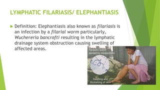
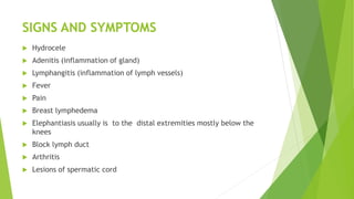
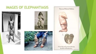
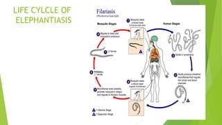
2. LYMPHATIC FILARIASIS/ ELEPHANTIASIS
Definition: Elephantiasis also known as filariasis is
an infection by a filarial worm particularly,
Wuchereria bancrofti resulting in the lymphatic
drainage system obstruction causing swelling of
affected areas.
3. INCIDENCE
It occurs globally but mostly in countries
with poor sanitation and very common in
West Africa. It is transmitted by a mosquito
bite. In Ghana Anopheles Gambei and an
funanestus species and man for
4. MODE OF TRANSMISSION
Transmitted through the bit of infected anopheles
/ culex periodicity
Reservoir: man
Vector: culex Anopheles mosquito, Anopheles
Gambei / funanestus
7. SIGNS AND SYMPTOMS
Hydrocele
Adenitis (inflammation of gland)
Lymphangitis (inflammation of lymph vessels)
Fever
Pain
Breast lymphedema
Elephantiasis usually is to the distal extremities mostly below the
knees
Block lymph duct
Arthritis
Lesions of spermatic cord
10. Life cycle cont’d
In West Africa, the disease is transmitted by culex and anopheles
species of mosquitoes. Infected microfilariae invade the lymphatic
system where they develop into adult worms within 3-12 months.
Adult worms produce more microfilariae that remain in the lymphatic
systems or are carried into the blood stream. The mosquito is
infected when it ingested microfilarie during a blood meal, the
microfilaria develop into effective forms which invades the mouth
parts of the mosquitoes.
An inflammatory process sets in due to the poor drainage caused by
the worms. Lymphatic inflammation leads to damage in the lymphatic
system leading to thickening and eventual blockage of lymph
drainage. The effected parts – the limbs, genitals – swells and
lymphatic becomes fibroses leading to elephantiasis.
11. DIAGNOSIS
Definitive diagnosis can be made only by demonstration of the parasite –
adult worm in the lymphatics (rare), micro filariea trapped in tissues, or
microfilariae in blood, urine, hydrocele fluid
Sampling for blood parasites should take into consideration the periodicity
(at night for nocturnally periodic parasites)
Parasitological (staining with geimsa or hematoxylin)
Molecular methods (amplification of Wu bancrofti specific DNA sequences
by PCR)
Serological methods ( indirect FAT, Kits)
Clinical (lymphangitis, manifestation of lymphstatis, eosinophilia etc
13. TREATMENT
Ivermectin, Albendazole
Dose.
Surgery (excision of adventitious tissue, opening up
pathways for lymph flow.
Antibiotics for bacterial infections
Elevation of the limb and use of elastic pressure
bandages
14. NURISNG MANAGEMENT
Monitor client’s vital signs particularly the
temperature
Assess skin color and integrity. Note for
wounds, bleeding or any skin changes.
Assess for any discomfort and pain.
Provide wound care
Elevate affected body area to reduce
swelling/
15. NURISNG MANAGEMENT CONT’D
Administer medications if ordered and discuss them to the
client.
Provide support to perform basic activities
Encourage range of motion and simple exercise of the
affected extremities to stimulate lymphatic flow.
Recognize client’s self –esteem needs
Provide health teaching and information for continuity of
care
16. COMPLICATIONS
Disfigurement of body part
Disability
Extreme swelling and necrosis
Blindness
NB. Secondary infection is always possible if care is not
taken.
17. REFERENCES
Lymphatic filariasis.(2018, February16). Retrieved from
http://www.who.int/news-
room/factsheets/detail/lymphatic-filariasis
Parasites (2013, June 14) Retrieved from
http://www.cdc.gov/parasite/lympaticfilariasis