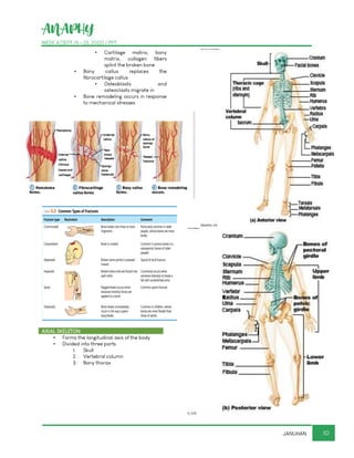
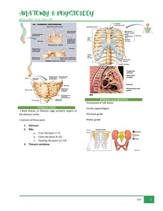
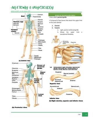
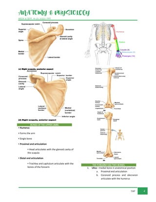
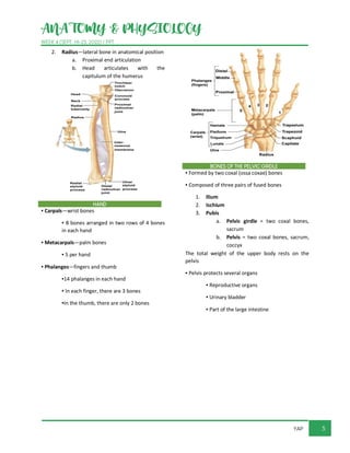
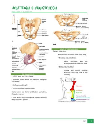
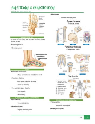
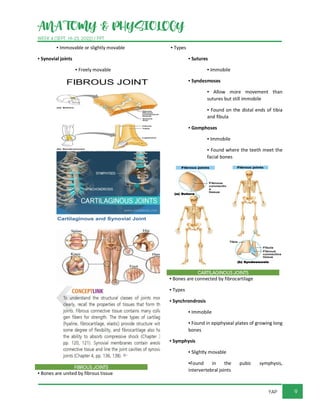
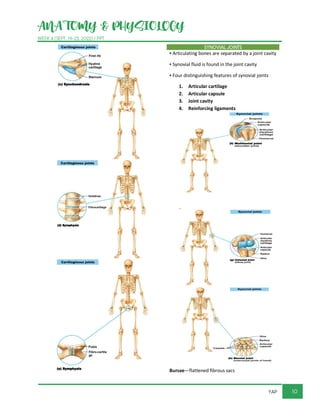
ANAPHY
WEEK 4 (SEPT 19 - 23, 2022) / PPT
1
JANUHAN
THE SKELETAL SYSTEM
TOPIC OUTLINE
1 PARTS OF THE SKELETAL SYSTEM
2 FUNCTIONS OF THE BONES
3 CLASSIFICATION OF BONES
4 STRUCTURE OF BONES
5 BONE FORMATION, GROWTH AND
REMODELING
6 BONE FRACTURES
7 AXIAL SKELETON
8 SKULL
9 VERTEBRAL COLUMN
10
11
12
13
PARTS OF THE SKELETAL SYSTEM
▪ Bones (skeleton)
▪ Joints
▪ Cartilages
▪ Ligaments
Two subdivisions of the skeleton
▪ Axial skeleton
▪ Appendicular skeleton
Joints are also called articulations are the sites where
2 or more bones meet. They have 2 functions
1. They hold the bones together securely
2. give the rigid skeleton mobility. Cartilage is a
resilient and smooth elastic tissue, a rubber-like
padding that covers and protects the ends of long
bones at the joints and nerves, and is a structural
component of the rib cage, the ear, the nose, the
bronchial tubes, the intervertebral discs, and many
other body components. Ligaments a short band of
tough, flexible fibrous connective tissue which connects
two bones or cartilages or holds together a joint.
FUNCTIONS OF THE BONES
▪ Support the body
▪ Protect soft organs
▪ Skull and vertebrae protect brain
and spinal cord
▪ Rib cage protects thoracic cavity
organs
▪ Attached skeletal muscles allow movement
▪ Store minerals and fats
▪ Calcium and phosphorus
▪ Fat in the internal marrow cavity
▪ Blood cell formation (hematopoiesis) –occurs
within the marrow cavities of certain bones.
▪ Therefore the bones of the skeleton are part
of the skeletal system which also includes
joints, cartilages and ligaments
ANAPHY
WEEK 4 (SEPT 19 - 23, 2022) / PPT
2
JANUHAN
• Store minerals and fats Calcium and
phosphorus Fat in the internal marrow cavity
.Blood cell formation (hematopoiesis)
CLASSIFICATION OF BONES
▪ The adult skeleton has 206 bones
▪ Two basic types of osseous (bone) tissue
1. Compact bone
▪ Dense, smooth, and
homogeneous
2. Spongy bone
▪ Small needlelike pieces of
bone
▪ Many open spaces
▪ Bones are classified on the basis of shape into
four groups
▪ Long
▪ Flat
▪ Short
▪ Irregular
Here is how they are classified according to shape.
Bones are classified on the basis of shape into four
groups
Long bones are typically longer in shape. Flat bones
are thin, flattened and usually curved. Most bones of
the skull, the ribs and the sternum (BREASTBONE) are
flat bones.
Short bones are generally cube-shaped. The bones of
the wrist and ankle are short bones. Sesamoid bones
within tendons are special type of short bone.
The best known example is the Patella
▪ Long bones
▪ Typically longer than they are wide
▪ Shaft with enlarged ends
▪ Contain mostly compact bone;
spongy bone at ends
▪ All of the bones of the limbs (except
wrist, ankle, and kneecap bones)
are long bones
▪ Examples:
▪ Femur
▪ Humerus
ANAPHY
WEEK 4 (SEPT 19 - 23, 2022) / PPT
3
JANUHAN
All of the bones of the limbs (except wrist, ankle, and
kneecap bones) are long bones
▪ Flat bones
▪ Thin, flattened, and usually curved
▪ Two thin layers of compact bone
sandwich a layer of spongy bone
between them
▪ Examples:
▪ Most bones of the skull
▪ Ribs
▪ Sternum
There are flat bones in the skull (occipital, parietal,
frontal, nasal, lacrimal, and vomer), the thoracic cage
(sternum and ribs), and the pelvis (ilium, ischium, and
pubis). The function of flat bones is to protect internal
organs such as the brain, heart, and pelvic organs.
▪ Short bones
▪ Generally cube-shaped
▪ Contain mostly spongy bone with an
outer layer of compact bone
▪ Sesamoid bones are a type of short
bone that form within tendons
(patella)
▪ Examples:
▪ Carpals (wrist bones)
▪ Tarsals (ankle bones)
The carpal bones are the eight small bones that make
up the wrist (or carpus) that connects the hand to the
forearm. The term "carpus" is derived from the Latin
carpus and the Greek καρπός (karpós), meaning
"wrist".
ANAPHY
WEEK 4 (SEPT 19 - 23, 2022) / PPT
4
JANUHAN
The tarsal bones consist of seven short bones located
at the proximal region of the foot. They are arranged in
proximal and distal rows. Calcaneus bone: The largest
tarsal bone that projects posteriorly as the heel.
Therefore What is tarsal region?
the tarsal region encompassing the ankle, the pedal
region encompassing the foot. the digital/phalangeal
region encompassing the toes.
▪ Irregular bones
▪ Irregular shape
▪ Do not fit into other bone
classification categories
▪ Examples:
▪ Vertebrae
▪ The vertebrae,
which make up
the spinal column.
▪ Hip bones
STRUCTURE OF BONES
▪ Long bone anatomy
▪ Diaphysis (shaft)
▪ Makes up most of bone’s
length
▪ Composed of compact
bone
▪ Periosteum
▪ Outside covering of the
diaphysis
▪ Fibrous connective tissue
membrane
▪ Perforating (Sharpey’s)
fibers secure periosteum to
underlying bone
ANAPHY
WEEK 4 (SEPT 19 - 23, 2022) / PPT
5
JANUHAN
Long bone anatomy
Diaphysis (shaft)-Makes up most of bone’s length and
Composed of compact bone
Periosteum-Outside covering of the diaphysis and
Fibrous connective tissue membrane
Perforating (Sharpey’s) fibers secure periosteum to
underlying bone
▪ Long bone anatomy (continued)
▪ Epiphysis (ends)
▪ Composed mostly of
spongy bone enclosed by
thin layer of compact bone
▪ Articular cartilage
▪ Covers the external
surface of the epiphyses
▪ Made of hyaline cartilage
▪ Decreases friction at joint
surfaces
▪ Epiphysis (ends) = Diaphysis ( the shaft or the
central part of a long bone)
There are 2 basic types of osseous, or bone.
COMPACT BONE- is dense and looks smooth and
homogeneous, whereas SPONGY BONE has a spiky,
open appearances like a sponge
▪ Long bone anatomy (continued)
▪ Epiphyseal line
▪ Remnant of the epiphyseal
plate
▪ Seen in adult bones
▪ Epiphyseal plate
▪ Flat plate of hyaline
cartilage seen in young,
growing bone
▪ Causes lengthwise growth
of a long bone
▪ Epiphyseal line -Remnant (Remains) # The
epiphyseal plate (or epiphysial plate, physis,
or growth plate) is a hyaline cartilage plate in
the metaphysis at each end of a long bone.
The metaphysis are the wide portions of long
bones and the regions of the bone where
growth occurs. Growth occurs at the section
of the metaphysis that is adjacent to the
growth plate (physis).
ANAPHY
WEEK 4 (SEPT 19 - 23, 2022) / PPT
6
JANUHAN
Epiphyseal line-Remnant of the epiphyseal plate-Seen
in adult bones #Epiphyseal plate-Flat plate of hyaline
cartilage seen in young, growing bone
Causes lengthwise growth of a long bone. The
epiphyseal plate is responsible for longitudinal bone
growth
▪ Epiphyseal plate
▪ Flat plate of hyaline
cartilage seen in young,
growing bone
▪ Causes lengthwise growth
of a long bone
Growth plates usually close near the end of puberty.
For girls, this usually is when they're 13–15; for boys, it's
when they're 15–17.
▪ Long bone anatomy (continued)
▪ Endosteum
▪ Lines the inner surface of
the shaft
▪ Made of connective tissue
▪ Medullary cavity
▪ Cavity inside the shaft
▪ Contains yellow marrow
(mostly fat) in adults
▪ Contains red marrow for
blood cell formation in
infants until age 6 or 7
The periosteum is a layer of connective tissue covering
most of your bones
Bone markings
▪ Sites of attachments for muscles,
tendons, and ligaments
▪ Passages for nerves and blood
vessels
Categories of bone markings
▪ Projections or processes—grow out
from the bone surface
▪ Terms often begin with “T”
▪ Depressions or cavities—
indentations
▪ Terms often begin with “F”
▪ Microscopic anatomy of spongy bone
▪ Composed of small, needlelike
pieces of bone called trabeculae
and open spaces
▪ Open spaces are filled by marrow,
blood vessels, and nerves
▪ Microscopic anatomy of spongy
bone
▪ Composed of small, needlelike
pieces of bone called trabeculae
and open spaces
▪ Open spaces are filled by marrow,
blood vessels, and nerves
ANAPHY
WEEK 4 (SEPT 19 - 23, 2022) / PPT
7
JANUHAN
▪ Microscopic anatomy of spongy bone
▪ Composed of small, needlelike
pieces of bone called trabeculae
and open spaces
▪ Open spaces are filled by marrow,
blood vessels, and nerves
• Haversian canals are a series of tubes around
narrow channels formed by lamellae. The
Haversian canals surround blood vessels and
nerve fibers throughout the bone and
communicate with osteocytes. The canals
and the surrounding lamellae are called a
Haversian system (or an osteon)
▪ Microscopic anatomy of compact bone
▪ Osteocytes
▪ Mature bone cells situated
in bone matrix
▪ Lacunae
▪ Cavities in bone matrix
that house osteocytes
▪ Lamellae
▪ Concentric circles of
lacunae situated around
the central (Haversian)
canal
ANAPHY
WEEK 4 (SEPT 19 - 23, 2022) / PPT
8
JANUHAN
▪ Microscopic anatomy of compact bone
(continued)
▪ Central (Haversian) canal
▪ Opening in the center of an
osteon (Haversian system)
▪ Runs lengthwise through
bone
▪ Carries blood vessels and
nerves
▪ Osteon (Haversian system)
▪ A unit of bone containing
central canal and matrix
rings
▪ Structural and functional
unit of compact bone
▪ Microscopic anatomy of compact bone
(continued)
▪ Canaliculi
▪ Tiny canals
▪ Radiate from the central
canal to lacunae
▪ Form a transport system
connecting all bone cells to
a nutrient supply
▪ Perforating (Volkmann’s) canal
▪ Canal perpendicular to the
central canal
▪ Carries blood vessels and
nerves
▪ Bone is relatively lightweight and resists
tension and other forces
▪ Organic parts (collagen fibers) of the bone
make bone flexible and have great tensile
strength
▪ Calcium salts deposited in the bone make
bone hard to resist compression
BONE FORMATION, GROWTH AND REMODELING
▪ Bone formation and growth
▪ Ossification is the process of bone
formation
▪ Occurs on hyaline cartilage models
or fibrous membranes
▪ Long bone growth involves two
major phases
▪ Two major phases of ossification in long
bones
▪ Osteoblasts (bone-forming cells)
cover hyaline cartilage model with
bone matrix
▪ In a fetus, the enclosed cartilage is
digested away, opening up a
medullary cavity
Bone formation and growth. Occurs on hyaline
cartilage models or fibrous membranes
ANAPHY
WEEK 4 (SEPT 19 - 23, 2022) / PPT
9
JANUHAN
▪ By birth, most cartilage is converted to bone
except for two regions in a long bone
1. Articular cartilages
2. Epiphyseal plates
▪ New cartilage is formed continuously on
external face of these two cartilages
▪ Old cartilage is broken down and replaced by
bony matrix
▪ Appositional growth
▪ Bones grow in width
▪ Osteoblasts in the periosteum add
bone matrix to the outside of the
diaphysis
▪ Osteoclasts in the endosteum
remove bone from the inner surface
of the diaphysis
▪ Bone growth is controlled by hormones, such
as growth hormone and sex hormones
▪ Bones are remodeled throughout life in
response to two factors
1. Calcium ion level in the blood
determines when bone matrix is to
be broken down or formed
2. Pull of gravity and muscles on the
skeleton determines where bone
matrix is to be broken down or
formed
▪ Calcium ion regulation
1. Parathyroid hormone (PTH)
▪ Released when calcium ion
levels in blood are low
▪ Activates osteoclasts
(bone-destroying cells)
▪ Osteoclasts break down
bone and release calcium
ions into the blood
2. Hypercalcemia (high blood calcium
levels) prompts calcium storage to
bones by osteoblasts
▪ Parathyroid hormone is secreted from four
parathyroid glands, which are small glands in
the neck, located behind the thyroid gland.
Parathyroid hormone regulates calcium
levels in the blood, largely by increasing the
levels when they are too low. OSTEOBLASTS
are the cells that form new bone.
BONE FRACTURES
▪ Fracture: break in a bone
▪ Types of bone fractures
▪ Closed (simple) fracture is a break
that does not penetrate the skin
▪ Open (compound) fracture is a
broken bone that penetrates
through the skin
▪ Bone fractures are treated by reduction and
immobilization
▪ Closed reduction: bones are
manually coaxed into position by
physician’s hands
▪ Open reduction: bones are secured
with pins or wires during surgery
▪ Healing time is 6–8 weeks
▪ Repair of bone fractures involves four major
events
▪ Hematoma (blood-filled swelling, or
bruise) is formed
▪ Fibrocartilage callus forms
ANAPHY
WEEK 4 (SEPT 19 - 23, 2022) / PPT
10
JANUHAN
▪ Cartilage matrix, bony
matrix, collagen fibers
splint the broken bone
▪ Bony callus replaces the
fibrocartilage callus
▪ Osteoblasts and
osteoclasts migrate in
▪ Bone remodeling occurs in response
to mechanical stresses
AXIAL SKELETON
▪ Forms the longitudinal axis of the body
▪ Divided into three parts
1. Skull
2. Vertebral column
3. Bony thorax
ANAPHY
WEEK 4 (SEPT 19 - 23, 2022) / PPT
11
JANUHAN
SKULL
▪ Two sets of bones form the skull
1. Cranium bones enclose the brain
2. Facial bones
▪ Hold eyes in anterior
position
▪ Allow facial muscles to
express feelings
▪ Bones are joined by sutures
▪ Only the mandible is attached by a freely
movable joint
▪ 8 cranial bones protect the brain
1 Frontal bone
2 Occipital bone
3 Ethmoid bone
4 Sphenoid bone
5, 6 Parietal bones (pair)
7, 8 Temporal bones (pair)
▪ 14 facial bones
1, 2 Maxillae (pair)
3, 4 Palatine bones (pair)
5, 6 Lacrimal bones (pair)
7, 8 Zygomatic bones (pair)
9, 10 Nasal bones (pair)
11 Vomer bone
12, 13 Inferior nasal conchae (pair)
14 Mandible
▪ Paranasal sinuses
▪ Hollow portions of bones
surrounding the nasal cavity
▪ Functions of paranasal sinuses
▪ Lighten the skull
▪ Amplify sounds made as
we speak
ANAPHY
WEEK 4 (SEPT 19 - 23, 2022) / PPT
12
JANUHAN
our sinuses are air spaces in your skull and facial
bones that make up the upper part of your respiratory
tract from your nose into your throat. The sinuses are
in your forehead (frontal sinuses), inside your
cheekbones (maxillary sinuses), and behind the nose
(ethmoid and sphenoid sinuses).
▪ Hyoid bone
▪ Closely related to mandible and
temporal bones
▪ The only bone that does not
articulate with another bone
▪ Serves as a movable base for the
tongue
▪ Aids in swallowing and speech
VERTEBRAL COLUMN
▪ Vertebral column provides axial support
▪ Extends from skull to the pelvis
▪ 26 vertebral bones are separated by
intervertebral discs
▪ 7 cervical vertebrae are in the neck
▪ 12 thoracic vertebrae are in the
chest region
▪ 5 lumbar vertebrae are associated
with the lower back
▪ Sacrum (formed by fusion of 5
vertebrae)
▪ Coccyx (formed by fusion of 3–5
vertebrae)
▪ Vertebrae. Vertebrae are the 33 individual
bones that interlock with each other to form
the spinal column. The vertebrae are
numbered and divided into regions: cervical,
thoracic, lumbar, sacrum, and coccyx .
Before birth, the spine consist of 33 separate
bones called vertebrae, but 9 of these
eventually fuse to form the 2 composite
bones, the sacrum, the coccyx, that construct
the inferior portion of the vertebral column
▪ Primary curvatures
▪ Spinal curvatures of the thoracic
and sacral regions
▪ Present from birth
▪ Form a C-shaped curvature in
newborns
▪ Secondary curvatures
▪ Spinal curvatures of the cervical and
lumbar regions
▪ Develop after birth
ANAPHY
WEEK 4 (SEPT 19 - 23, 2022) / PPT
13
JANUHAN
▪ Form an S-shaped curvature in
adults
ANATOMY & PHYSIOLOGY
WEEK 4 (SEPT. 19-23, 2022) / PPT
1
YAP
VERTEBRAL COLUMN (SPINE)
PARTS OF A TYPICAL VERTEBRA
▪ Body (centrum)
▪ Vertebral arch
▪ Pedicle
▪ Lamina
▪ Vertebral foramen
▪ Transverse processes
▪ Spinous process
▪ Superior and inferior articular processes
ANATOMY & PHYSIOLOGY
WEEK 4 (SEPT. 19-23, 2022) / PPT
2
YAP
THORACIC CAGE
▪ Bony thorax, or thoracic cage, protects organs of
the thoracic cavity
▪ Consists of three parts
1. Sternum
2. Ribs
a. True ribs (pairs 1–7)
b. False ribs (pairs 8–12)
c. Floating ribs (pairs 11–12)
3. Thoracic vertebrae
APPENDICULAR SKELETON
▪Composed of 126 bones
▪Limbs (appendages)
▪Pectoral girdle
▪Pelvic girdle
ANATOMY & PHYSIOLOGY
WEEK 4 (SEPT. 19-23, 2022) / PPT
3
YAP
BONES OF THE SHOULDER GIRDLE
▪ Also called pectoral girdle
▪ Composed of two bones that attach the upper limb
to the axial skeletal
1. Clavicle
2. Scapula
a. Light, poorly reinforced girdle
b. Allows the upper limb a
exceptional flexibility
ANATOMY & PHYSIOLOGY
WEEK 4 (SEPT. 19-23, 2022) / PPT
4
YAP
BONES OF THE UPPER LIMBS
▪ Humerus
▪ Forms the arm
▪ Single bone
▪ Proximal end articulation
▪ Head articulates with the glenoid cavity of
the scapula
▪ Distal end articulation
▪ Trochlea and capitulum articulate with the
bones of the forearm
THE FOREARM HAS TWO BONES:
1. Ulna—medial bone in anatomical position
a. Proximal end articulation
b. Coronoid process and olecranon
articulate with the humerus
ANATOMY & PHYSIOLOGY
WEEK 4 (SEPT. 19-23, 2022) / PPT
5
YAP
2. Radius—lateral bone in anatomical position
a. Proximal end articulation
b. Head articulates with the
capitulum of the humerus
HAND
▪ Carpals—wrist bones
▪ 8 bones arranged in two rows of 4 bones
in each hand
▪ Metacarpals—palm bones
▪ 5 per hand
▪ Phalanges—fingers and thumb
▪14 phalanges in each hand
▪ In each finger, there are 3 bones
▪In the thumb, there are only 2 bones
BONES OF THE PELVIC GIRDLE
▪ Formed by two coxal (ossa coxae) bones
▪ Composed of three pairs of fused bones
1. Ilium
2. Ischium
3. Pubis
a. Pelvic girdle = two coxal bones,
sacrum
b. Pelvis = two coxal bones, sacrum,
coccyx
The total weight of the upper body rests on the
pelvis
▪ Pelvis protects several organs
▪ Reproductive organs
▪ Urinary bladder
▪ Part of the large intestine
ANATOMY & PHYSIOLOGY
WEEK 4 (SEPT. 19-23, 2022) / PPT
6
YAP
THE FEMALE PELVIS
▪ Inlet is larger and more circular
▪ Shallower, on the whole, and the bones are lighter
and thinner
▪ Ilia flare more laterally
▪ Sacrum is shorter and less curved
▪Ischial spines are shorter and farther apart; thus,
the outlet is larger
▪ Pubic arch is more rounded because the angle of
the pubic arch is greater
BONES OF THE LOWER LIMBS
▪ Femur—thigh bone
▪ The heaviest, strongest bone in the body
▪ Proximal end articulation
▪Head articulates with the
acetabulum of the coxal (hip) bone
▪ Distal end articulation
▪Lateral and medial condyles
articulate with the tibia in the
lower leg
ANATOMY & PHYSIOLOGY
WEEK 4 (SEPT. 19-23, 2022) / PPT
7
YAP
THE LOWER LEG HAS TWO BONES:
1. Tibia—shinbone; larger and medially
oriented
a. Proximal end articulation
b. Medial and lateral condyles
articulate with the femur to form
the knee joint
c. Distal end articulation
d. Medial malleolus forms the inner
part of the ankle
2. Fibula—thin and sticklike; lateral to the
tibia
a. Has no role in forming the knee
joint
b. Distal end articulation
c. Lateral malleolus forms the outer
part of the ankle
FOOT
▪Tarsals—7 bones
▪ Two largest tarsals are the:
▪Calcaneus(heel bone)
▪ Talus
▪ Metatarsals—5 bones form the sole of the foot
▪ Phalanges—14 bones form the toes
ANATOMY & PHYSIOLOGY
WEEK 4 (SEPT. 19-23, 2022) / PPT
8
YAP
ARCHES OF THE FEET
▪ Bones of the foot are arranged to form three
strong arches
▪ Two longitudinal
▪ One transverse
JOINTS
▪ Joints are articulations
▪ Occur where two or more bones meet
▪ Functions of joints
▪Hold bones together securely
▪ Allow for mobility
▪Two ways joints are classified
▪ Functionally
▪ Structurally
FUNCTIONAL JOINT CLASSIFICATIONS
▪ Synarthroses
▪ Immovable joints
▪ Amphiarthroses
▪ Slightly movable joints
▪ Diarthroses
▪ Freely movable joints
STRUCTURAL JOINT CLASSIFICA8
▪Fibrous joints
▪Generally immovable
▪ Cartilaginous joints
ANATOMY & PHYSIOLOGY
WEEK 4 (SEPT. 19-23, 2022) / PPT
9
YAP
▪ Immovable or slightly movable
▪ Synovial joints
▪ Freely movable
FIBROUS JOINTS
▪ Bones are united by fibrous tissue
▪ Types
▪ Sutures
▪ Immobile
▪ Syndesmoses
▪ Allow more movement than
sutures but still immobile
▪ Found on the distal ends of tibia
and fibula
▪ Gomphoses
▪ Immobile
▪ Found where the teeth meet the
facial bones
CARTILAGINOUS JOINTS
▪ Bones are connected by fibrocartilage
▪ Types
▪ Synchrondrosis
▪ Immobile
▪ Found in epiphyseal plates of growing long
bones
▪ Symphysis
▪ Slightly movable
▪Found in the pubic symphysis,
intervertebral joints
ANATOMY & PHYSIOLOGY
WEEK 4 (SEPT. 19-23, 2022) / PPT
10
YAP
SYNOVIAL JOINTS
▪ Articulating bones are separated by a joint cavity
▪ Synovial fluid is found in the joint cavity
▪ Four distinguishing features of synovial joints
1. Articular cartilage
2. Articular capsule
3. Joint cavity
4. Reinforcing ligaments
Bursae—flattened fibrous sacs
ANATOMY & PHYSIOLOGY
WEEK 4 (SEPT. 19-23, 2022) / PPT
11
YAP
▪ Lined with synovial membranes
▪ Filled with synovial fluid
▪ Not actually part of the joint
▪ Tendon sheath
▪Elongated bursa that wraps around a
tendon
TYPES OF SYNOVIAL JOINTS BASED ON SHAPE
▪ Plane joint
▪ Hinge joint
▪ Pivot joint
▪ Condylar joint
▪ Saddle joint
▪ Ball-and-socket joint DEVELOPMENTAL ASPECTS OF THE SKELETON
▪ Birth to adulthood
▪ First “long bones” of a fetus are hyaline cartilage
▪ Earliest “flat bones” of the fetal skull are fibrous
membranes
▪ As fetus grows, all bone models are converted to
bone
FETAL SKULL
▪Fontanels are fibrous membranes connecting the
cranial bones
▪ Known as “soft spots”
▪ Allow skull compression during birth
▪ Allow the brain to grow during later pregnancy and
infancy
▪ Usually ossify by 2 years of age
ANATOMY & PHYSIOLOGY
WEEK 4 (SEPT. 19-23, 2022) / PPT
12
YAP
GROWTH OF CRANIUM AFTER BIRTH IS RELATED TO
BRAIN GROWTH
▪ Increase in size of the facial skeleton follows tooth
development and enlargement of the respiratory
passageways
SIZE OF CRANIUM IN RELATIONSHIP TO BODY
▪ 2 years old—skull is three-fourths the size of adult
skull
▪ 8 or 9 years old—skull is near adult in size and
proportion
▪ Between ages 6 and 11, the face grows out from
the skull
SKELETAL CHANGES
▪ At birth, the head and trunk are proportionately
much longer than the lower limbs
▪ During puberty:
▪ Female pelvis broadens
▪ Entire male skeleton becomes more robust
▪ By the end of adolescence:
▪ Epiphyseal plates become fully ossified
OLDER ADULTS
▪ Osteoporosis
▪ Bone-thinning disease afflicting:
▪ 50 percent of women over age 65
▪ 20 percent of men over age 70
▪ Disease makes bones fragile, and bones can easily
fracture
▪ Vertebral collapse results in kyphosis (also known
as “dowager’s hump”)
▪ Estrogen aids in health and normal density of a
female skeleton

NURSING TRANS NOTES 1.05 ANAPHY-SKELETAL-FINAL.pdf

  • 1.
    ANAPHY WEEK 4 (SEPT19 - 23, 2022) / PPT 1 JANUHAN THE SKELETAL SYSTEM TOPIC OUTLINE 1 PARTS OF THE SKELETAL SYSTEM 2 FUNCTIONS OF THE BONES 3 CLASSIFICATION OF BONES 4 STRUCTURE OF BONES 5 BONE FORMATION, GROWTH AND REMODELING 6 BONE FRACTURES 7 AXIAL SKELETON 8 SKULL 9 VERTEBRAL COLUMN 10 11 12 13 PARTS OF THE SKELETAL SYSTEM ▪ Bones (skeleton) ▪ Joints ▪ Cartilages ▪ Ligaments Two subdivisions of the skeleton ▪ Axial skeleton ▪ Appendicular skeleton Joints are also called articulations are the sites where 2 or more bones meet. They have 2 functions 1. They hold the bones together securely 2. give the rigid skeleton mobility. Cartilage is a resilient and smooth elastic tissue, a rubber-like padding that covers and protects the ends of long bones at the joints and nerves, and is a structural component of the rib cage, the ear, the nose, the bronchial tubes, the intervertebral discs, and many other body components. Ligaments a short band of tough, flexible fibrous connective tissue which connects two bones or cartilages or holds together a joint. FUNCTIONS OF THE BONES ▪ Support the body ▪ Protect soft organs ▪ Skull and vertebrae protect brain and spinal cord ▪ Rib cage protects thoracic cavity organs ▪ Attached skeletal muscles allow movement ▪ Store minerals and fats ▪ Calcium and phosphorus ▪ Fat in the internal marrow cavity ▪ Blood cell formation (hematopoiesis) –occurs within the marrow cavities of certain bones. ▪ Therefore the bones of the skeleton are part of the skeletal system which also includes joints, cartilages and ligaments
  • 2.
    ANAPHY WEEK 4 (SEPT19 - 23, 2022) / PPT 2 JANUHAN • Store minerals and fats Calcium and phosphorus Fat in the internal marrow cavity .Blood cell formation (hematopoiesis) CLASSIFICATION OF BONES ▪ The adult skeleton has 206 bones ▪ Two basic types of osseous (bone) tissue 1. Compact bone ▪ Dense, smooth, and homogeneous 2. Spongy bone ▪ Small needlelike pieces of bone ▪ Many open spaces ▪ Bones are classified on the basis of shape into four groups ▪ Long ▪ Flat ▪ Short ▪ Irregular Here is how they are classified according to shape. Bones are classified on the basis of shape into four groups Long bones are typically longer in shape. Flat bones are thin, flattened and usually curved. Most bones of the skull, the ribs and the sternum (BREASTBONE) are flat bones. Short bones are generally cube-shaped. The bones of the wrist and ankle are short bones. Sesamoid bones within tendons are special type of short bone. The best known example is the Patella ▪ Long bones ▪ Typically longer than they are wide ▪ Shaft with enlarged ends ▪ Contain mostly compact bone; spongy bone at ends ▪ All of the bones of the limbs (except wrist, ankle, and kneecap bones) are long bones ▪ Examples: ▪ Femur ▪ Humerus
  • 3.
    ANAPHY WEEK 4 (SEPT19 - 23, 2022) / PPT 3 JANUHAN All of the bones of the limbs (except wrist, ankle, and kneecap bones) are long bones ▪ Flat bones ▪ Thin, flattened, and usually curved ▪ Two thin layers of compact bone sandwich a layer of spongy bone between them ▪ Examples: ▪ Most bones of the skull ▪ Ribs ▪ Sternum There are flat bones in the skull (occipital, parietal, frontal, nasal, lacrimal, and vomer), the thoracic cage (sternum and ribs), and the pelvis (ilium, ischium, and pubis). The function of flat bones is to protect internal organs such as the brain, heart, and pelvic organs. ▪ Short bones ▪ Generally cube-shaped ▪ Contain mostly spongy bone with an outer layer of compact bone ▪ Sesamoid bones are a type of short bone that form within tendons (patella) ▪ Examples: ▪ Carpals (wrist bones) ▪ Tarsals (ankle bones) The carpal bones are the eight small bones that make up the wrist (or carpus) that connects the hand to the forearm. The term "carpus" is derived from the Latin carpus and the Greek καρπός (karpós), meaning "wrist".
  • 4.
    ANAPHY WEEK 4 (SEPT19 - 23, 2022) / PPT 4 JANUHAN The tarsal bones consist of seven short bones located at the proximal region of the foot. They are arranged in proximal and distal rows. Calcaneus bone: The largest tarsal bone that projects posteriorly as the heel. Therefore What is tarsal region? the tarsal region encompassing the ankle, the pedal region encompassing the foot. the digital/phalangeal region encompassing the toes. ▪ Irregular bones ▪ Irregular shape ▪ Do not fit into other bone classification categories ▪ Examples: ▪ Vertebrae ▪ The vertebrae, which make up the spinal column. ▪ Hip bones STRUCTURE OF BONES ▪ Long bone anatomy ▪ Diaphysis (shaft) ▪ Makes up most of bone’s length ▪ Composed of compact bone ▪ Periosteum ▪ Outside covering of the diaphysis ▪ Fibrous connective tissue membrane ▪ Perforating (Sharpey’s) fibers secure periosteum to underlying bone
  • 5.
    ANAPHY WEEK 4 (SEPT19 - 23, 2022) / PPT 5 JANUHAN Long bone anatomy Diaphysis (shaft)-Makes up most of bone’s length and Composed of compact bone Periosteum-Outside covering of the diaphysis and Fibrous connective tissue membrane Perforating (Sharpey’s) fibers secure periosteum to underlying bone ▪ Long bone anatomy (continued) ▪ Epiphysis (ends) ▪ Composed mostly of spongy bone enclosed by thin layer of compact bone ▪ Articular cartilage ▪ Covers the external surface of the epiphyses ▪ Made of hyaline cartilage ▪ Decreases friction at joint surfaces ▪ Epiphysis (ends) = Diaphysis ( the shaft or the central part of a long bone) There are 2 basic types of osseous, or bone. COMPACT BONE- is dense and looks smooth and homogeneous, whereas SPONGY BONE has a spiky, open appearances like a sponge ▪ Long bone anatomy (continued) ▪ Epiphyseal line ▪ Remnant of the epiphyseal plate ▪ Seen in adult bones ▪ Epiphyseal plate ▪ Flat plate of hyaline cartilage seen in young, growing bone ▪ Causes lengthwise growth of a long bone ▪ Epiphyseal line -Remnant (Remains) # The epiphyseal plate (or epiphysial plate, physis, or growth plate) is a hyaline cartilage plate in the metaphysis at each end of a long bone. The metaphysis are the wide portions of long bones and the regions of the bone where growth occurs. Growth occurs at the section of the metaphysis that is adjacent to the growth plate (physis).
  • 6.
    ANAPHY WEEK 4 (SEPT19 - 23, 2022) / PPT 6 JANUHAN Epiphyseal line-Remnant of the epiphyseal plate-Seen in adult bones #Epiphyseal plate-Flat plate of hyaline cartilage seen in young, growing bone Causes lengthwise growth of a long bone. The epiphyseal plate is responsible for longitudinal bone growth ▪ Epiphyseal plate ▪ Flat plate of hyaline cartilage seen in young, growing bone ▪ Causes lengthwise growth of a long bone Growth plates usually close near the end of puberty. For girls, this usually is when they're 13–15; for boys, it's when they're 15–17. ▪ Long bone anatomy (continued) ▪ Endosteum ▪ Lines the inner surface of the shaft ▪ Made of connective tissue ▪ Medullary cavity ▪ Cavity inside the shaft ▪ Contains yellow marrow (mostly fat) in adults ▪ Contains red marrow for blood cell formation in infants until age 6 or 7 The periosteum is a layer of connective tissue covering most of your bones Bone markings ▪ Sites of attachments for muscles, tendons, and ligaments ▪ Passages for nerves and blood vessels Categories of bone markings ▪ Projections or processes—grow out from the bone surface ▪ Terms often begin with “T” ▪ Depressions or cavities— indentations ▪ Terms often begin with “F” ▪ Microscopic anatomy of spongy bone ▪ Composed of small, needlelike pieces of bone called trabeculae and open spaces ▪ Open spaces are filled by marrow, blood vessels, and nerves ▪ Microscopic anatomy of spongy bone ▪ Composed of small, needlelike pieces of bone called trabeculae and open spaces ▪ Open spaces are filled by marrow, blood vessels, and nerves
  • 7.
    ANAPHY WEEK 4 (SEPT19 - 23, 2022) / PPT 7 JANUHAN ▪ Microscopic anatomy of spongy bone ▪ Composed of small, needlelike pieces of bone called trabeculae and open spaces ▪ Open spaces are filled by marrow, blood vessels, and nerves • Haversian canals are a series of tubes around narrow channels formed by lamellae. The Haversian canals surround blood vessels and nerve fibers throughout the bone and communicate with osteocytes. The canals and the surrounding lamellae are called a Haversian system (or an osteon) ▪ Microscopic anatomy of compact bone ▪ Osteocytes ▪ Mature bone cells situated in bone matrix ▪ Lacunae ▪ Cavities in bone matrix that house osteocytes ▪ Lamellae ▪ Concentric circles of lacunae situated around the central (Haversian) canal
  • 8.
    ANAPHY WEEK 4 (SEPT19 - 23, 2022) / PPT 8 JANUHAN ▪ Microscopic anatomy of compact bone (continued) ▪ Central (Haversian) canal ▪ Opening in the center of an osteon (Haversian system) ▪ Runs lengthwise through bone ▪ Carries blood vessels and nerves ▪ Osteon (Haversian system) ▪ A unit of bone containing central canal and matrix rings ▪ Structural and functional unit of compact bone ▪ Microscopic anatomy of compact bone (continued) ▪ Canaliculi ▪ Tiny canals ▪ Radiate from the central canal to lacunae ▪ Form a transport system connecting all bone cells to a nutrient supply ▪ Perforating (Volkmann’s) canal ▪ Canal perpendicular to the central canal ▪ Carries blood vessels and nerves ▪ Bone is relatively lightweight and resists tension and other forces ▪ Organic parts (collagen fibers) of the bone make bone flexible and have great tensile strength ▪ Calcium salts deposited in the bone make bone hard to resist compression BONE FORMATION, GROWTH AND REMODELING ▪ Bone formation and growth ▪ Ossification is the process of bone formation ▪ Occurs on hyaline cartilage models or fibrous membranes ▪ Long bone growth involves two major phases ▪ Two major phases of ossification in long bones ▪ Osteoblasts (bone-forming cells) cover hyaline cartilage model with bone matrix ▪ In a fetus, the enclosed cartilage is digested away, opening up a medullary cavity Bone formation and growth. Occurs on hyaline cartilage models or fibrous membranes
  • 9.
    ANAPHY WEEK 4 (SEPT19 - 23, 2022) / PPT 9 JANUHAN ▪ By birth, most cartilage is converted to bone except for two regions in a long bone 1. Articular cartilages 2. Epiphyseal plates ▪ New cartilage is formed continuously on external face of these two cartilages ▪ Old cartilage is broken down and replaced by bony matrix ▪ Appositional growth ▪ Bones grow in width ▪ Osteoblasts in the periosteum add bone matrix to the outside of the diaphysis ▪ Osteoclasts in the endosteum remove bone from the inner surface of the diaphysis ▪ Bone growth is controlled by hormones, such as growth hormone and sex hormones ▪ Bones are remodeled throughout life in response to two factors 1. Calcium ion level in the blood determines when bone matrix is to be broken down or formed 2. Pull of gravity and muscles on the skeleton determines where bone matrix is to be broken down or formed ▪ Calcium ion regulation 1. Parathyroid hormone (PTH) ▪ Released when calcium ion levels in blood are low ▪ Activates osteoclasts (bone-destroying cells) ▪ Osteoclasts break down bone and release calcium ions into the blood 2. Hypercalcemia (high blood calcium levels) prompts calcium storage to bones by osteoblasts ▪ Parathyroid hormone is secreted from four parathyroid glands, which are small glands in the neck, located behind the thyroid gland. Parathyroid hormone regulates calcium levels in the blood, largely by increasing the levels when they are too low. OSTEOBLASTS are the cells that form new bone. BONE FRACTURES ▪ Fracture: break in a bone ▪ Types of bone fractures ▪ Closed (simple) fracture is a break that does not penetrate the skin ▪ Open (compound) fracture is a broken bone that penetrates through the skin ▪ Bone fractures are treated by reduction and immobilization ▪ Closed reduction: bones are manually coaxed into position by physician’s hands ▪ Open reduction: bones are secured with pins or wires during surgery ▪ Healing time is 6–8 weeks ▪ Repair of bone fractures involves four major events ▪ Hematoma (blood-filled swelling, or bruise) is formed ▪ Fibrocartilage callus forms
  • 10.
    ANAPHY WEEK 4 (SEPT19 - 23, 2022) / PPT 10 JANUHAN ▪ Cartilage matrix, bony matrix, collagen fibers splint the broken bone ▪ Bony callus replaces the fibrocartilage callus ▪ Osteoblasts and osteoclasts migrate in ▪ Bone remodeling occurs in response to mechanical stresses AXIAL SKELETON ▪ Forms the longitudinal axis of the body ▪ Divided into three parts 1. Skull 2. Vertebral column 3. Bony thorax
  • 11.
    ANAPHY WEEK 4 (SEPT19 - 23, 2022) / PPT 11 JANUHAN SKULL ▪ Two sets of bones form the skull 1. Cranium bones enclose the brain 2. Facial bones ▪ Hold eyes in anterior position ▪ Allow facial muscles to express feelings ▪ Bones are joined by sutures ▪ Only the mandible is attached by a freely movable joint ▪ 8 cranial bones protect the brain 1 Frontal bone 2 Occipital bone 3 Ethmoid bone 4 Sphenoid bone 5, 6 Parietal bones (pair) 7, 8 Temporal bones (pair) ▪ 14 facial bones 1, 2 Maxillae (pair) 3, 4 Palatine bones (pair) 5, 6 Lacrimal bones (pair) 7, 8 Zygomatic bones (pair) 9, 10 Nasal bones (pair) 11 Vomer bone 12, 13 Inferior nasal conchae (pair) 14 Mandible ▪ Paranasal sinuses ▪ Hollow portions of bones surrounding the nasal cavity ▪ Functions of paranasal sinuses ▪ Lighten the skull ▪ Amplify sounds made as we speak
  • 12.
    ANAPHY WEEK 4 (SEPT19 - 23, 2022) / PPT 12 JANUHAN our sinuses are air spaces in your skull and facial bones that make up the upper part of your respiratory tract from your nose into your throat. The sinuses are in your forehead (frontal sinuses), inside your cheekbones (maxillary sinuses), and behind the nose (ethmoid and sphenoid sinuses). ▪ Hyoid bone ▪ Closely related to mandible and temporal bones ▪ The only bone that does not articulate with another bone ▪ Serves as a movable base for the tongue ▪ Aids in swallowing and speech VERTEBRAL COLUMN ▪ Vertebral column provides axial support ▪ Extends from skull to the pelvis ▪ 26 vertebral bones are separated by intervertebral discs ▪ 7 cervical vertebrae are in the neck ▪ 12 thoracic vertebrae are in the chest region ▪ 5 lumbar vertebrae are associated with the lower back ▪ Sacrum (formed by fusion of 5 vertebrae) ▪ Coccyx (formed by fusion of 3–5 vertebrae) ▪ Vertebrae. Vertebrae are the 33 individual bones that interlock with each other to form the spinal column. The vertebrae are numbered and divided into regions: cervical, thoracic, lumbar, sacrum, and coccyx . Before birth, the spine consist of 33 separate bones called vertebrae, but 9 of these eventually fuse to form the 2 composite bones, the sacrum, the coccyx, that construct the inferior portion of the vertebral column ▪ Primary curvatures ▪ Spinal curvatures of the thoracic and sacral regions ▪ Present from birth ▪ Form a C-shaped curvature in newborns ▪ Secondary curvatures ▪ Spinal curvatures of the cervical and lumbar regions ▪ Develop after birth
  • 13.
    ANAPHY WEEK 4 (SEPT19 - 23, 2022) / PPT 13 JANUHAN ▪ Form an S-shaped curvature in adults
  • 14.
    ANATOMY & PHYSIOLOGY WEEK4 (SEPT. 19-23, 2022) / PPT 1 YAP VERTEBRAL COLUMN (SPINE) PARTS OF A TYPICAL VERTEBRA ▪ Body (centrum) ▪ Vertebral arch ▪ Pedicle ▪ Lamina ▪ Vertebral foramen ▪ Transverse processes ▪ Spinous process ▪ Superior and inferior articular processes
  • 15.
    ANATOMY & PHYSIOLOGY WEEK4 (SEPT. 19-23, 2022) / PPT 2 YAP THORACIC CAGE ▪ Bony thorax, or thoracic cage, protects organs of the thoracic cavity ▪ Consists of three parts 1. Sternum 2. Ribs a. True ribs (pairs 1–7) b. False ribs (pairs 8–12) c. Floating ribs (pairs 11–12) 3. Thoracic vertebrae APPENDICULAR SKELETON ▪Composed of 126 bones ▪Limbs (appendages) ▪Pectoral girdle ▪Pelvic girdle
  • 16.
    ANATOMY & PHYSIOLOGY WEEK4 (SEPT. 19-23, 2022) / PPT 3 YAP BONES OF THE SHOULDER GIRDLE ▪ Also called pectoral girdle ▪ Composed of two bones that attach the upper limb to the axial skeletal 1. Clavicle 2. Scapula a. Light, poorly reinforced girdle b. Allows the upper limb a exceptional flexibility
  • 17.
    ANATOMY & PHYSIOLOGY WEEK4 (SEPT. 19-23, 2022) / PPT 4 YAP BONES OF THE UPPER LIMBS ▪ Humerus ▪ Forms the arm ▪ Single bone ▪ Proximal end articulation ▪ Head articulates with the glenoid cavity of the scapula ▪ Distal end articulation ▪ Trochlea and capitulum articulate with the bones of the forearm THE FOREARM HAS TWO BONES: 1. Ulna—medial bone in anatomical position a. Proximal end articulation b. Coronoid process and olecranon articulate with the humerus
  • 18.
    ANATOMY & PHYSIOLOGY WEEK4 (SEPT. 19-23, 2022) / PPT 5 YAP 2. Radius—lateral bone in anatomical position a. Proximal end articulation b. Head articulates with the capitulum of the humerus HAND ▪ Carpals—wrist bones ▪ 8 bones arranged in two rows of 4 bones in each hand ▪ Metacarpals—palm bones ▪ 5 per hand ▪ Phalanges—fingers and thumb ▪14 phalanges in each hand ▪ In each finger, there are 3 bones ▪In the thumb, there are only 2 bones BONES OF THE PELVIC GIRDLE ▪ Formed by two coxal (ossa coxae) bones ▪ Composed of three pairs of fused bones 1. Ilium 2. Ischium 3. Pubis a. Pelvic girdle = two coxal bones, sacrum b. Pelvis = two coxal bones, sacrum, coccyx The total weight of the upper body rests on the pelvis ▪ Pelvis protects several organs ▪ Reproductive organs ▪ Urinary bladder ▪ Part of the large intestine
  • 19.
    ANATOMY & PHYSIOLOGY WEEK4 (SEPT. 19-23, 2022) / PPT 6 YAP THE FEMALE PELVIS ▪ Inlet is larger and more circular ▪ Shallower, on the whole, and the bones are lighter and thinner ▪ Ilia flare more laterally ▪ Sacrum is shorter and less curved ▪Ischial spines are shorter and farther apart; thus, the outlet is larger ▪ Pubic arch is more rounded because the angle of the pubic arch is greater BONES OF THE LOWER LIMBS ▪ Femur—thigh bone ▪ The heaviest, strongest bone in the body ▪ Proximal end articulation ▪Head articulates with the acetabulum of the coxal (hip) bone ▪ Distal end articulation ▪Lateral and medial condyles articulate with the tibia in the lower leg
  • 20.
    ANATOMY & PHYSIOLOGY WEEK4 (SEPT. 19-23, 2022) / PPT 7 YAP THE LOWER LEG HAS TWO BONES: 1. Tibia—shinbone; larger and medially oriented a. Proximal end articulation b. Medial and lateral condyles articulate with the femur to form the knee joint c. Distal end articulation d. Medial malleolus forms the inner part of the ankle 2. Fibula—thin and sticklike; lateral to the tibia a. Has no role in forming the knee joint b. Distal end articulation c. Lateral malleolus forms the outer part of the ankle FOOT ▪Tarsals—7 bones ▪ Two largest tarsals are the: ▪Calcaneus(heel bone) ▪ Talus ▪ Metatarsals—5 bones form the sole of the foot ▪ Phalanges—14 bones form the toes
  • 21.
    ANATOMY & PHYSIOLOGY WEEK4 (SEPT. 19-23, 2022) / PPT 8 YAP ARCHES OF THE FEET ▪ Bones of the foot are arranged to form three strong arches ▪ Two longitudinal ▪ One transverse JOINTS ▪ Joints are articulations ▪ Occur where two or more bones meet ▪ Functions of joints ▪Hold bones together securely ▪ Allow for mobility ▪Two ways joints are classified ▪ Functionally ▪ Structurally FUNCTIONAL JOINT CLASSIFICATIONS ▪ Synarthroses ▪ Immovable joints ▪ Amphiarthroses ▪ Slightly movable joints ▪ Diarthroses ▪ Freely movable joints STRUCTURAL JOINT CLASSIFICA8 ▪Fibrous joints ▪Generally immovable ▪ Cartilaginous joints
  • 22.
    ANATOMY & PHYSIOLOGY WEEK4 (SEPT. 19-23, 2022) / PPT 9 YAP ▪ Immovable or slightly movable ▪ Synovial joints ▪ Freely movable FIBROUS JOINTS ▪ Bones are united by fibrous tissue ▪ Types ▪ Sutures ▪ Immobile ▪ Syndesmoses ▪ Allow more movement than sutures but still immobile ▪ Found on the distal ends of tibia and fibula ▪ Gomphoses ▪ Immobile ▪ Found where the teeth meet the facial bones CARTILAGINOUS JOINTS ▪ Bones are connected by fibrocartilage ▪ Types ▪ Synchrondrosis ▪ Immobile ▪ Found in epiphyseal plates of growing long bones ▪ Symphysis ▪ Slightly movable ▪Found in the pubic symphysis, intervertebral joints
  • 23.
    ANATOMY & PHYSIOLOGY WEEK4 (SEPT. 19-23, 2022) / PPT 10 YAP SYNOVIAL JOINTS ▪ Articulating bones are separated by a joint cavity ▪ Synovial fluid is found in the joint cavity ▪ Four distinguishing features of synovial joints 1. Articular cartilage 2. Articular capsule 3. Joint cavity 4. Reinforcing ligaments Bursae—flattened fibrous sacs
  • 24.
    ANATOMY & PHYSIOLOGY WEEK4 (SEPT. 19-23, 2022) / PPT 11 YAP ▪ Lined with synovial membranes ▪ Filled with synovial fluid ▪ Not actually part of the joint ▪ Tendon sheath ▪Elongated bursa that wraps around a tendon TYPES OF SYNOVIAL JOINTS BASED ON SHAPE ▪ Plane joint ▪ Hinge joint ▪ Pivot joint ▪ Condylar joint ▪ Saddle joint ▪ Ball-and-socket joint DEVELOPMENTAL ASPECTS OF THE SKELETON ▪ Birth to adulthood ▪ First “long bones” of a fetus are hyaline cartilage ▪ Earliest “flat bones” of the fetal skull are fibrous membranes ▪ As fetus grows, all bone models are converted to bone FETAL SKULL ▪Fontanels are fibrous membranes connecting the cranial bones ▪ Known as “soft spots” ▪ Allow skull compression during birth ▪ Allow the brain to grow during later pregnancy and infancy ▪ Usually ossify by 2 years of age
  • 25.
    ANATOMY & PHYSIOLOGY WEEK4 (SEPT. 19-23, 2022) / PPT 12 YAP GROWTH OF CRANIUM AFTER BIRTH IS RELATED TO BRAIN GROWTH ▪ Increase in size of the facial skeleton follows tooth development and enlargement of the respiratory passageways SIZE OF CRANIUM IN RELATIONSHIP TO BODY ▪ 2 years old—skull is three-fourths the size of adult skull ▪ 8 or 9 years old—skull is near adult in size and proportion ▪ Between ages 6 and 11, the face grows out from the skull SKELETAL CHANGES ▪ At birth, the head and trunk are proportionately much longer than the lower limbs ▪ During puberty: ▪ Female pelvis broadens ▪ Entire male skeleton becomes more robust ▪ By the end of adolescence: ▪ Epiphyseal plates become fully ossified OLDER ADULTS ▪ Osteoporosis ▪ Bone-thinning disease afflicting: ▪ 50 percent of women over age 65 ▪ 20 percent of men over age 70 ▪ Disease makes bones fragile, and bones can easily fracture ▪ Vertebral collapse results in kyphosis (also known as “dowager’s hump”) ▪ Estrogen aids in health and normal density of a female skeleton