"Anatomy and Physiology of Bone"- Sheersha Pramanik
The document provides an extensive overview of bone anatomy, including types, functions, and classification of bones, as well as the cellular components involved in bone formation and remodeling. It discusses the differences between major bone types such as long, short, flat, irregular, pneumatic, and sesamoid bones, as well as the structural characteristics of compact and cancellous bone. Additionally, key cellular processes and signaling pathways related to osteoblasts, osteocytes, and osteoclasts are elaborated, alongside the mechanisms of ossification.
"Anatomy and Physiology of Bone"- Sheersha Pramanik
1.
ANATOMY OF BONE
PRESENTEDBY: SHEERSHA
PRAMANIK(NIPERA1719MD10)
COURSE INSTRUCTOR: DR. AKSHAY SRIVASTAVA
2.
BONE – ANINTRODUCTION
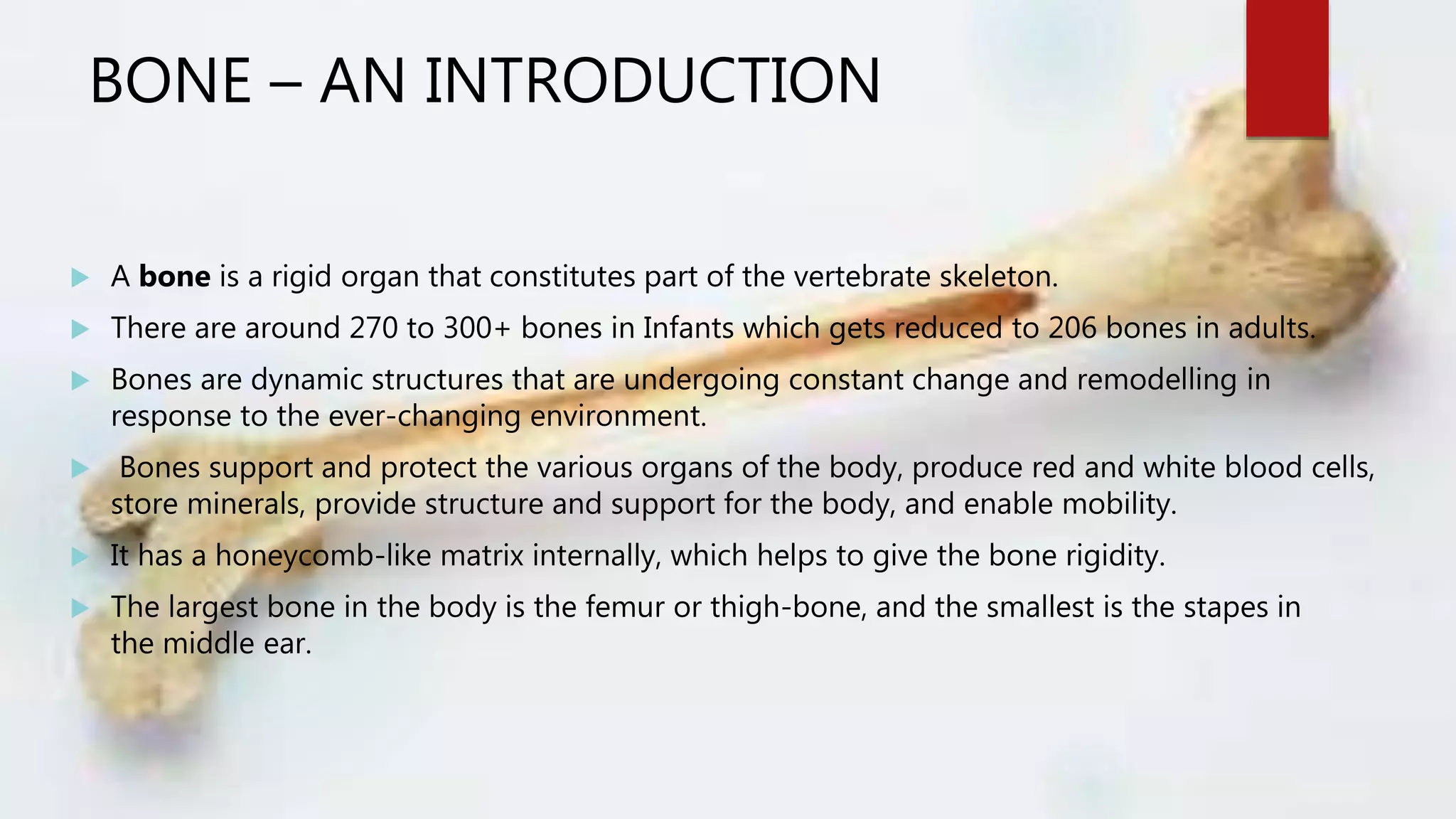
A bone is a rigid organ that constitutes part of the vertebrate skeleton.
There are around 270 to 300+ bones in Infants which gets reduced to 206 bones in adults.
Bones are dynamic structures that are undergoing constant change and remodelling in
response to the ever-changing environment.
Bones support and protect the various organs of the body, produce red and white blood cells,
store minerals, provide structure and support for the body, and enable mobility.
It has a honeycomb-like matrix internally, which helps to give the bone rigidity.
The largest bone in the body is the femur or thigh-bone, and the smallest is the stapes in
the middle ear.
3.
CLASSIFICATION OF BONE
According to position :
1. Axial Skeleton = The axial skeleton is the part of the skeleton
that consists of the bones of the head and trunk of a vertebrate.
https://en.wikipedia.org/wiki/Axial_skeleton
4.
Contd.
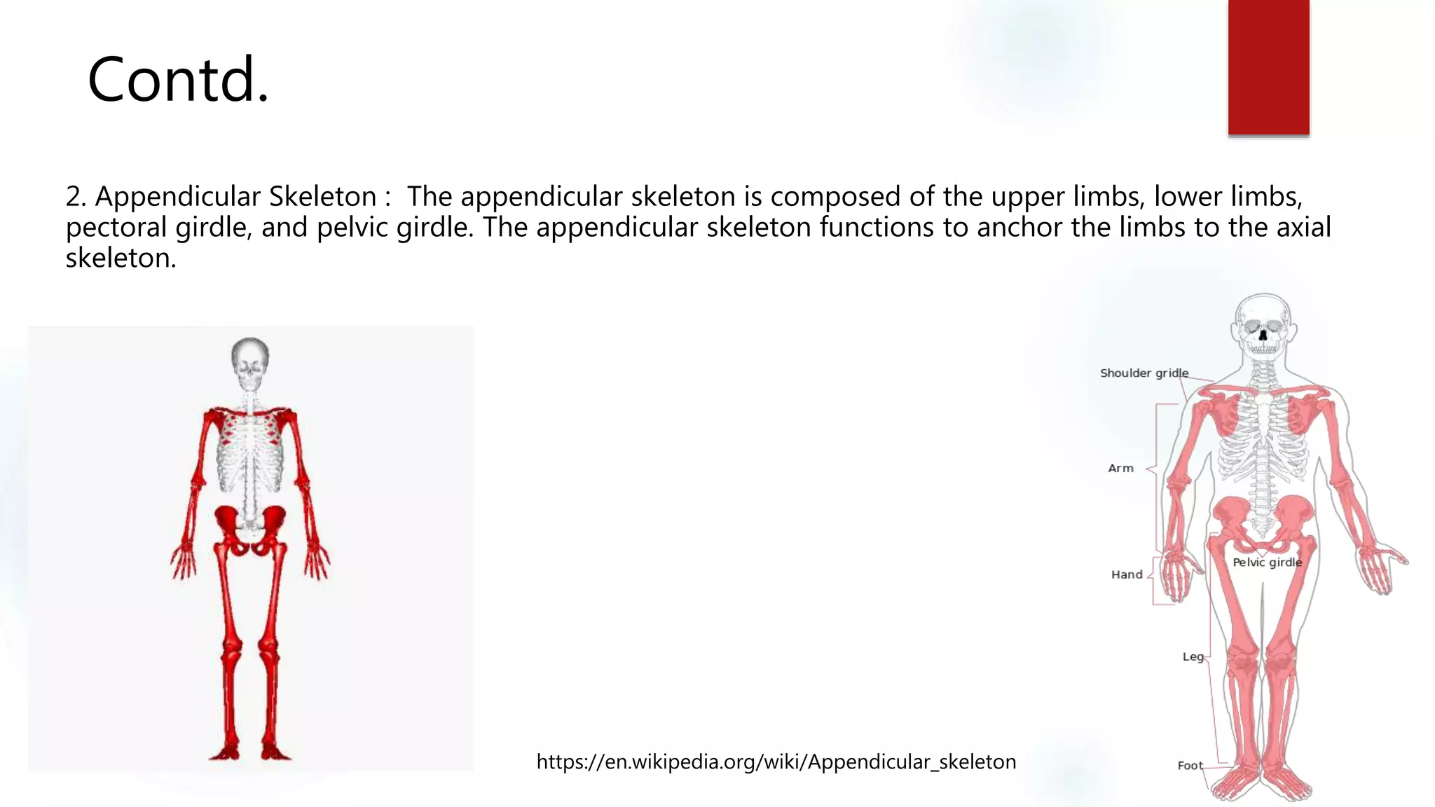
2. Appendicular Skeleton: The appendicular skeleton is composed of the upper limbs, lower limbs,
pectoral girdle, and pelvic girdle. The appendicular skeleton functions to anchor the limbs to the axial
skeleton.
https://en.wikipedia.org/wiki/Appendicular_skeleton
5.
CONTD.
2. Accordingto Size and Shape :
Long bones –
A. DIAPHYSIS: Portion of long bone between two cartilaginous
ends.
- Shaft of long bone.
Consist of Adipose tissue and bone marrow.
Consist of Nutrient Foramen directed away from the growing end.
- Primary Ossification occurs in this region.
https://en.wikipedia.org/wiki/Diaphysis
6.
CONTD.
B. EPIPHYSIS: The epiphysis is the rounded end of a long bone,
at its joint with adjacent bone(s).
Epiphyseal Line: The epiphyseal plate ( growth plate) is a hyaline
cartilage plate in the metaphysis at each end of a long bone. It is the part
of a long bone where new bone growth takes place.
Ends of epiphyses are covered with hyaline cartilage("articular cartilage").
C. METAPHYSIS : Metaphysis is the narrow portion of a long bone
between the epiphysis and the diaphysis.
- It consists the growth plate.
https://en.wikipedia.org/wiki/Long_bone
CONTD.
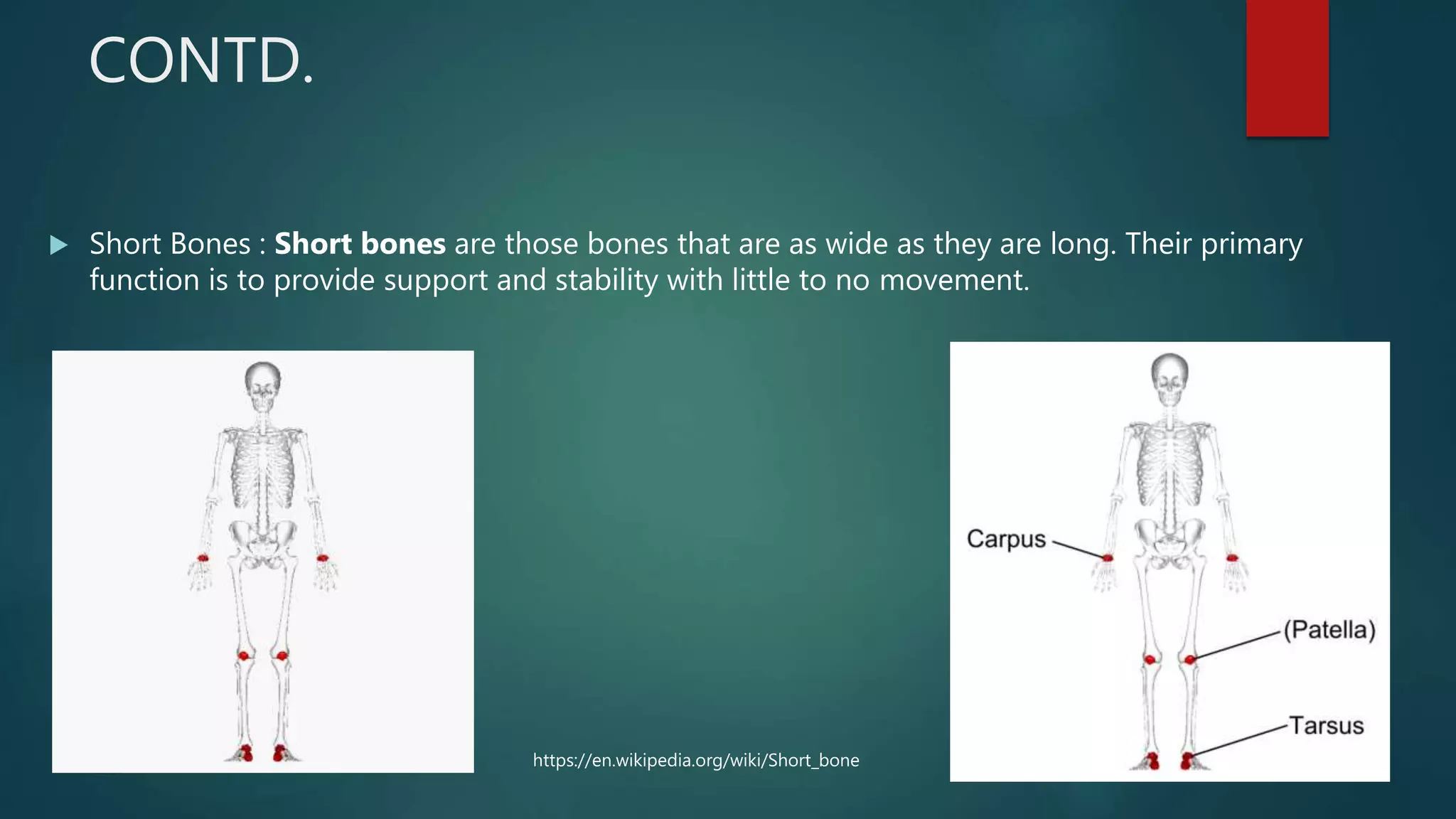
Short Bones: Short bones are those bones that are as wide as they are long. Their primary
function is to provide support and stability with little to no movement.
https://en.wikipedia.org/wiki/Short_bone
9.
CONTD.
Flat bones: Flat bones are bones whose principal function is either extensive protection or the
provision of broad surfaces for muscular attachment.
They are thin with parallel surface.
Present between two compact bone.
https://en.wikipedia.org/wiki/Flat_bone
10.
CONTD.
Irregular Bones: The irregular bones are bones which form their peculiar form.
-Have complex shapes.
Irregular bones serve various purposes in the body, such as protection of nervous tissue (such as
the vertebrae protect the spinal cord), and maintaining pharynx and trachea support, and tongue attachment
(such as the hyoid bone).
https://en.wikipedia.org/wiki/Irregular_bone
11.
CONTD.
Pneumatic Bones: Certain irregular bones contain large air spaces lined by epithelium.
- Make the skull light in weight,
- Helps in resonance of voice.
- Act as air conditioning chambers for the inspired air.
Examples : Maxilla, Sphenoid etc.
http://infinitespider.com/pneumatic-bones-birds-and-you/
12.
CONTD.
Sesamoid Bones: It is the bone which is embedded within a tendon or muscle.
Sesamoids act like pulleys, providing a smooth surface for tendons to slide over, increasing the
tendon's ability to transmit muscular forces.
https://en.wikipedia.org/wiki/Sesamoid_bone
STRUCTURAL CLASSIFICATION (Macroscopically)
1.Compact Bone : Cortical bone, also known as compact bone, forms the hard outer shell of all bones. It is the
strongest and densest form of bone in the body.
- Strong dense (80% of the skeleton)
- Best developed in the cortex of long bones
- The functional unit is Osteon (Haversian System) which contains osteoblasts and arteriole supplying the osteon.
OSTEONS : They are cylindrical, parallel to bone, and are group of hollow tube. Each osteon consists of concentric
layers (Lamellae), of compact bone tissue that surround a central canal, the Haversian canal.
https://www.studyblue.com/notes/note/n/6-skeletal-system/deck/7962817 https://www.dreamstime.com/
16.
Microscopically
1. HAVERSIANCANAL : Haversian canals are a series of microscopic tubes in the outermost
region of bone called cortical bone that allow blood vessels and nerves to travel through them.
- Each Haversian canal generally contains one or two capillaries and nerve fibres.
- The channels are formed by concentric layers called lamellae.
2. LACUNAE : Small spaces between lamellae, each containing a bone cell.
A lacuna never contains more than one osteocyte.
Example : Sinuses
https://www.embibe.com/
17.
CONTD.
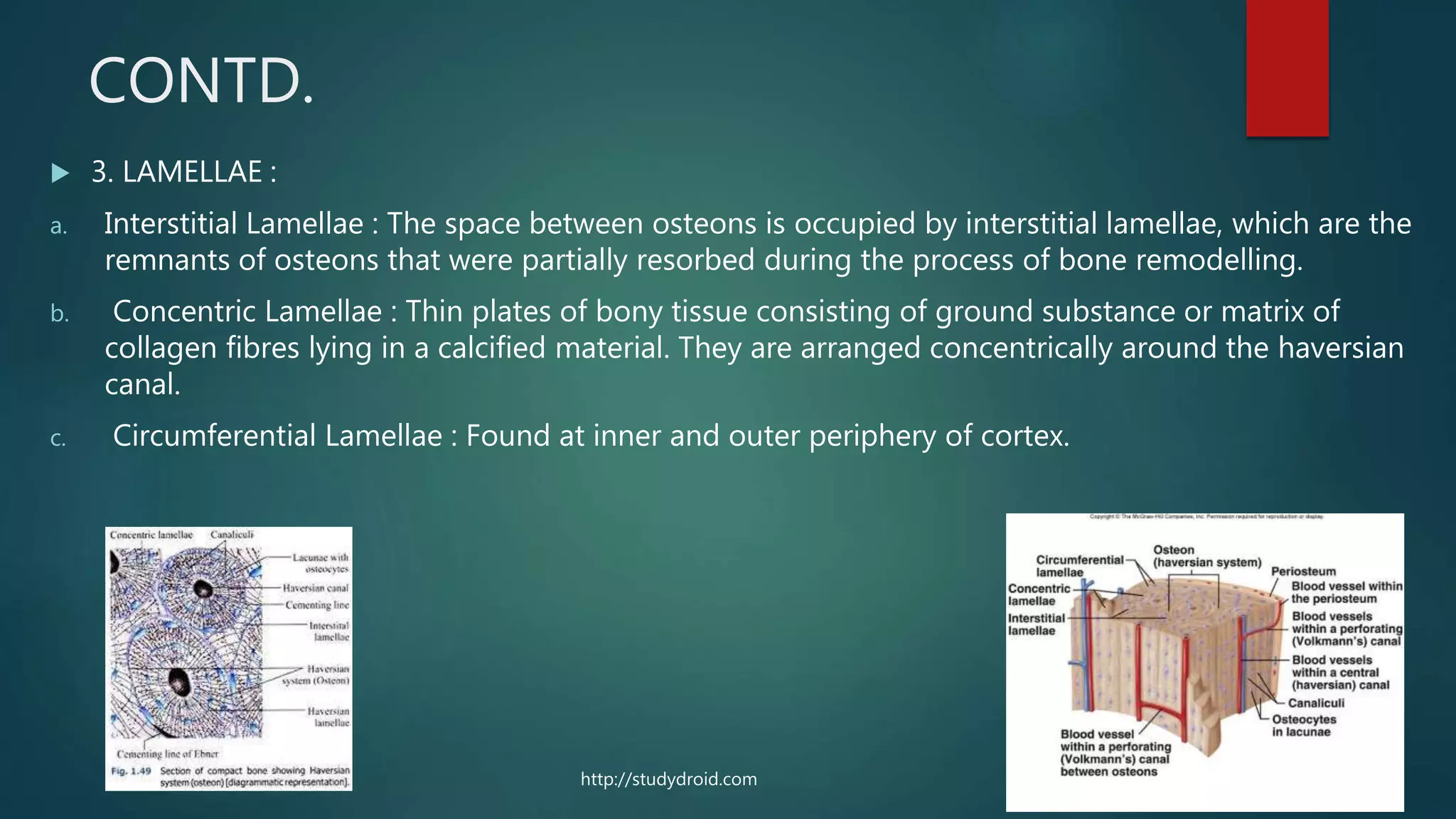
3. LAMELLAE:
a. Interstitial Lamellae : The space between osteons is occupied by interstitial lamellae, which are the
remnants of osteons that were partially resorbed during the process of bone remodelling.
b. Concentric Lamellae : Thin plates of bony tissue consisting of ground substance or matrix of
collagen fibres lying in a calcified material. They are arranged concentrically around the haversian
canal.
c. Circumferential Lamellae : Found at inner and outer periphery of cortex.
http://studydroid.com
18.
CONTD.
4. BoneCanaliculi : Bone canaliculi are microscopic canals between the
lacunae of ossified bone.
- They are the fine radiating channels which connects lacunae with each other and
Central Haversian Canal.
- Osteocytes do not entirely fill up the canaliculi. The remaining space is known as
the periosteocytic space, which is filled with periosteocytic fluid.
https://fatunmbi.wordpress.com
http://antranik.org/cartilage-and-bones/
19.
CONTD.
5. Volkmann'sCanal : Volkmann's canals, also known as perforating holes or channels, are atomic
arrangements in cortical bones.
- Oblique canals running at right angles to the long axis of the bone.
- Contains the neurovascular bundle and connect Haversian canals with the medullary cavity and
surface of the bone.
http://slideplayer.com
20.
CONTD.
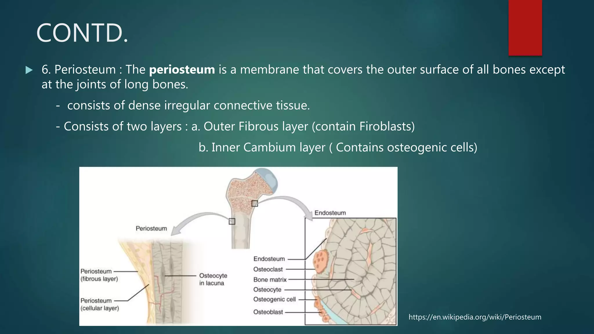
6. Periosteum: The periosteum is a membrane that covers the outer surface of all bones except
at the joints of long bones.
- consists of dense irregular connective tissue.
- Consists of two layers : a. Outer Fibrous layer (contain Firoblasts)
b. Inner Cambium layer ( Contains osteogenic cells)
https://en.wikipedia.org/wiki/Periosteum
21.
CONTD.
7. Endosteum: Endosteum (plural endostea) is a thin vascular membrane of connective
tissue that lines the inner surface of the bony tissue that forms the medullary cavity of
long bones.
- To prevent the bone from becoming unnecessarily thick, osteoclasts resorb the bone from the
endosteal side.
https://en.wikipedia.org/wiki/Endosteum
22.
CANCELLOUS BONE
Itis the internal tissue of the skeletal bone and is an open cell porous network.
- Cancellous bone has a higher surface-area-to-volume ratio than cortical bone because it is less dense.
- This makes it softer, and weaker but more flexible. The greater surface area also makes it suitable for
metabolic activities such as the exchange of calcium ions.
- Does not have osteons.
- The primary anatomical and functional unit of
cancellous bone is the trabecula.
- Trabeculae has no blood vessels.
https://image.slidesharecdn.com
23.
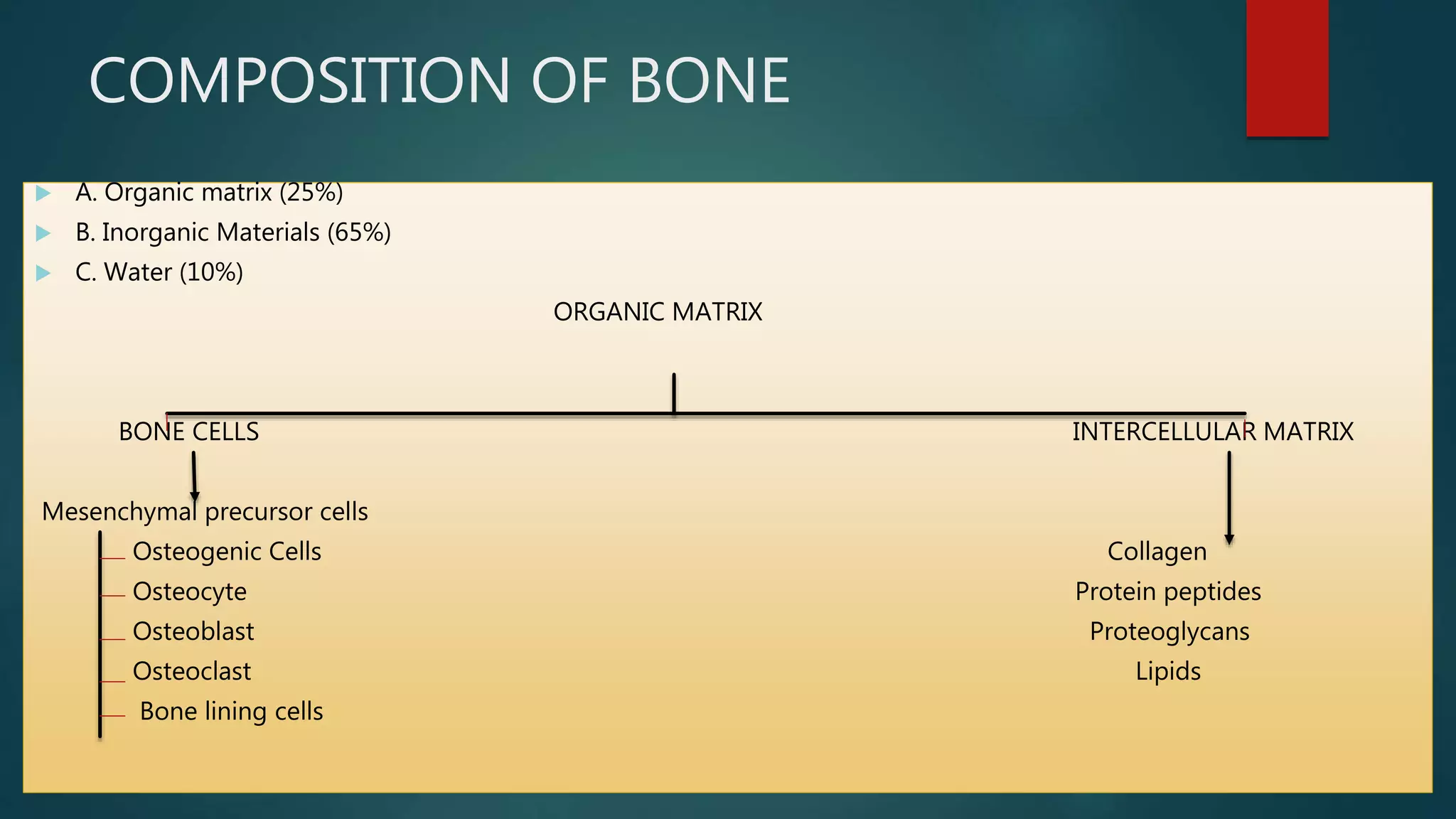
COMPOSITION OF BONE
A. Organic matrix (25%)
B. Inorganic Materials (65%)
C. Water (10%)
ORGANIC MATRIX
BONE CELLS INTERCELLULAR MATRIX
Mesenchymal precursor cells
Osteogenic Cells Collagen
Osteocyte Protein peptides
Osteoblast Proteoglycans
Osteoclast Lipids
Bone lining cells
25.
OSTEOPROGENITOR CELLS
Theseare the mesenchymal stem cells (MSC) that divide to form osteoblasts in bone marrow.
Runx2 (which may also be known as Cbfa1), and Osx (a zinc finger containing transcription
factor) are necessary for osteochondroprogenitor cells to differentiate into the osteoblast cell
lineage.
Runx2 : Runt-related transcription factor 2 (RUNX2) also known as core-binding factor
subunit alpha-1 (CBF-alpha-1) is a protein that in humans is encoded by the RUNX2 gene.
OSX : Transcription factor Sp7, also called Osterix (Osx), is a protein that in humans is encoded
by the SP7 gene.
These cells are present in endosteum, periosteum, stromal component of Bone matrix.
26.
CONTD.
The pathwayswhich are responsible for osteoblast differentiation are :
WNT SIGNALLING
BMP PATHWAY
TGF-β PATHWAY
FGF PATHWAY
PDGF PATHWAY
IGF PATHWAY
27.
SIGNALLING PATHWAYS
1.WNT SIGNALLING : The Wnt signaling pathways are a group of signal transduction pathways
made of proteins that pass signals into a cell through cell surface receptors.
The canonical pathway is responsible for the osteoblast differentiation.
Accumulation of β-catenin in cytoplasm
DSH becomes activated via phosphorylation and its
DIX and PDZ domains inhibit the GSK3 activity of the destruction complex
Act as transcriptional co- activator
of transcription factors (TCF/LEF Fam)
Axin becomes de-phosphorylated and its stability and levels decrease
WNT causes the translocation of the negative WNT regulator, Axin
WNT binds to FZ and its co receptor LRP 5/6
Accumulation of β-catenin in cytoplasm Translocation to the nucleus
Act as a transcriptional co-activator
Of transcription factors (TCF/LEF FAM)
28.
BMP PATHWAY
Homomeric dimersof type II
BMP receptors binds to
homomeric dimers of type I
BMP receptors
Induce trans
phosphorylation of type 1
receptors
Induce Signal transduction
through SMAD AND MAPK
Activates transcription of
target genes
29.
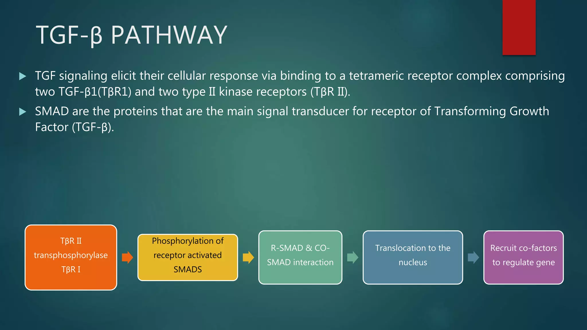
TGF-β PATHWAY
TGFsignaling elicit their cellular response via binding to a tetrameric receptor complex comprising
two TGF-β1(TβR1) and two type II kinase receptors (TβR II).
SMAD are the proteins that are the main signal transducer for receptor of Transforming Growth
Factor (TGF-β).
TβR II
transphosphorylase
TβR I
Phosphorylation of
receptor activated
SMADS
R-SMAD & CO-
SMAD interaction
Translocation to the
nucleus
Recruit co-factors
to regulate gene
30.
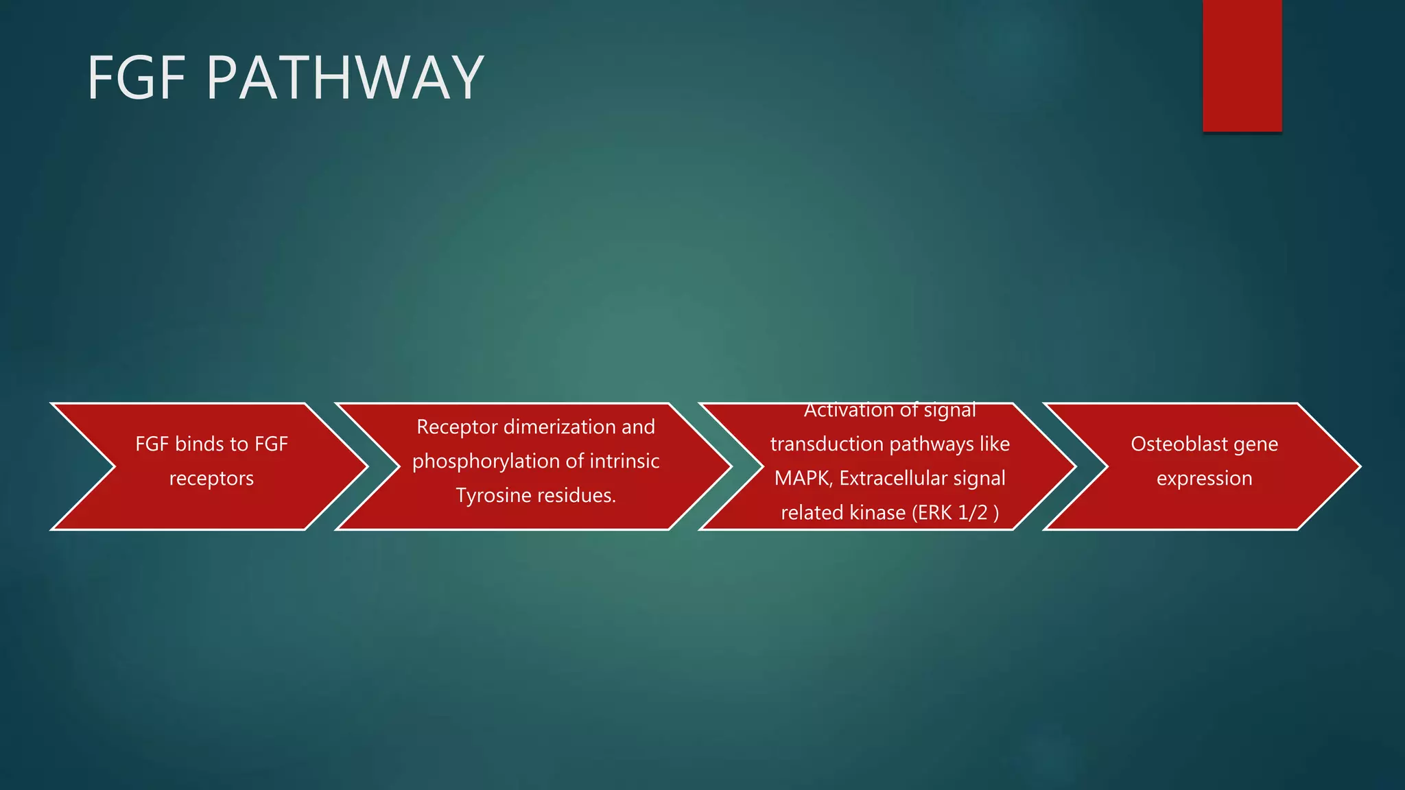
FGF PATHWAY
FGF bindsto FGF
receptors
Receptor dimerization and
phosphorylation of intrinsic
Tyrosine residues.
Activation of signal
transduction pathways like
MAPK, Extracellular signal
related kinase (ERK 1/2 )
Osteoblast gene
expression
31.
PDGF PATHWAY
ThePlatelet derived growth factor has two receptors – α type and β type.
The alpha type binds to PDGF-AA, PDGF-BB and PDGF-AB, whereas the beta type PDGFR binds
with high affinity to PDGF-BB and PDGF-AB.
PDGF activates the receptor causing
dimerization of the receptors
"switched on" by auto-
phosphorylation of several sites
on their cytosolic domains
serve to mediate binding of cofactors
and subsequently activate signal
transduction, through PIK3 Pathway
Regulates the gene expression
32.
IGF PATHWAY
IGF bindsto IGF
1R (Type II
Tyrosine Kinase)
Auto phosphorylation
of Tyr residues in
kinase domain
Phosphorylation
of Tyr 950 in
juxtamembrane
domain.
It activates Insulin
receptor substrate
(IRS) and Shc by
tyrosine
phosphorylation
In IGF-1 induction, IRF-1
activates PI3K, MAPK/ERK ,
by binding to Shc and Grb2
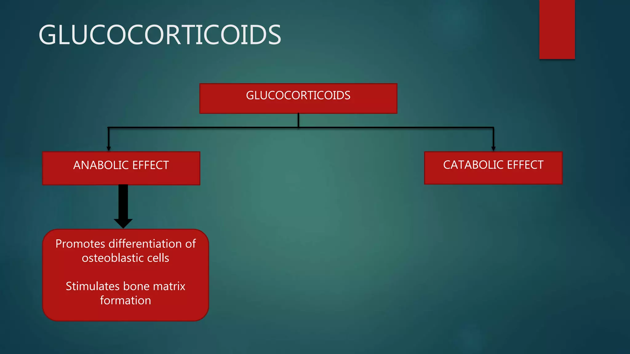
VITAMIN D3
VITAMIN D3
ANABOLICEFFECT CATABOLIC EFFECT
primary function in Ca
absorption from intestine
Stimulate bone resorption
Supresses Collagen
production
THYROID HORMONE
THYROIDHORMONE – ANABOLIC EFFECT – affects the endochondral bone formation by its
action on cartilage formation.
37.
OSTEOBLAST
Osteoblast arethe cells with a single nucleus that synthesizes bone.
Osteoblasts are specialized, terminally differentiated products of mesenchymal stem cells.
They synthesize dense, crosslinked collagen and specialized proteins in much smaller quantities,
including osteocalcin, osteonectin, osteopontin, which compose the organic matrix of bone.
As Osteocalcin {bone gamma-carboxyglutamic acid-containing protein (BGLAP)} is produced
by osteoblasts, it is often used as a marker for the bone formation process.
Osteopontin (OPN), also known as bone sialoprotein I (BSP-1 or BNSP), secreted phosphoprotein
1 (SPP1), is a protein that in humans is encoded by the SPP1 gene.
Osteonectin (ON) also known as secreted protein acidic and rich in cysteine (SPARC) is
a protein that in humans is encoded by the SPARC gene.
Before the organic matrix is mineralized, it is called the osteoid.
38.
OSTEOCYTE
Osteocytes arethe cells that generally helps in bone remodeling and detect micro damage in
bone.
When osteoblasts become trapped in the matrix that they secrete, they become osteocytes.
Osteocytes are networked to each other via long cytoplasmic extensions that occupy tiny
canals called canaliculi, which are used for exchange of nutrients and waste through gap
junctions.
It also helps to maintain the protein and mineral content of the matrix.
39.
OSTEOCLAST
Osteoclasts arethe cells that helps in bone resorption or the cells that break down the bone
tissue.
Osteoclasts are found in pits in the bone surface which are called resorption bays,
or Howship's Lacunae.
40.
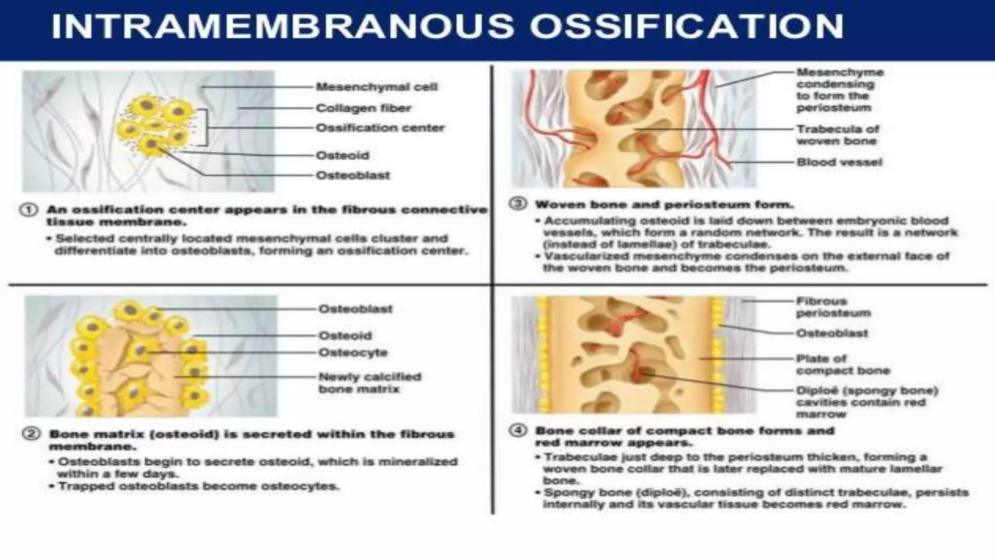
FORMATION OF BONE
Mainly there are two types of ossification :
1. Intramembranous ossification
2. Endochondral ossification
REFERENCES
1. Morris,W., American heritage dictionary of the English language. 1969: American heritage.
2. Steele, D.G. and C.A. Bramblett, The anatomy and biology of the human skeleton. 1988: Texas
A&M University Press.
3. Tözeren, A., Human body dynamics: classical mechanics and human movement. 1999: Springer
Science & Business Media.
4. Feng, X., Chemical and biochemical basis of cell-bone matrix interaction in health and disease.
Current chemical biology, 2009. 3(2): p. 189-196.
5. Marieb, E.N. and K. Hoehn, Human anatomy & physiology. 2007: Pearson Education.
6. Waugh, A. and A. Grant, Ross & Wilson Anatomy and Physiology in Health and Illness E-Book.
2010: Elsevier Health Sciences.
7. Hall, J.E., Guyton and Hall textbook of medical physiology e-Book. 2015: Elsevier Health Sciences.
8. White, T.D., M.T. Black, and P.A. Folkens, Human osteology. 2011: Academic press.
9. Gray, H., ANTOMY OF THE HUMAN BODY. Annals of Surgery, 1918. 68(5): p. 564-566.
10. Silverstein, J., J. Moeller, and M. Hutchinson, Common issues in orthopedics. Textbook of Family
Medicine. 9th ed. Philadelphia, PA: Elsevier Saunders, 2016.
44.
CONTD.
11. Pickrell,K.D., Miller-Keane encyclopedia and dictionary of medicine, nursing, and allied health. Hospitals &
Health Networks, 2003. 77(8): p. 70.
12. Clarke, B., Normal bone anatomy and physiology. Clinical journal of the American Society of Nephrology,
2008. 3(Supplement 3): p. S131-S139.
13. Grigoriou, E., A. Trocle, and J.P. Dormans, The Growth Plate: Embryologic Origin, Structure, and Function, in
Fetal and Neonatal Physiology (Fifth Edition). 2017, Elsevier. p. 1421-1429. e2.
14. Stedman, T., Medical dictionary for the health professions and nursing (p. 2339). Walters Kluwer Health. 2012,
Lippincott Williams & Wilkins.
15. Netter, F.H., Musculoskeletal system: anatomy, physiology, and metabolic disorders. 1990: CIBA Medical
Education Division.
16. Standring, S., Gray's anatomy e-book: the anatomical basis of clinical practice. 2015: Elsevier Health Sciences.
17. Ashalatha, P. and G. Deepa, Textbook of Anatomy & Physiology for Nurses. 2012: JP Medical Ltd.
18. NEW, L.I., The Oxford English Dictionary. 1989.
19. Saladin, K.S. and L. Miller, Anatomy & physiology. 1998: WCB/McGraw-Hill New York (NY).
20. Singh, V., General Anatomy-E-book. 2015: Elsevier Health Sciences.
45.
CONTD.
21. Dictionary,A., The American Heritage Medical Dictionary. 2007, Houghton Mifflin Company.
22. Guyton, A.C. and J.E. Hall, Medical physiology. 1961: Saunders.
23. Junqueira, L.C. and J. Carneiro, Basic histology: text and atlas. 2005: McGraw-Hill Professional.
24. Komori, T., Regulation of osteoblast differentiation by Runx2, in Osteoimmunology. 2009, Springer. p.
43-49.
25. Nakashima, K., et al., The novel zinc finger-containing transcription factor osterix is required for
osteoblast differentiation and bone formation. Cell, 2002. 108(1): p. 17-29.
26. Komiya, Y. and R. Habas, Wnt signal transduction pathways. Organogenesis, 2008. 4(2): p. 68-75.
27. Reddi, A.H. and A. Reddi, Bone morphogenetic proteins (BMPs): from morphogens to metabologens.
2009, Pergamon.
28. Kaminska, B., A. Wesolowska, and M. Danilkiewicz, TGF beta signalling and its role in tumour
pathogenesis. ACTA BIOCHIMICA POLONICA-ENGLISH EDITION-, 2005. 52(2): p. 329.
29. Erynck, R., Y. Zhang, and X. Feng, Smads: Trancriptional activators of TGF-β response. Cell, 1998. 95(6):
p. 737.
46.
CONTD.
30. Wu,J.-W., et al., Crystal structure of a phosphorylated Smad2: Recognition of
phosphoserine by the MH2 domain and insights on Smad function in TGF-β signaling.
Molecular cell, 2001. 8(6): p. 1277-1289.
31. Massagué, J., TGFβ signalling in context. Nature reviews Molecular cell biology, 2012.
13(10): p. 616.
32. Shi, Y., et al., A structural basis for mutational inactivation of the tumour suppressor
Smad4. Nature, 1997. 388(6637): p. 87.
33. Ornitz, D.M. and N. Itoh, The fibroblast growth factor signaling pathway. Wiley
Interdisciplinary Reviews: Developmental Biology, 2015. 4(3): p. 215-266.