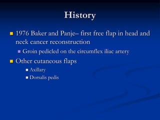
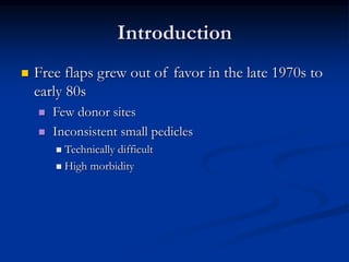
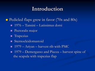
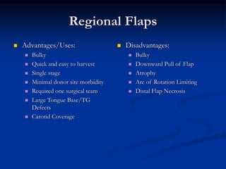
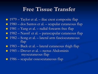
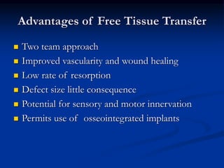
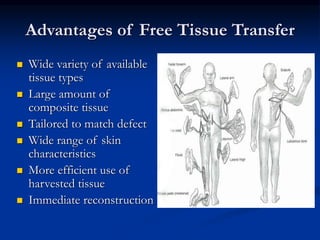
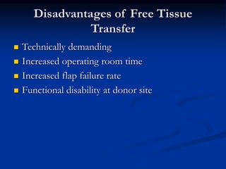
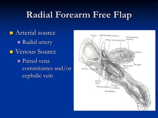
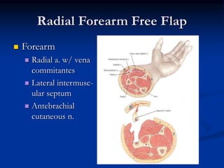
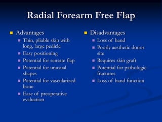
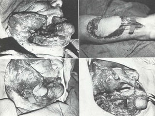
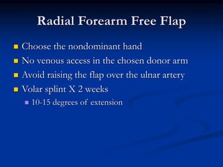
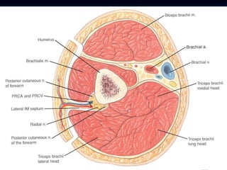
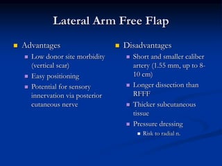
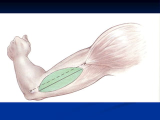
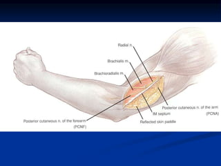
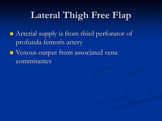
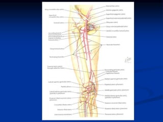
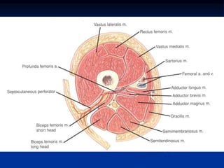
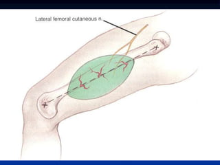
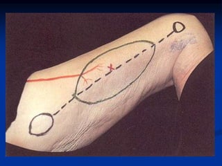
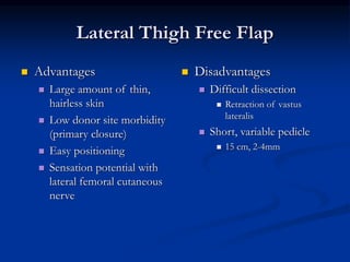
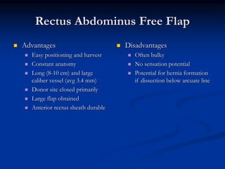
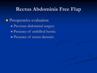
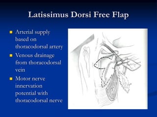
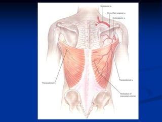
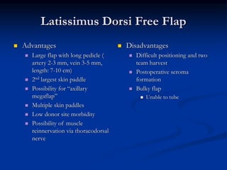
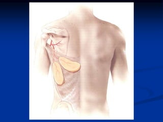
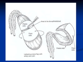
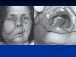
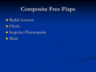
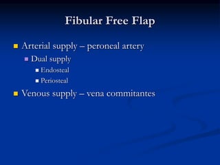
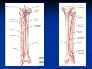
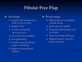
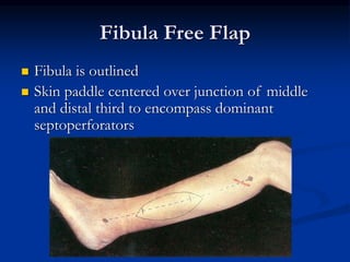
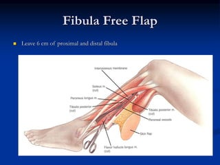
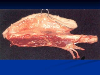
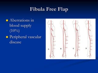
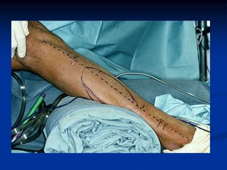
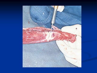
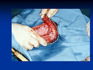
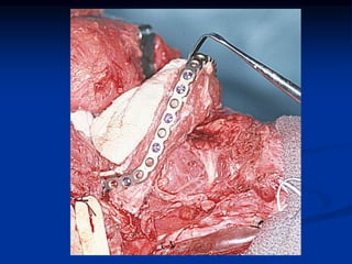
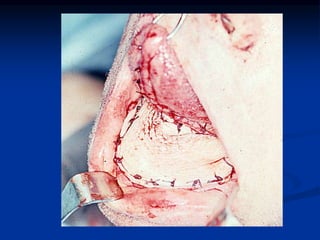
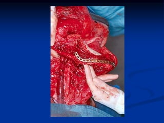
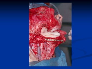
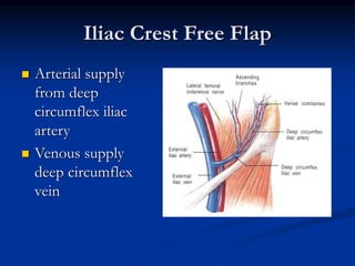
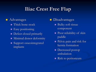
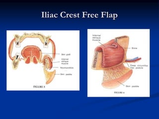
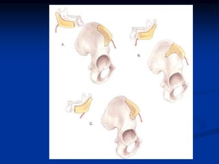
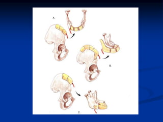
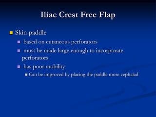
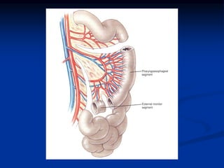
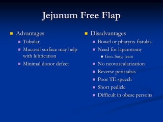
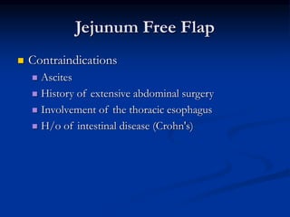
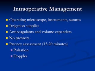
Free Flap Reconstruction of
Head and Neck Defects
Parag Parikh, MD
UC-Irvine
April 7, 2004
Introduction
 Prior to 3 Decades Ago
 Majority of Head and Neck Defects closed with
 Local Tissue
 Local Skin Flaps from other sites to the H/N
 Forehead Flap…Indians then McGregor in 1963
 1965 Bakamjian…Deltopectoral Flap
 Limited Reach
Introduction
 Early 1900’s Alexis Carrel
 Free tissue transfer in animals (jejunum to neck)
 1950’s Jacobsen and Suarez-- first anastomoses
in animal
 1959 Seidenberg– free jejunum segments to
repair pharyngoesophageal defects
 1973 Daniels and Taylor– “free flap”
 First free cutaneous flap
History
 1976 Baker and Panje– first free flap in head and
neck cancer reconstruction
 Groin pedicled on the circumflex iliac artery
 Other cutaneous flaps
 Axillary
 Dorsalis pedis
Introduction
 Free flaps grew out of favor in the late 1970s to
early 80s
 Few donor sites
 Inconsistent small pedicles
 Technically difficult
 High morbidity
Introduction
 Pedicled flaps grew in favor (70s and 80s)
 1976 – Tansini – Latissimus dorsi
 Pectoralis major
 Trapezius
 Sternocleidomastoid
 1979 – Ariyan – harvest rib with PMC
 1979 – Demergasso and Piazza – harvest spine of
the scapula with trapezius flap
Regional Flaps
 Advantages/Uses:
 Bulky
 Quick and easy to harvest
 Single stage
 Minimal donor site morbidity
 Required one surgical team
 Large Tongue Base/TG
Defects
 Carotid Coverage
 Disadvantages:
 Bulky
 Downward Pull of Flap
 Atrophy
 Arc of Rotation Limiting
 Distal Flap Necrosis
Free Tissue Transfer
 1979 – Taylor et al. – iliac crest composite flap
 1980 – dos Santos et al. – scapular cutaneous flap
 1981 – Yang et al. – radial forearm free flap
 1982 – Nassif et al. – parascapular cutaneous flap
 1982 – Song et al. – lateral arm fasciocutaneous
flap
 1983 – Baek et al. – lateral cutaneous thigh flap
 1985 – Drever et al. – rectus Abdominis
myocutaneous flap
 1986 – scapular osseocutaneous flap
Advantages of Free Tissue Transfer
 Two team approach
 Improved vascularity and wound healing
 Low rate of resorption
 Defect size little consequence
 Potential for sensory and motor innervation
 Permits use of osseointegrated implants
Advantages of Free Tissue Transfer
 Wide variety of available
tissue types
 Large amount of
composite tissue
 Tailored to match defect
 Wide range of skin
characteristics
 More efficient use of
harvested tissue
 Immediate reconstruction
Disadvantages of Free Tissue
Transfer
 Technically demanding
 Increased operating room time
 Increased flap failure rate
 Functional disability at donor site
Preoperative Planning
 Amount and type of tissue required
 Bone, soft tissue bulk, external vs. internal lining
 Anticipated functional gains
 History of previous surgery or injury around the donor site
 Donor morbidity
 Patient positioning and donor location
 Operative time
 Need for carotid coverage
 Patient factors
 General medical status
 Wishes and expectations
Preoperative Planning
 Patient selection
 Age
 Diabetes
 Arteriosclerosis/Cardiac
 Tobacco use
 Collagen vascular disease
 Coagulopathies
 Hypercoagulable states
Reconstructive Planning
 Must consider all options for particular defect
and patient
 Options
 Secondary intent
 Primary closure
 Skin grafts
 Local flaps
 Myocutaneous flaps
 Free flaps
Fasciocutaneous Free Flaps
 Radial forearm
 Lateral arm
 Lateral thigh
Radial Forearm Free Flap
 Arterial source
 Radial artery
 Venous Source
 Paired vena
commitantes and/or
cephalic vein
Radial Forearm Free Flap
 Forearm
 Radial a. w/ vena
commitantes
 Lateral intermusc-
ular septum
 Antebrachial
cutaneous n.
Radial Forearm Free Flap
 Advantages
 Thin, pliable skin with
long, large pedicle
 Easy positioning
 Potential for sensate flap
 Potential for unusual
shapes
 Potential for vascularized
bone
 Ease of preoperative
evaluation
 Disadvantages
 Loss of hand
 Poorly aesthetic donor
site
 Requires skin graft
 Potential for pathologic
fractures
 Loss of hand function
Radial Forearm Free Flap
 Choose the nondominant hand
 No venous access in the chosen donor arm
 Avoid raising the flap over the ulnar artery
 Volar splint X 2 weeks
 10-15 degrees of extension
Lateral Arm Free Flap
 Arterial supply
 Posterior radial collateral artery from profunda
brachii artery
 Venous supply
 Vena commitantes in spiral groove of humerus
Lateral Arm Free Flap
 Advantages
 Low donor site morbidity
(vertical scar)
 Easy positioning
 Potential for sensory
innervation via posterior
cutaneous nerve
 Disadvantages
 Short and smaller caliber
artery (1.55 mm, up to 8-
10 cm)
 Longer dissection than
RFFF
 Thicker subcutaneous
tissue
 Pressure dressing
 Risk to radial n.
Lateral Thigh Free Flap
 Arterial supply is from third perforator of
profunda femoris artery
 Venous output from associated vena
commitantes
Lateral Thigh Free Flap
 Advantages
 Large amount of thin,
hairless skin
 Low donor site morbidity
(primary closure)
 Easy positioning
 Sensation potential with
lateral femoral cutaneous
nerve
 Disadvantages
 Difficult dissection
 Retraction of vastus
lateralis
 Short, variable pedicle
 15 cm, 2-4mm
Muscle and Musculocutaneous Free
Flaps
 Rectus abdominis
 Latissimus dorsi
Rectus Abdominus Free Flap
 Arterial supply based
on deep inferior
epigastric artery
 Venous supply form
vena commitantes
joining external iliac
vein
Rectus Abdominis Free Flap
 Versatility of the inf
epig. a.
 Periumbilical perforators
 A. Transverse
 B. Extended
 C. Extended
 Less muscle
 D. Longitudinal
 Thick
 E. Subarcuate
 Thinner
Rectus Abdominus Free Flap
 Advantages
 Easy positioning and harvest
 Constant anatomy
 Long (8-10 cm) and large
caliber vessel (avg 3.4 mm)
 Donor site closed primarily
 Large flap obtained
 Anterior rectus sheath durable
 Disadvantages
 Often bulky
 No sensation potential
 Potential for hernia formation
if dissection below arcuate line
Rectus Abdominis Free Flap
 Preoperative evaluation
 Previous abdominal surgery
 Presence of umbilical hernia
 Presence of rectus diastasis
Latissimus Dorsi Free Flap
 Arterial supply
based on
thoracodorsal artery
 Venous drainage
from thoracodorsal
vein
 Motor nerve
innervation
potential with
thoracodorsal nerve
Latissimus Dorsi Free Flap
 Advantages
 Large flap with long pedicle (
artery 2-3 mm, vein 3-5 mm,
length: 7-10 cm)
 2nd largest skin paddle
 Possibility for “axillary
megaflap”
 Multiple skin paddles
 Low donor site morbidity
 Possibility of muscle
reinnervation via thoracodorsal
nerve
 Disadvantages
 Difficult positioning and two
team harvest
 Postoperative seroma
formation
 Bulky flap
 Unable to tube
Composite Free Flaps
 Radial forearm
 Fibula
 Scapular/Parascapular
 Ilium
Fibular Free Flap
 Arterial supply – peroneal artery
 Dual supply
 Endosteal
 Periosteal
 Venous supply – vena commitantes
Fibular Free Flap
 Advantages
 Longest and strongest bone
stock (25 cm of bone)
 Pedicle 12 cm
 Can be a sensate flap
 Lateral sural n.
 Low donor site morbidity
 Easy positioning
 Excellent periosteal blood
supply (contouring)
 Support osseointegrated
implants
 Disadvantages
 High incidence of peripheral
vascular disease
 Small cutaneous paddle
 Decreased ankle strength and
toe flexion
 Small risk chronic ankle pain
 Requires invasive study for
preop. evaluation
Fibula Free Flap
 Fibula is outlined
 Skin paddle centered over junction of middle
and distal third to encompass dominant
septoperforators
Fibula Free Flap
 Leave 6 cm of proximal and distal fibula
Fibula Free Flap
 Aberrations in
blood supply
(10%)
 Peripheral vascular
disease
Iliac Crest Free Flap
 Arterial supply
from deep
circumflex iliac
artery
 Venous supply
deep circumflex
vein
Iliac Crest Free Flap
 Advantages
 Thick bone stock
 Easy positioning
 Defect closed primarily
 Minimal donor deformity
 Support osseointegrated
implants
 Disadvantages
 Bulky soft tissue
component
 Poor reliability of skin
paddle
 Pelvic pain and risk for
hernia formation
 Decreased postop
ambulation
 Risk to peritoneum
Iliac Crest Free Flap
 Most commonly used for mandibular defects in
the head and neck
 best for angle/body defects
 can be used for symphyseal and
parasymphyseal defects
Iliac Crest Free Flap
 Skin paddle
 based on cutaneous perforators
 must be made large enough to incorporate
perforators
 has poor mobility
 Can be improved by placing the paddle more cephalad
Iliac Crest Free Flap
 Postoperative care
 Progressive mobilization
 Assisted ambulation POD # 3 or 4
 Stair climbing 3 weeks
Scapular/Parascapular Free Flap
 Arterial supply
 Circumflex scapular
 Venous Supply
 Vena commitantes
Scapular/Parascapular Free Flap
 Advantages
 Large skin paddle
 Easy to harvest
 Low donor site morbidity
(closes primarily)
 Availability for bone
 Disadvantages
 Thick skin
 Difficult positioning
Jejunum Free Flap
 Seidenberg (1959) - First case report in a human
 Roberts and Douglas (1961) – first patient to
survive
 Primarily use for reconstruction of
pharyngoesophageal defects
Jejunum Free Flap
 Arterial supply
from portion of
superior mesenteric
arterial arcade (2nd
or 3rd arcade)
 Venous supply
from venous
branches along
arcade
Jejunum Free Flap
 Advantages
 Tubular
 Mucosal surface may help
with lubrication
 Minimal donor defect
 Disadvantages
 Bowel or pharynx fistulas
 Need for laparotomy
 Gen. Surg. team
 No neovascularization
 Reverse peristalsis
 Poor TE speech
 Short pedicle
 Difficult in obese persons
Jejunum Free Flap
 Contraindications
 Ascites
 History of extensive abdominal surgery
 Involvement of the thoracic esophagus
 H/o of intestinal disease (Crohn's)
Intraoperative Management
 Operating microscope, instruments, sutures
 Irrigation supplies
 Anticoagulants and volume expanders
 No pressors
 Patency assessment (15-20 minutes)
 Pulsation
 Doppler
Postoperative Management
 Skilled nursing important
 No pressure on pedicle (no ties on neck)
 Eliminate cooling of flap
 Keep head in neutral position
 No pressors– keep BP stable
 Hematocrit important
 Frequent inspections and doppler pedicle
Postoperative Management
 Inspection and prick test
 Arterial vs. venous insufficiency
 Pharmacotherapy
 Heparin, dextran, aspirin
Oral Cavity and Oropharynx
Reconstruction
 Thin pliable mucosa
 Possibilities
 Radial Forearm
 Scapular/Parascapular
 Lateral Arm
 Lateral thigh
Tongue Reconstruction
 Reconstruction aimed at preserving what has not been resected
 Less than 1/3-1/2– primary closure vs. STSG
 Over ½--consider free free flap if expected contracture makes
speech/bolus transit difficult (sensate)
 Anterior 2/3–consider coned RFFF (sensate
Tongue Reconstruction
 For tongue base and total glossectomy defects—
need adequate oral mound to approximate with
palate for speech and bolus transit
 May consider rectus abdominus and latissimus dorsi
free flaps
Hypopharynx and Cervical Esophageal
Reconstruction
 Must be prepared for possibility of complete
circumferential pharyngeal defect
 Over 3 cm remains– primary closure
 Less than 3 cm—pec flap vs. RFFF
 Total loss above thoracic inlet– tubed pec flap,
RFFF, scapular FF, lateral thigh free flap, or free
jejunum flap
 Total loss below thoracic inlet– gastric pull-up
Mandibular Reconstruction
 Loss of anterior mandibular arch
 Loss of chin/lip support
 Sensory loss
 Malocclusion
 Retrognathia
 Lack of oral competence/eating/speaking
 Consider osteocutaneous free flaps-- fibula, iliac
crest, scapula, radius
Mandibular Reconstruction
 Loss of lateral mandible
 Concavity of cheek
 Mandible rotation to defect side with cross bite
 Remnant rotation superiorly and medially
 Mental nerve loss
 Easier for patient to adjust
 Consider osteocutaneous free flap
9162_free-flap-reconstruction-of-head-and-neck-defects.ppt

9162_free-flap-reconstruction-of-head-and-neck-defects.ppt

  • 1.
    Free Flap Reconstructionof Head and Neck Defects Parag Parikh, MD UC-Irvine April 7, 2004
  • 3.
    Introduction  Prior to3 Decades Ago  Majority of Head and Neck Defects closed with  Local Tissue  Local Skin Flaps from other sites to the H/N  Forehead Flap…Indians then McGregor in 1963  1965 Bakamjian…Deltopectoral Flap  Limited Reach
  • 4.
    Introduction  Early 1900’sAlexis Carrel  Free tissue transfer in animals (jejunum to neck)  1950’s Jacobsen and Suarez-- first anastomoses in animal  1959 Seidenberg– free jejunum segments to repair pharyngoesophageal defects  1973 Daniels and Taylor– “free flap”  First free cutaneous flap
  • 5.
    History  1976 Bakerand Panje– first free flap in head and neck cancer reconstruction  Groin pedicled on the circumflex iliac artery  Other cutaneous flaps  Axillary  Dorsalis pedis
  • 6.
    Introduction  Free flapsgrew out of favor in the late 1970s to early 80s  Few donor sites  Inconsistent small pedicles  Technically difficult  High morbidity
  • 7.
    Introduction  Pedicled flapsgrew in favor (70s and 80s)  1976 – Tansini – Latissimus dorsi  Pectoralis major  Trapezius  Sternocleidomastoid  1979 – Ariyan – harvest rib with PMC  1979 – Demergasso and Piazza – harvest spine of the scapula with trapezius flap
  • 8.
    Regional Flaps  Advantages/Uses: Bulky  Quick and easy to harvest  Single stage  Minimal donor site morbidity  Required one surgical team  Large Tongue Base/TG Defects  Carotid Coverage  Disadvantages:  Bulky  Downward Pull of Flap  Atrophy  Arc of Rotation Limiting  Distal Flap Necrosis
  • 9.
    Free Tissue Transfer 1979 – Taylor et al. – iliac crest composite flap  1980 – dos Santos et al. – scapular cutaneous flap  1981 – Yang et al. – radial forearm free flap  1982 – Nassif et al. – parascapular cutaneous flap  1982 – Song et al. – lateral arm fasciocutaneous flap  1983 – Baek et al. – lateral cutaneous thigh flap  1985 – Drever et al. – rectus Abdominis myocutaneous flap  1986 – scapular osseocutaneous flap
  • 10.
    Advantages of FreeTissue Transfer  Two team approach  Improved vascularity and wound healing  Low rate of resorption  Defect size little consequence  Potential for sensory and motor innervation  Permits use of osseointegrated implants
  • 11.
    Advantages of FreeTissue Transfer  Wide variety of available tissue types  Large amount of composite tissue  Tailored to match defect  Wide range of skin characteristics  More efficient use of harvested tissue  Immediate reconstruction
  • 12.
    Disadvantages of FreeTissue Transfer  Technically demanding  Increased operating room time  Increased flap failure rate  Functional disability at donor site
  • 13.
    Preoperative Planning  Amountand type of tissue required  Bone, soft tissue bulk, external vs. internal lining  Anticipated functional gains  History of previous surgery or injury around the donor site  Donor morbidity  Patient positioning and donor location  Operative time  Need for carotid coverage  Patient factors  General medical status  Wishes and expectations
  • 14.
    Preoperative Planning  Patientselection  Age  Diabetes  Arteriosclerosis/Cardiac  Tobacco use  Collagen vascular disease  Coagulopathies  Hypercoagulable states
  • 15.
    Reconstructive Planning  Mustconsider all options for particular defect and patient  Options  Secondary intent  Primary closure  Skin grafts  Local flaps  Myocutaneous flaps  Free flaps
  • 16.
    Fasciocutaneous Free Flaps Radial forearm  Lateral arm  Lateral thigh
  • 17.
    Radial Forearm FreeFlap  Arterial source  Radial artery  Venous Source  Paired vena commitantes and/or cephalic vein
  • 19.
    Radial Forearm FreeFlap  Forearm  Radial a. w/ vena commitantes  Lateral intermusc- ular septum  Antebrachial cutaneous n.
  • 20.
    Radial Forearm FreeFlap  Advantages  Thin, pliable skin with long, large pedicle  Easy positioning  Potential for sensate flap  Potential for unusual shapes  Potential for vascularized bone  Ease of preoperative evaluation  Disadvantages  Loss of hand  Poorly aesthetic donor site  Requires skin graft  Potential for pathologic fractures  Loss of hand function
  • 22.
    Radial Forearm FreeFlap  Choose the nondominant hand  No venous access in the chosen donor arm  Avoid raising the flap over the ulnar artery  Volar splint X 2 weeks  10-15 degrees of extension
  • 23.
    Lateral Arm FreeFlap  Arterial supply  Posterior radial collateral artery from profunda brachii artery  Venous supply  Vena commitantes in spiral groove of humerus
  • 25.
    Lateral Arm FreeFlap  Advantages  Low donor site morbidity (vertical scar)  Easy positioning  Potential for sensory innervation via posterior cutaneous nerve  Disadvantages  Short and smaller caliber artery (1.55 mm, up to 8- 10 cm)  Longer dissection than RFFF  Thicker subcutaneous tissue  Pressure dressing  Risk to radial n.
  • 28.
    Lateral Thigh FreeFlap  Arterial supply is from third perforator of profunda femoris artery  Venous output from associated vena commitantes
  • 33.
    Lateral Thigh FreeFlap  Advantages  Large amount of thin, hairless skin  Low donor site morbidity (primary closure)  Easy positioning  Sensation potential with lateral femoral cutaneous nerve  Disadvantages  Difficult dissection  Retraction of vastus lateralis  Short, variable pedicle  15 cm, 2-4mm
  • 34.
    Muscle and MusculocutaneousFree Flaps  Rectus abdominis  Latissimus dorsi
  • 35.
    Rectus Abdominus FreeFlap  Arterial supply based on deep inferior epigastric artery  Venous supply form vena commitantes joining external iliac vein
  • 36.
    Rectus Abdominis FreeFlap  Versatility of the inf epig. a.  Periumbilical perforators  A. Transverse  B. Extended  C. Extended  Less muscle  D. Longitudinal  Thick  E. Subarcuate  Thinner
  • 38.
    Rectus Abdominus FreeFlap  Advantages  Easy positioning and harvest  Constant anatomy  Long (8-10 cm) and large caliber vessel (avg 3.4 mm)  Donor site closed primarily  Large flap obtained  Anterior rectus sheath durable  Disadvantages  Often bulky  No sensation potential  Potential for hernia formation if dissection below arcuate line
  • 39.
    Rectus Abdominis FreeFlap  Preoperative evaluation  Previous abdominal surgery  Presence of umbilical hernia  Presence of rectus diastasis
  • 40.
    Latissimus Dorsi FreeFlap  Arterial supply based on thoracodorsal artery  Venous drainage from thoracodorsal vein  Motor nerve innervation potential with thoracodorsal nerve
  • 43.
    Latissimus Dorsi FreeFlap  Advantages  Large flap with long pedicle ( artery 2-3 mm, vein 3-5 mm, length: 7-10 cm)  2nd largest skin paddle  Possibility for “axillary megaflap”  Multiple skin paddles  Low donor site morbidity  Possibility of muscle reinnervation via thoracodorsal nerve  Disadvantages  Difficult positioning and two team harvest  Postoperative seroma formation  Bulky flap  Unable to tube
  • 47.
    Composite Free Flaps Radial forearm  Fibula  Scapular/Parascapular  Ilium
  • 48.
    Fibular Free Flap Arterial supply – peroneal artery  Dual supply  Endosteal  Periosteal  Venous supply – vena commitantes
  • 50.
    Fibular Free Flap Advantages  Longest and strongest bone stock (25 cm of bone)  Pedicle 12 cm  Can be a sensate flap  Lateral sural n.  Low donor site morbidity  Easy positioning  Excellent periosteal blood supply (contouring)  Support osseointegrated implants  Disadvantages  High incidence of peripheral vascular disease  Small cutaneous paddle  Decreased ankle strength and toe flexion  Small risk chronic ankle pain  Requires invasive study for preop. evaluation
  • 51.
    Fibula Free Flap Fibula is outlined  Skin paddle centered over junction of middle and distal third to encompass dominant septoperforators
  • 52.
    Fibula Free Flap Leave 6 cm of proximal and distal fibula
  • 55.
    Fibula Free Flap Aberrations in blood supply (10%)  Peripheral vascular disease
  • 63.
    Iliac Crest FreeFlap  Arterial supply from deep circumflex iliac artery  Venous supply deep circumflex vein
  • 64.
    Iliac Crest FreeFlap  Advantages  Thick bone stock  Easy positioning  Defect closed primarily  Minimal donor deformity  Support osseointegrated implants  Disadvantages  Bulky soft tissue component  Poor reliability of skin paddle  Pelvic pain and risk for hernia formation  Decreased postop ambulation  Risk to peritoneum
  • 65.
    Iliac Crest FreeFlap  Most commonly used for mandibular defects in the head and neck  best for angle/body defects  can be used for symphyseal and parasymphyseal defects
  • 66.
  • 69.
    Iliac Crest FreeFlap  Skin paddle  based on cutaneous perforators  must be made large enough to incorporate perforators  has poor mobility  Can be improved by placing the paddle more cephalad
  • 70.
    Iliac Crest FreeFlap  Postoperative care  Progressive mobilization  Assisted ambulation POD # 3 or 4  Stair climbing 3 weeks
  • 71.
    Scapular/Parascapular Free Flap Arterial supply  Circumflex scapular  Venous Supply  Vena commitantes
  • 75.
    Scapular/Parascapular Free Flap Advantages  Large skin paddle  Easy to harvest  Low donor site morbidity (closes primarily)  Availability for bone  Disadvantages  Thick skin  Difficult positioning
  • 76.
    Jejunum Free Flap Seidenberg (1959) - First case report in a human  Roberts and Douglas (1961) – first patient to survive  Primarily use for reconstruction of pharyngoesophageal defects
  • 77.
    Jejunum Free Flap Arterial supply from portion of superior mesenteric arterial arcade (2nd or 3rd arcade)  Venous supply from venous branches along arcade
  • 79.
    Jejunum Free Flap Advantages  Tubular  Mucosal surface may help with lubrication  Minimal donor defect  Disadvantages  Bowel or pharynx fistulas  Need for laparotomy  Gen. Surg. team  No neovascularization  Reverse peristalsis  Poor TE speech  Short pedicle  Difficult in obese persons
  • 80.
    Jejunum Free Flap Contraindications  Ascites  History of extensive abdominal surgery  Involvement of the thoracic esophagus  H/o of intestinal disease (Crohn's)
  • 81.
    Intraoperative Management  Operatingmicroscope, instruments, sutures  Irrigation supplies  Anticoagulants and volume expanders  No pressors  Patency assessment (15-20 minutes)  Pulsation  Doppler
  • 82.
    Postoperative Management  Skillednursing important  No pressure on pedicle (no ties on neck)  Eliminate cooling of flap  Keep head in neutral position  No pressors– keep BP stable  Hematocrit important  Frequent inspections and doppler pedicle
  • 83.
    Postoperative Management  Inspectionand prick test  Arterial vs. venous insufficiency  Pharmacotherapy  Heparin, dextran, aspirin
  • 84.
    Oral Cavity andOropharynx Reconstruction  Thin pliable mucosa  Possibilities  Radial Forearm  Scapular/Parascapular  Lateral Arm  Lateral thigh
  • 85.
    Tongue Reconstruction  Reconstructionaimed at preserving what has not been resected  Less than 1/3-1/2– primary closure vs. STSG  Over ½--consider free free flap if expected contracture makes speech/bolus transit difficult (sensate)  Anterior 2/3–consider coned RFFF (sensate
  • 86.
    Tongue Reconstruction  Fortongue base and total glossectomy defects— need adequate oral mound to approximate with palate for speech and bolus transit  May consider rectus abdominus and latissimus dorsi free flaps
  • 87.
    Hypopharynx and CervicalEsophageal Reconstruction  Must be prepared for possibility of complete circumferential pharyngeal defect  Over 3 cm remains– primary closure  Less than 3 cm—pec flap vs. RFFF  Total loss above thoracic inlet– tubed pec flap, RFFF, scapular FF, lateral thigh free flap, or free jejunum flap  Total loss below thoracic inlet– gastric pull-up
  • 88.
    Mandibular Reconstruction  Lossof anterior mandibular arch  Loss of chin/lip support  Sensory loss  Malocclusion  Retrognathia  Lack of oral competence/eating/speaking  Consider osteocutaneous free flaps-- fibula, iliac crest, scapula, radius
  • 89.
    Mandibular Reconstruction  Lossof lateral mandible  Concavity of cheek  Mandible rotation to defect side with cross bite  Remnant rotation superiorly and medially  Mental nerve loss  Easier for patient to adjust  Consider osteocutaneous free flap