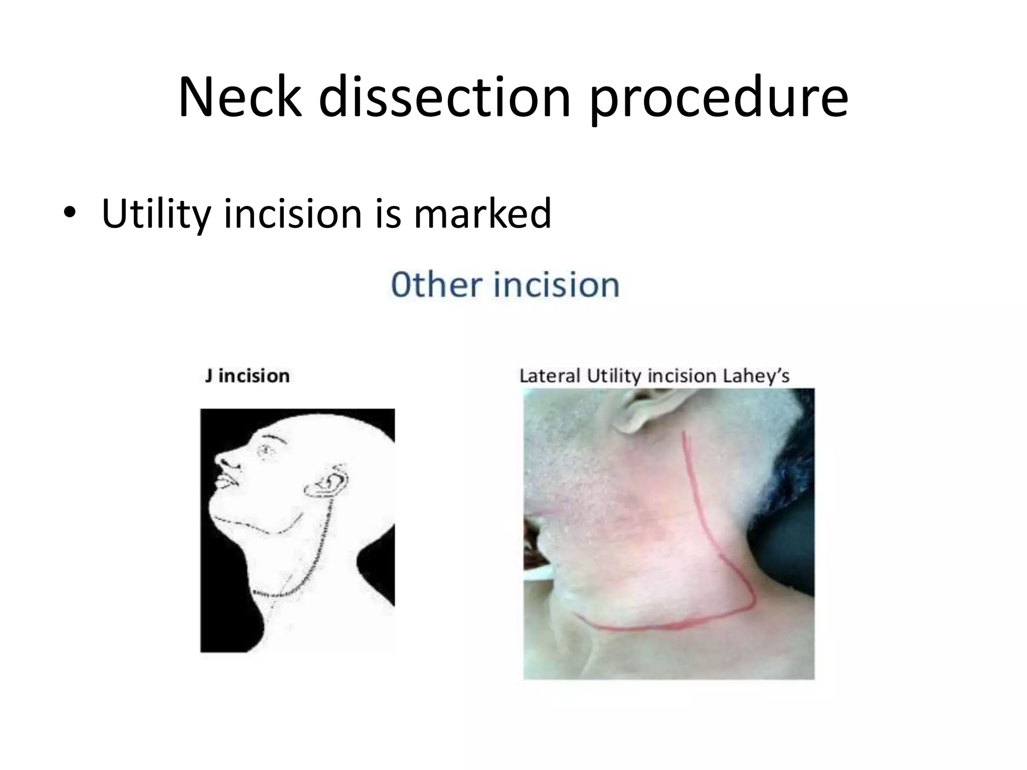
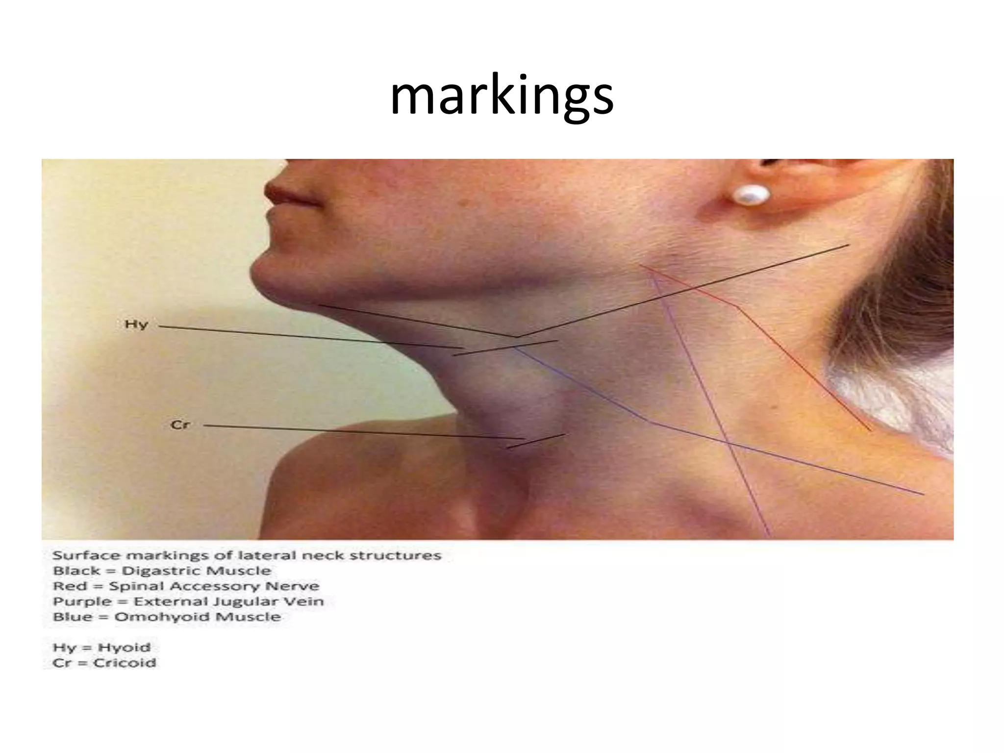
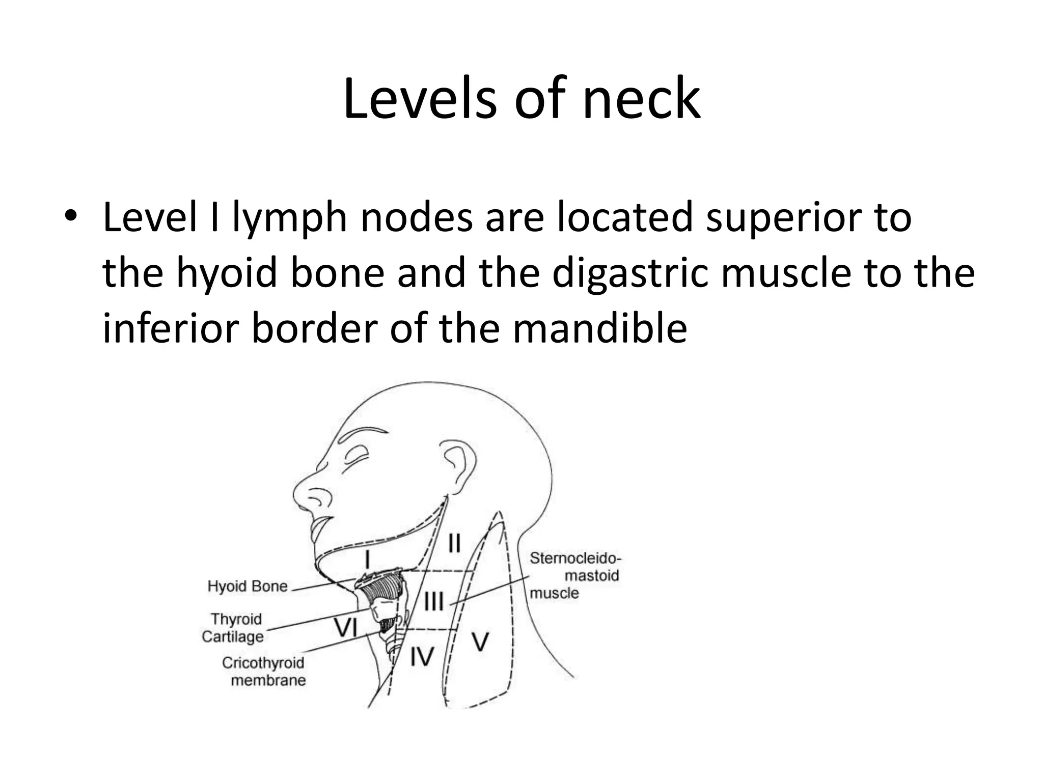
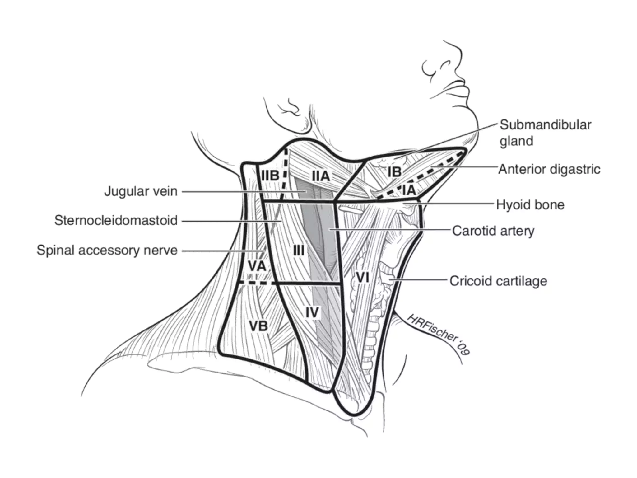
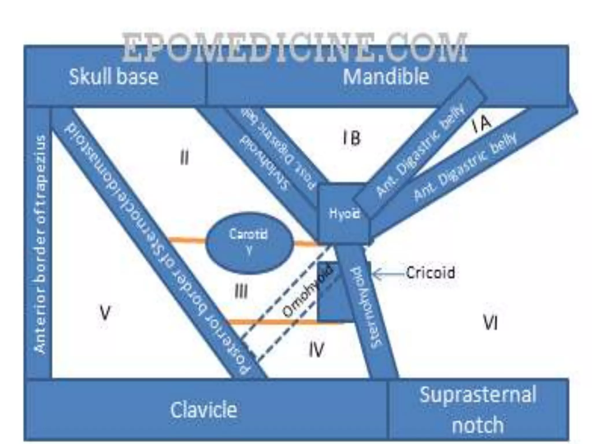
NECK DISSECTION PROCEDURE
JAMEEL KIFAYATULLAH
Neck dissection procedure
• Utility incision is marked
Landmarks
• Mastoid process
• Inferior border of the mandible
• The clavicle
• Midline chin
Incision
• Incision centered in a skin crease between the
inferior border of mandible and the clavicle
Marking of external jugular vein
• The external jugular vein is marked because
this structure is preserved for lateral
microvascular anastomosis and it commonly
serves as a landmark for locating the greater
auricular nerve, which usually lies in the same
plane within 1 cm posterior to the external
jugular vein
markings
anaesthesia
• the skin is infiltrated with epinephrine
containing local anesthetic.
Elevation of subplatysmal flaps
• the skin incision is made and carried through
the platysma. Subplatysmal flaps are elevated
to the level of the inferior border of the
mandible superiorly and the clavicle inferiorly
• Once the flaps are elevated, they are retracted
with stay sutures or hooks
Freeing of SCM
• For a radical neck dissection or modified
radical neck dissection with sacrifice of the
SCM, the SCM is freed from its attachments to
the mastoid and clavicle
LYMPH NODES DISSECTION
• Then, level V lymph nodes are dissected and
the spinal accessory nerve is preserved if
possible. Level V lymph nodes along with the
SCM, level II, III, and IV lymph nodes are
dissected in an anterior direction.
Lymph nodes dissection
• During dissection of levels III and IV, care is
taken to avoid injury to the lymphatic ducts,
phrenic nerve, vagus nerve, or brachial plexus
Internal jugular vein
• If the IJV is to be sacrificed, it is ligated
proximally and distally and then dissected in
continuity with the specimen. If the IJV is to
be left in place, careful removal of the fascia
and lymphatic tissue circumferentially around
the IJV is performed while avoiding excessive
torsion on the vein. Branches emanating from
the vein on the anterior and medial surface
are ligated and divided
Dissection of specimen
• The entire specimen is dissected further off of
the carotid artery, taking care not to
excessively thin the carotid artery or injure the
vagus nerve.
Level I node dissection
• Next, level I lymph nodes are dissected
starting in level IA and moving posterior
• The facial nerve is identified and dissected
free of the fascia and retracted superiorly; this
allows complete dissection of level IB and the
perifacial lymph nodes while protecting the
facial nerve from inadvertent injury
Level 1 dissection
• The mylohyoid muscle is identified and
retracted anteriorly, and the submandibular
duct, lingual nerve, and hypoglossal nerves
are identified. The duct is ligated and divided,
and the gland along with the nodal contents
of the submandibular triangle is then
delivered inferiorly.
Delivery of specimen
• By this point, the specimen is held only by
some attachments immediately anterior to
the carotid artery. These attachments are
carefully divided taking care not to injure
branches of the external carotid artery. Once
this is complete, the specimen is delivered
from the neck.
Selective neck dissection
• In a selective neck dissection, once the
subplatysmal flaps are elevated, the fascia over
the SCM is incised and the SCM is retracted
laterally to expose level II, III, and IV lymph nodes
• These levels are then dissected from the fascia
over the splenius capitus and levator scapulae
muscles, taking care not to injure the spinal
accessory nerve, phrenic nerve, brachial plexus,
or lymphatic ducts. The specimen is brought
anterior as the dissection proceeds
Selective neck dissection
• Dissection of the IJV proceeds with care not to
injure the vein, the vagus nerve, or carotid
artery
• Dissection of level I proceeds in the same
manner as for a modified radical or radical
neck dissection.
Selective neck dissection
• Once the specimen is removed, it is oriented
for the pathologist and the levels are marked
with skin staples or sutures
• Drains are placed in the wound and the
wound is closed, reapproximating platysma
with Vicryl suture and skin with staples.
Levels of neck
• Level I lymph nodes are located superior to
the hyoid bone and the digastric muscle to the
inferior border of the mandible
Levels of neck
• Level 1 further subdivided into submental (IA)
and submandibular (IB) groups
• Submental group lymph nodes are located
anterior to the anterior belly of the digastric
muscle and cephalad to the hyoid bone up to
the inferior border of the mandible
SUBMENTAL( IA) GROUP
Submandibular (IB) Group
Lymph nodes located posterior to the anterior
belly of the digastric muscle and superior to
the anterior and posterior bellies of the
digastric muscle up to the inferior border of
the mandible are the submandibular group
Level II lymph nodes
• Level II lymph nodes are located around the
upper portion of the IJV and the upper portion
of the spinal accessory nerve extending from
the base of the skull downwards to the
bifurcation of the carotid artery.
Level 3 lymph nodes
• Level III lymph nodes are around the middle
portion of the IJV starting from the inferior
border of level II extending downwards to the
omohyoid muscle.
Neck levels
Level IV group
• Level IV lymph nodes are the lower portion of
the IJV extending from the inferior border of
level III downwards to the clavicle
Levels of neck
• The posterior triangle group or level V is
bound by the triangle formed by the clavicle,
posterior border of the SCM, and the anterior
border of the trapezius muscle .
Level VI AND VII
• The central compartment of the neck is
broken down into level VI and VII. Level VI
lymph nodes are from the hyoid bone to the
suprasternal notch and between the medial
borders of the carotid sheath. Level VII, the
superior mediastinal group, are the lymph
nodes located in the anterior mediastinum
extending from the innominate artery
inferiorly to the suprasternal notch.
Neck dissection procedure

Neck dissection procedure

  • 1.
  • 2.
    Neck dissection procedure •Utility incision is marked
  • 3.
    Landmarks • Mastoid process •Inferior border of the mandible • The clavicle • Midline chin
  • 4.
    Incision • Incision centeredin a skin crease between the inferior border of mandible and the clavicle
  • 5.
    Marking of externaljugular vein • The external jugular vein is marked because this structure is preserved for lateral microvascular anastomosis and it commonly serves as a landmark for locating the greater auricular nerve, which usually lies in the same plane within 1 cm posterior to the external jugular vein
  • 6.
  • 7.
    anaesthesia • the skinis infiltrated with epinephrine containing local anesthetic.
  • 8.
    Elevation of subplatysmalflaps • the skin incision is made and carried through the platysma. Subplatysmal flaps are elevated to the level of the inferior border of the mandible superiorly and the clavicle inferiorly • Once the flaps are elevated, they are retracted with stay sutures or hooks
  • 9.
    Freeing of SCM •For a radical neck dissection or modified radical neck dissection with sacrifice of the SCM, the SCM is freed from its attachments to the mastoid and clavicle
  • 10.
    LYMPH NODES DISSECTION •Then, level V lymph nodes are dissected and the spinal accessory nerve is preserved if possible. Level V lymph nodes along with the SCM, level II, III, and IV lymph nodes are dissected in an anterior direction.
  • 11.
    Lymph nodes dissection •During dissection of levels III and IV, care is taken to avoid injury to the lymphatic ducts, phrenic nerve, vagus nerve, or brachial plexus
  • 12.
    Internal jugular vein •If the IJV is to be sacrificed, it is ligated proximally and distally and then dissected in continuity with the specimen. If the IJV is to be left in place, careful removal of the fascia and lymphatic tissue circumferentially around the IJV is performed while avoiding excessive torsion on the vein. Branches emanating from the vein on the anterior and medial surface are ligated and divided
  • 13.
    Dissection of specimen •The entire specimen is dissected further off of the carotid artery, taking care not to excessively thin the carotid artery or injure the vagus nerve.
  • 15.
    Level I nodedissection • Next, level I lymph nodes are dissected starting in level IA and moving posterior • The facial nerve is identified and dissected free of the fascia and retracted superiorly; this allows complete dissection of level IB and the perifacial lymph nodes while protecting the facial nerve from inadvertent injury
  • 16.
    Level 1 dissection •The mylohyoid muscle is identified and retracted anteriorly, and the submandibular duct, lingual nerve, and hypoglossal nerves are identified. The duct is ligated and divided, and the gland along with the nodal contents of the submandibular triangle is then delivered inferiorly.
  • 17.
    Delivery of specimen •By this point, the specimen is held only by some attachments immediately anterior to the carotid artery. These attachments are carefully divided taking care not to injure branches of the external carotid artery. Once this is complete, the specimen is delivered from the neck.
  • 19.
    Selective neck dissection •In a selective neck dissection, once the subplatysmal flaps are elevated, the fascia over the SCM is incised and the SCM is retracted laterally to expose level II, III, and IV lymph nodes • These levels are then dissected from the fascia over the splenius capitus and levator scapulae muscles, taking care not to injure the spinal accessory nerve, phrenic nerve, brachial plexus, or lymphatic ducts. The specimen is brought anterior as the dissection proceeds
  • 20.
    Selective neck dissection •Dissection of the IJV proceeds with care not to injure the vein, the vagus nerve, or carotid artery • Dissection of level I proceeds in the same manner as for a modified radical or radical neck dissection.
  • 21.
    Selective neck dissection •Once the specimen is removed, it is oriented for the pathologist and the levels are marked with skin staples or sutures • Drains are placed in the wound and the wound is closed, reapproximating platysma with Vicryl suture and skin with staples.
  • 22.
    Levels of neck •Level I lymph nodes are located superior to the hyoid bone and the digastric muscle to the inferior border of the mandible
  • 23.
    Levels of neck •Level 1 further subdivided into submental (IA) and submandibular (IB) groups • Submental group lymph nodes are located anterior to the anterior belly of the digastric muscle and cephalad to the hyoid bone up to the inferior border of the mandible
  • 24.
  • 25.
    Submandibular (IB) Group Lymphnodes located posterior to the anterior belly of the digastric muscle and superior to the anterior and posterior bellies of the digastric muscle up to the inferior border of the mandible are the submandibular group
  • 27.
    Level II lymphnodes • Level II lymph nodes are located around the upper portion of the IJV and the upper portion of the spinal accessory nerve extending from the base of the skull downwards to the bifurcation of the carotid artery.
  • 29.
    Level 3 lymphnodes • Level III lymph nodes are around the middle portion of the IJV starting from the inferior border of level II extending downwards to the omohyoid muscle.
  • 30.
  • 31.
    Level IV group •Level IV lymph nodes are the lower portion of the IJV extending from the inferior border of level III downwards to the clavicle
  • 32.
    Levels of neck •The posterior triangle group or level V is bound by the triangle formed by the clavicle, posterior border of the SCM, and the anterior border of the trapezius muscle .
  • 33.
    Level VI ANDVII • The central compartment of the neck is broken down into level VI and VII. Level VI lymph nodes are from the hyoid bone to the suprasternal notch and between the medial borders of the carotid sheath. Level VII, the superior mediastinal group, are the lymph nodes located in the anterior mediastinum extending from the innominate artery inferiorly to the suprasternal notch.