Akc
•Download as DOCX, PDF•
0 likes•1,123 views

Atopic Keratoconjunctivitis, etiology, Signs and symptoms, Differential diagnosis, management

Recommended
More Related Content
What's hot
What's hot (20)
Viewers also liked
Viewers also liked (20)
Similar to Akc
Similar to Akc (20)
More from Raju Kaiti
More from Raju Kaiti (20)
Recently uploaded
Recently uploaded (20)
Akc
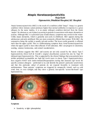
- 1. Atopic Keratoconjunctivitis Raju Kaiti Optometrist, Dhulikhel Hospital, KU Hospital Atopic keratoconjunctivitis (AKC) is the result of a condition called "atopy". Atopy is a genetic condition where immune system produces higher than normal antibodies in response to a given allergen. As the name implies, it is an atopic (allergic) condition (derived from the Greek ‘atopos’ for aberrant or out of place) occurring in patients in association with atopic dermatitis or eczema. Although AKC is a perennial (year round) disease, symptoms may worsen in the winter. Unlike atopic dermatitis, which is generally seen early in childhood, AKC appears during late adolescence and early adulthood. Men are more commonly affected than women. With AKC, the conjunctiva lining the eyelids is usually red and swollen. The lower eyelid generally is affected more than the upper eyelid. This is a differentiating symptom from vernal keratoconjunctivitis where the upper eyelid is most often affected. If left untreated, AKC can progress to ulceration, scarring, cataract, keratoconus, and corneal vascularization. Recent evidence suggests that AKC and eczema are not only caused by the classic Type I hypersensitivity (from Gell & Coomb’s original classification system) but also by Type IV (delayed-type) hypersensitivity. This is supported by the clinical findings that some patients have neither peripheral eosinophilia nor high IgE levels (seen in classic Type I hypersensitivities), have negative RAST tests (radio-immunoallergosorption testing that measures IgE levels for specific common allergens – performed to try and identify the patient’s particular sensitivities) and that this particular subset of patients do not respond favorably to treatment with antihistamines. These patients’ symptoms are triggered by nonspecific stimuli, such as cold wind, dust, or even sunlight, and they show a general, non-specific ‘across-the-board’ type hypersensitivity. Symptoms Sensitivity to light (photophobia)
- 2. Itching Burning Tearing Red and hardened eyelids Blurred vision White stringy mucoid discharge Onset of ocular symptoms may occur several years after onset of atopy. Almost all patients eventually develop bilateral involvement, though it often starts off unilateral and asymmetrical. Signs: Eyelids may be thickened, crusted and fissured Associated chronic staphylococcal blepharitis Pale, edematous, boggy and featureless conjunctiva in the early stages, progressing to papillary hypertrophy, subepithelial fibrosis, fornix foreshortening, and a progressive chronic cicatrizing conjunctivitis. Limbal inflammation Late complications include the development of trichiasis, entropion, madarosis and the resulting corneal sequelae. Keratopathy is the main cause of visual impairment in patients with AKC Corneal involvement is common and may be sight-threatening: beginning with punctate epitheliopathy that may progress to macro-erosion, plaque formation (usually upper half), progressive corneal sub epithelial scarring, neovascularization and thinning. These patients are prone to develop herpes simplex keratitis, corneal ectasia such as keratoconus, atopic (anterior or posterior polar) cataracts, retinal detachment. Etiologies AKC is a genetic condition. Risk Factors A family history of multiple allergies Atopic dermatitis Eczema
- 3. Asthma Tests and Diagnosis AKC usually is diagnosed by clinical exam and a medical and family history, although a conjunctival biopsy may be helpful in distinguishing AKC from other conditions. In histopathological specimens, increased numbers of mast cells and eosinophil are seen infiltrating the conjunctival epithelium and the substantia propria. Squamous metaplasia and increased numbers of fibroblasts and collagen deposition are also seen. Eosinophil is directly responsible for the corneal complications, as their toxic granule contents (eosinophil major basic protein, eosinophil derived neurotoxin, eosinophil lymphotoxin, and eosinophil protein X) have been detected in the beds of stromal ulcers and epithelial defects. The tears of patients with AKC contain high levels of IL-4 and 5, RANTES, ECP that selectively recruit, stimulate and encourage terminal differentiation of eosinophil. High numbers of IgE, B cells, T helper cells, IL 2 and the IL2 receptor have also been described in tears and in sera. Differential Diagnoses Blepharitis, Adult Cicatricial Pemphigoid Conjunctivitis, Allergic Conjunctivitis, Giant Papillary Conjunctivitis, Viral Trachoma Treatment and Drugs The treatment of AKC should include involvement of an allergist for identification of the provoking allergen(s) and education about avoidance of triggers. The triggering antigen may be identified in patients with skin patch testing against a panel of commonly occurring antigens, or in a more sophisticated manner by RAST testing. Combinations of oral and topical antihistamines and mast cell stabilizers usually are effective in controlling symptoms. In more severe cases, there is potential for damage to the eye caused by scratching and rubbing. Topical treatment with antibiotics for the associated staphylococcal blepharitis may be needed, and patients may also benefit from topical mupirocin treatment for their skin and eyelid eczema. Lubrication with artificial tears may be helpful; along with a dual acting antihistamine/mast cell stabilizer. Topical corticosteroids may be periodically needed for flare-ups, but their use should be closely monitored. Only in severe cases should topical steroid therapy be considered. In the most severe cases, systemic treatment with signal transduction inhibitors such as (steroid-sparing agents) cyclosporine or tacrolimus may be needed to control the systemic immune dysfunction that leads to the ocular and dermatological manifestations. Systemic antihistamines can also benefit patients and reduce the itching, especially if taken at night, also help the patient to sleep better.
- 4. An eye care practitioner may advise to wear cotton gloves at night to prevent unintentional damage to the ocular surface. Cold compresses and saline irrigation to lower the elevated tear pH also may be helpful. AKC, a potentially blinding condition, is also responsible for causing much ocular morbidity in patients from a peculiar set of other ocular conditions. Its chronic, unremitting (without treatment) course has much to do with its eventual blinding potential, as opposed to the other self-limiting conjunctivitis such as vernal keratoconjunctivitis, seasonal allergic conjunctivitis, or giant papillary conjunctivitis. Management by Optometrist: 1. Non pharmacological: Lid hygiene and treatment of associated staphylococcal blepharitis Cold compresses Advise avoidance of specific allergens if known, e.g. elimination of pets and carpeting, where necessary; instillation of air filtering devices and alterations to bedding materials 2. Pharmacological: Systemic antihistamines e.g. cetirizine Topical mast cell stabilizers e.g. Sodium cromoglycate 2%, 4% lodoxamide 0.1%, nedocromil sodium 2%, or dual acting agents e.g. olopatadine 0.1%, may also provide symptomatic relief 3. Appropriate Referral