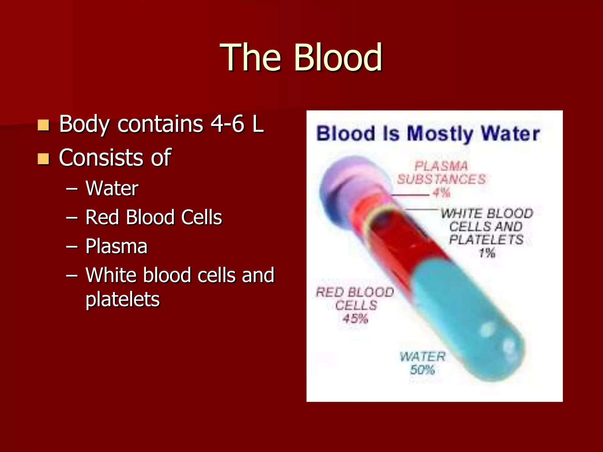
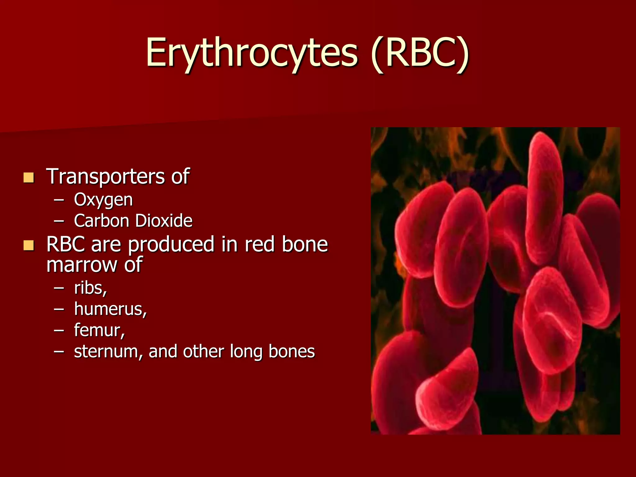
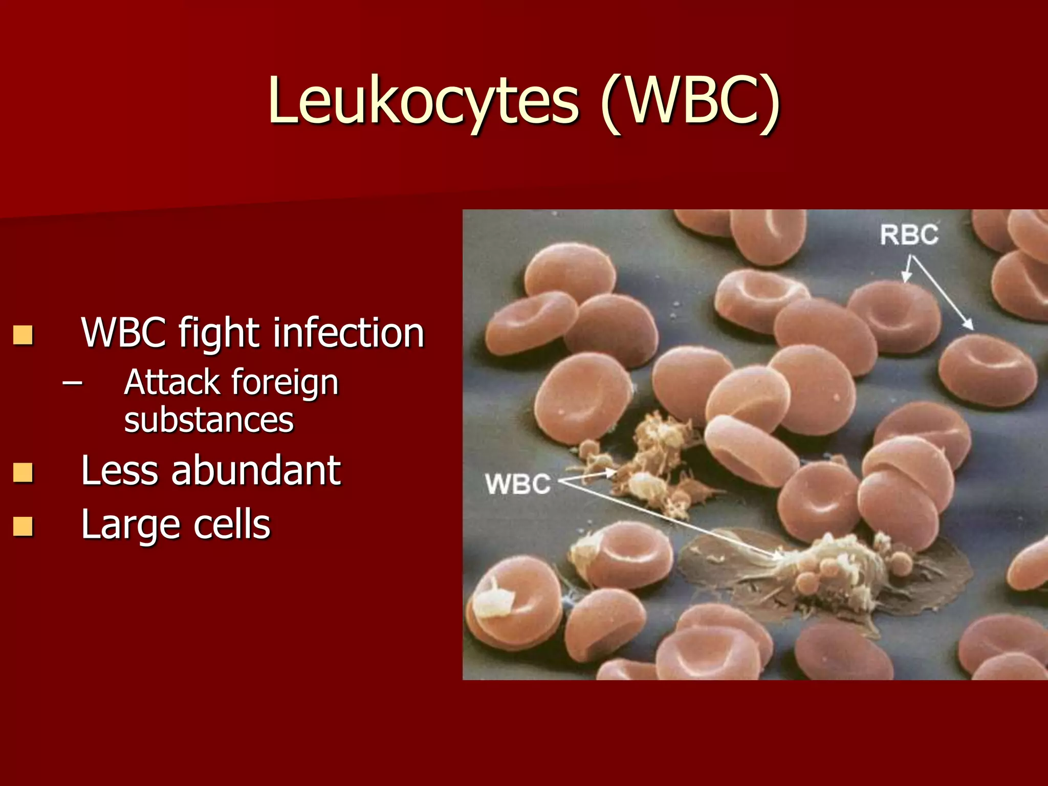
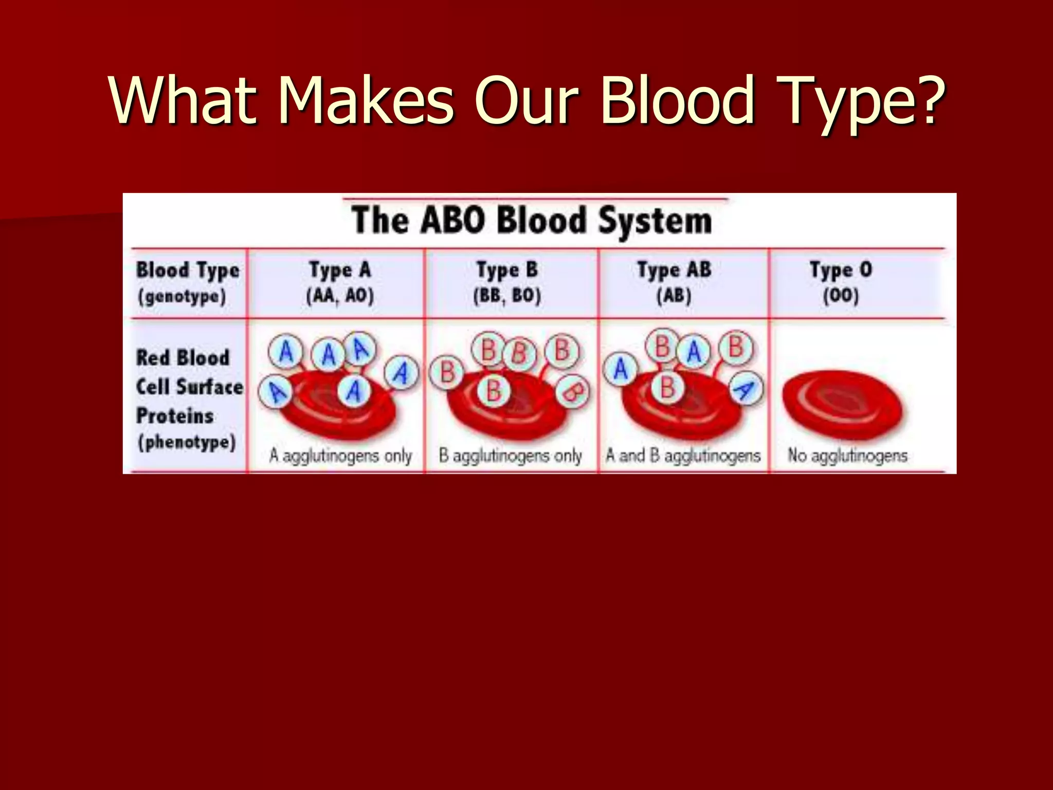
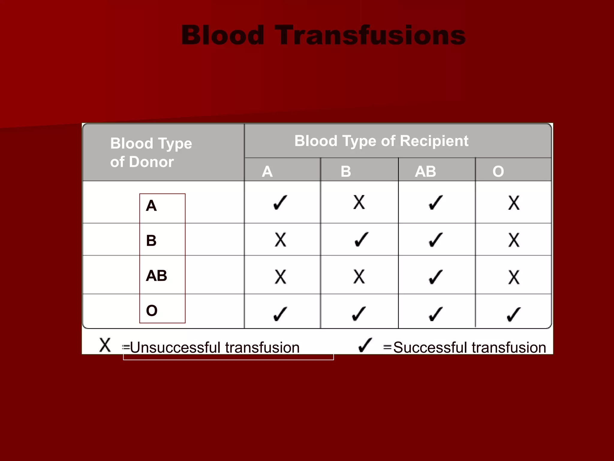
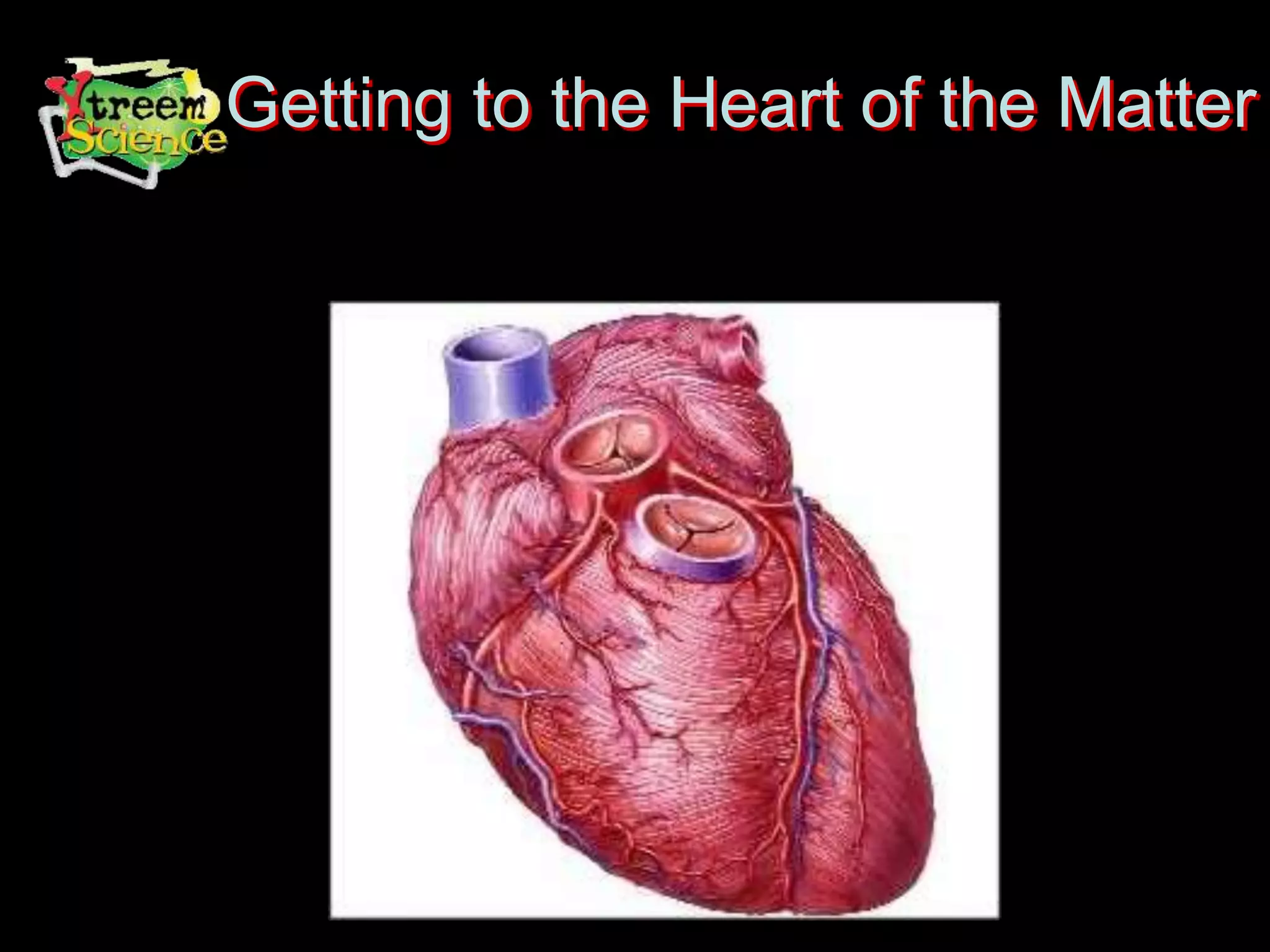
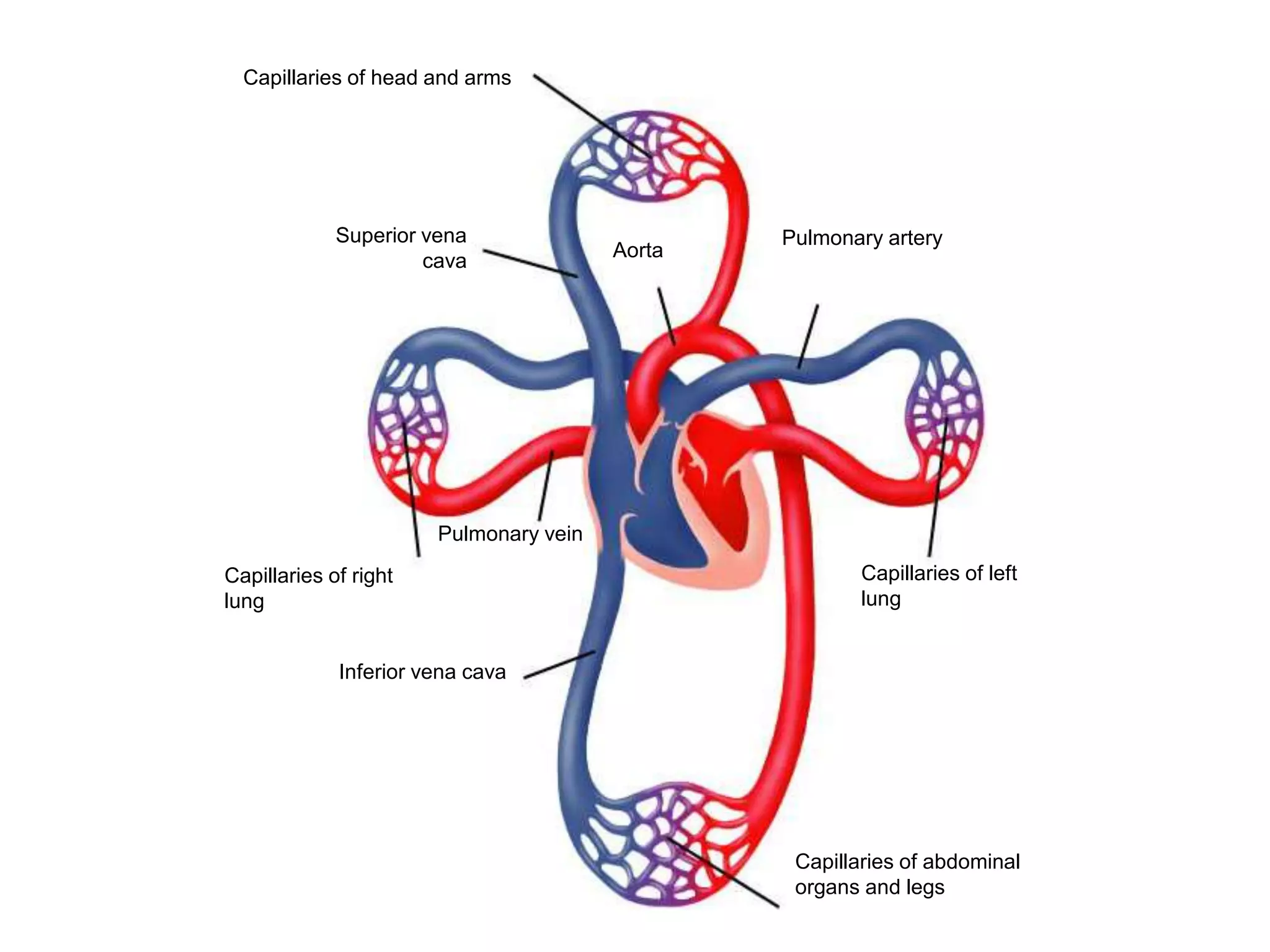
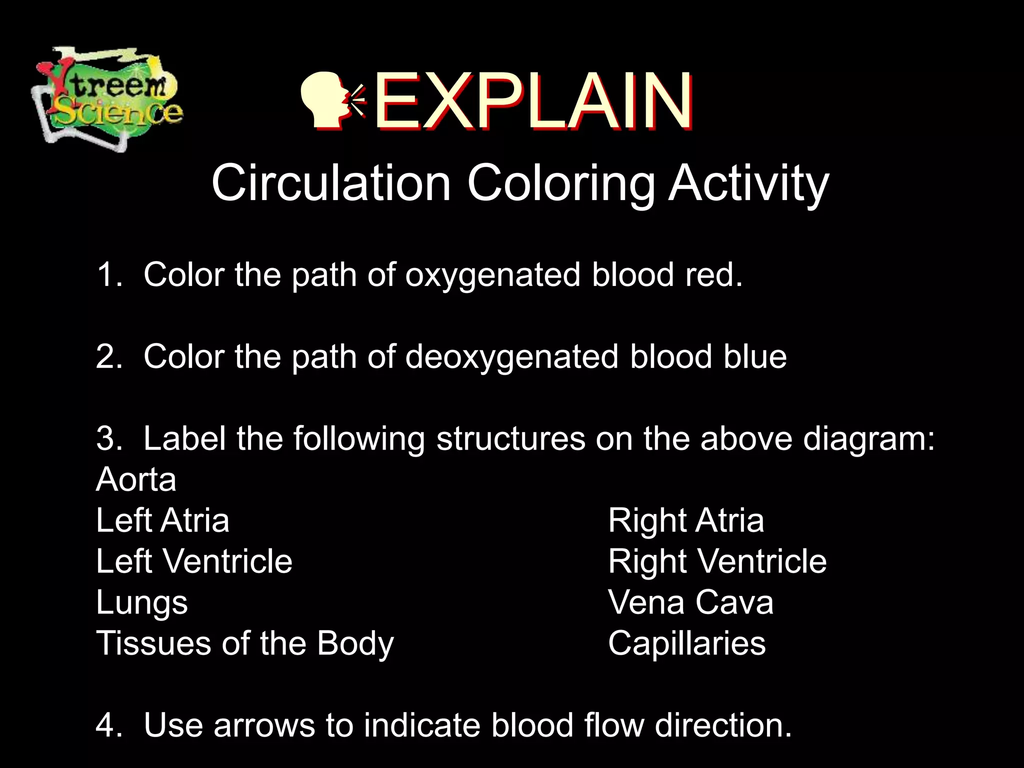
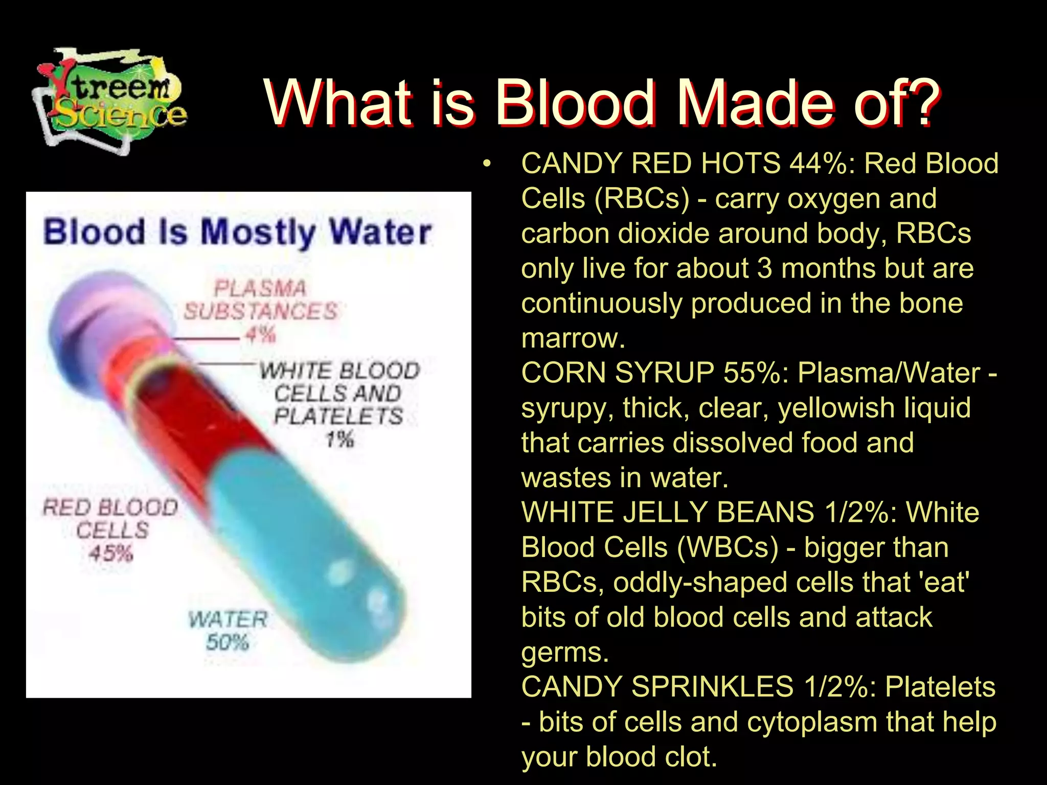
Science Module
7th Grade
Body Systems
Circulatory System
7th Grade Science
TAKS 2
TEKS 7.9(A)
TAKS Objective 2
The student will demonstrate an
understanding of living systems and the
environment – Interdependence occurs
among living systems
TEKS Science Concept
• TEKS 7.9 (A) identify the systems of the
human organism and describe their
functions.
Student Prior Knowledge
• TEKS 6.10 (C) identify how structure
complements function at different levels of
organization including organ and organ
systems.
Background
Circulatory System
Structures
• Heart
• Blood Vessels
– Arteries
– Veins
– Capillaries
• Blood
Circulatory System
Function
• The overall function of the circulatory system is
to transport materials throughout the body
toward and away from particular target organs
and tissues.
Two Pathways
• Pulmonary Circulation
– Carries blood to lungs and back to the heart
• Systemic Circulation
– Carries blood to body and back to the heart
Capillaries of head and arms
Capillaries of abdominal
organs and legs
Inferior vena cava
Pulmonary vein
Capillaries of right
lung
Superior vena
cava
Aorta
Pulmonary artery
Capillaries of left
lung
Your Blood Vessels:
Pathway of Circulation
 3 types of vessels
– Arteries
– Capillaries
– Veins
Artery vs. Vein
Arteries:
carries blood Away from heart
– Large
– Thick-walled, Muscular
– Elastic
– Oxygenated blood
 Exception Pulmonary Artery
– Carried under great pressure
– Steady pulsating
Arterioles: smaller vessels, enter tissue
Capillaries
– Smallest vessel
– Microscopic
– Walls one cell thick
– Nutrients and gases diffuse here
Veins:
Carries blood to heart
– Carries blood that contains
waste and CO2
 Exception pulmonary vein
– Blood not under much
pressure
– Valves to prevent much
gravity pull
Venules: larger than capillaries
Varicose Veins
Damaged Valves in Veins
Your Heart: The Vital Pump
 At REST, the heart
pumps about 5
QUARTS of blood a
minute.
 During EXTREME
EXERTION (exercise)
it can pump 40 quarts
a minute.
Heart:
Structure and Function
 Keeps blood moving
 Large organ
composed of Cardiac
muscle
Structure of Heart
 Four chambers
– Two upper (Atria)
 Right Atria
 Left Atria
– Two lower
(Ventricles)
 Right Ventricle
 Left Ventricle
Bloods Path Through the Heart
 Both Atria fill at same time
– Rt atrium receives oxygen POOR blood from
body via the vena cavas
– Left atrium receives oxygen RICH blood from
lungs through four pulmonary veins
 After filled with blood atria contract,
pushing blood into ventricle
Both ventricles contract
Right ventricle contracts and pushes
oxygen-poor blood toward lungs,
 against gravity,
 through pulmonary arteries
Left ventricle contracts and forces oxygen
rich blood
 out of heart through
 aorta (largest vessel)
Bloods Path Through the Heart
(cont)
The Blood
 Body contains 4-6 L
 Consists of
– Water
– Red Blood Cells
– Plasma
– White blood cells and
platelets
Erythrocytes (RBC)
 Transporters of
– Oxygen
– Carbon Dioxide
 RBC are produced in red bone
marrow of
– ribs,
– humerus,
– femur,
– sternum, and other long bones
Leukocytes (WBC)
 WBC fight infection
– Attack foreign
substances
 Less abundant
 Large cells
Platelets
 PLATELETS are for CLOTTING blood
 Cell fragments
 Produced in bone marrow
 Fibrin (sticky network of protein
fibers)
– Form a web trapping blood cells
Section 37-2
Blood Clotting
Break in Capillary Wall
Blood vessels injured.
Clumping of Platelets
Platelets clump at the site
and release thromboplastin.
Thromboplastin converts
prothrombin into thrombin..
Clot Forms
Thrombin converts
fibrinogen into fibrin, which
causes a clot. The clot
prevents further loss of
blood..
Blood Types
 Massive loss of blood requires a transfusion
 Four Types
– A
– B
– AB
– O
 Inherited from your parents
Blood Types
What happens when you mix blood
types?
 Plasma contains proteins that
correspond to the shape of the
different antigens
 If you mix one type with the
wrong one, you get CLUMPING
 Type O is the universal donor
 Type AB is the universal acceptor
What Makes Our Blood Type?
Blood Type
of Donor
A
B
AB
O
Blood Type of Recipient
A B AB O
Unsuccessful transfusion Successful transfusion
Blood Transfusions
Rh Factor
 Rhesus factor (Rh), also inherited
– Rh+ (have antigen)
– Rh- (NO antigen)
 Can cause complications in pregnancies
– mother Rh- 1st baby Rh+ : blood mixes with
mother; mother’s body makes anti-Rh+
antibodies
– 2nd Rh + body attacks baby
– Now have medicine to prevent antibody
formation
Getting to the Heart of the Matter
ENGAGE
1. Walt Disney’s 1957 “Hemo the Magnificent”
2. Play song from St. Joseph’s Aspirin Commercial
(originally in Happy Days episode) at:
http://www.stjosephaspirin.com/page.jhtml?id=/stjoseph/include/5_2.inc
Lyrics
• Pump, pump, pumps your Blood.
• The right atrium’s where the process begins, where the
CO2 Blood enters the heart.
• Through the tricuspid valve, to the right ventricle, the
pulmonary artery, and lungs.
• Once inside the lungs, it dumps its carbon dioxide and
picks up its oxygen supply.
• Then it’s back to the heart through the pulmonary vein,
through the atrium and left ventricle.
• Pump, pump, pumps your Blood.
EXPLORE
• Circulatory System
Simulation
Capillaries of head and arms
Capillaries of abdominal
organs and legs
Inferior vena cava
Pulmonary vein
Capillaries of right
lung
Superior vena
cava
Aorta
Pulmonary artery
Capillaries of left
lung
EXPLAIN
Circulation Coloring Activity
1. Color the path of oxygenated blood red.
2. Color the path of deoxygenated blood blue
3. Label the following structures on the above diagram:
Aorta
Left Atria Right Atria
Left Ventricle Right Ventricle
Lungs Vena Cava
Tissues of the Body Capillaries
4. Use arrows to indicate blood flow direction.
ELABORATE
Circulation Relay
What is Blood Made of?
• CANDY RED HOTS 44%: Red Blood
Cells (RBCs) - carry oxygen and
carbon dioxide around body, RBCs
only live for about 3 months but are
continuously produced in the bone
marrow.
CORN SYRUP 55%: Plasma/Water -
syrupy, thick, clear, yellowish liquid
that carries dissolved food and
wastes in water.
WHITE JELLY BEANS 1/2%: White
Blood Cells (WBCs) - bigger than
RBCs, oddly-shaped cells that 'eat'
bits of old blood cells and attack
germs.
CANDY SPRINKLES 1/2%: Platelets
- bits of cells and cytoplasm that help
your blood clot.
EVALUATE
• Given a drawing the student will label and describe the
functions of the four major parts of the circulatory system:
Heart, arteries, veins and capillaries.
• After participating the circulatory relay simulation, the
learner will travel the correct circulation pathway beginning
at the left ventricle and ending at the left atrium.
• After participating in the blood activity, the learner will list
the following four components of the blood: RBC, WBC,
Plasma and Platelets and describe the function of blood.

7th Circulatory System.ppt

  • 1.
  • 2.
    Body Systems Circulatory System 7thGrade Science TAKS 2 TEKS 7.9(A)
  • 3.
    TAKS Objective 2 Thestudent will demonstrate an understanding of living systems and the environment – Interdependence occurs among living systems
  • 4.
    TEKS Science Concept •TEKS 7.9 (A) identify the systems of the human organism and describe their functions.
  • 5.
    Student Prior Knowledge •TEKS 6.10 (C) identify how structure complements function at different levels of organization including organ and organ systems.
  • 6.
  • 7.
    Circulatory System Structures • Heart •Blood Vessels – Arteries – Veins – Capillaries • Blood
  • 8.
    Circulatory System Function • Theoverall function of the circulatory system is to transport materials throughout the body toward and away from particular target organs and tissues.
  • 9.
    Two Pathways • PulmonaryCirculation – Carries blood to lungs and back to the heart • Systemic Circulation – Carries blood to body and back to the heart
  • 10.
    Capillaries of headand arms Capillaries of abdominal organs and legs Inferior vena cava Pulmonary vein Capillaries of right lung Superior vena cava Aorta Pulmonary artery Capillaries of left lung
  • 11.
    Your Blood Vessels: Pathwayof Circulation  3 types of vessels – Arteries – Capillaries – Veins
  • 12.
  • 13.
    Arteries: carries blood Awayfrom heart – Large – Thick-walled, Muscular – Elastic – Oxygenated blood  Exception Pulmonary Artery – Carried under great pressure – Steady pulsating Arterioles: smaller vessels, enter tissue
  • 15.
    Capillaries – Smallest vessel –Microscopic – Walls one cell thick – Nutrients and gases diffuse here
  • 16.
    Veins: Carries blood toheart – Carries blood that contains waste and CO2  Exception pulmonary vein – Blood not under much pressure – Valves to prevent much gravity pull Venules: larger than capillaries
  • 17.
  • 19.
    Your Heart: TheVital Pump  At REST, the heart pumps about 5 QUARTS of blood a minute.  During EXTREME EXERTION (exercise) it can pump 40 quarts a minute.
  • 20.
    Heart: Structure and Function Keeps blood moving  Large organ composed of Cardiac muscle
  • 21.
    Structure of Heart Four chambers – Two upper (Atria)  Right Atria  Left Atria – Two lower (Ventricles)  Right Ventricle  Left Ventricle
  • 23.
    Bloods Path Throughthe Heart  Both Atria fill at same time – Rt atrium receives oxygen POOR blood from body via the vena cavas – Left atrium receives oxygen RICH blood from lungs through four pulmonary veins  After filled with blood atria contract, pushing blood into ventricle
  • 24.
    Both ventricles contract Rightventricle contracts and pushes oxygen-poor blood toward lungs,  against gravity,  through pulmonary arteries
  • 25.
    Left ventricle contractsand forces oxygen rich blood  out of heart through  aorta (largest vessel) Bloods Path Through the Heart (cont)
  • 27.
    The Blood  Bodycontains 4-6 L  Consists of – Water – Red Blood Cells – Plasma – White blood cells and platelets
  • 28.
    Erythrocytes (RBC)  Transportersof – Oxygen – Carbon Dioxide  RBC are produced in red bone marrow of – ribs, – humerus, – femur, – sternum, and other long bones
  • 29.
    Leukocytes (WBC)  WBCfight infection – Attack foreign substances  Less abundant  Large cells
  • 30.
    Platelets  PLATELETS arefor CLOTTING blood  Cell fragments  Produced in bone marrow  Fibrin (sticky network of protein fibers) – Form a web trapping blood cells
  • 31.
    Section 37-2 Blood Clotting Breakin Capillary Wall Blood vessels injured. Clumping of Platelets Platelets clump at the site and release thromboplastin. Thromboplastin converts prothrombin into thrombin.. Clot Forms Thrombin converts fibrinogen into fibrin, which causes a clot. The clot prevents further loss of blood..
  • 32.
    Blood Types  Massiveloss of blood requires a transfusion  Four Types – A – B – AB – O  Inherited from your parents
  • 33.
  • 34.
    What happens whenyou mix blood types?  Plasma contains proteins that correspond to the shape of the different antigens  If you mix one type with the wrong one, you get CLUMPING  Type O is the universal donor  Type AB is the universal acceptor
  • 35.
    What Makes OurBlood Type?
  • 36.
    Blood Type of Donor A B AB O BloodType of Recipient A B AB O Unsuccessful transfusion Successful transfusion Blood Transfusions
  • 37.
    Rh Factor  Rhesusfactor (Rh), also inherited – Rh+ (have antigen) – Rh- (NO antigen)  Can cause complications in pregnancies – mother Rh- 1st baby Rh+ : blood mixes with mother; mother’s body makes anti-Rh+ antibodies – 2nd Rh + body attacks baby – Now have medicine to prevent antibody formation
  • 38.
    Getting to theHeart of the Matter
  • 39.
    ENGAGE 1. Walt Disney’s1957 “Hemo the Magnificent” 2. Play song from St. Joseph’s Aspirin Commercial (originally in Happy Days episode) at: http://www.stjosephaspirin.com/page.jhtml?id=/stjoseph/include/5_2.inc
  • 40.
    Lyrics • Pump, pump,pumps your Blood. • The right atrium’s where the process begins, where the CO2 Blood enters the heart. • Through the tricuspid valve, to the right ventricle, the pulmonary artery, and lungs. • Once inside the lungs, it dumps its carbon dioxide and picks up its oxygen supply. • Then it’s back to the heart through the pulmonary vein, through the atrium and left ventricle. • Pump, pump, pumps your Blood.
  • 41.
  • 42.
    Capillaries of headand arms Capillaries of abdominal organs and legs Inferior vena cava Pulmonary vein Capillaries of right lung Superior vena cava Aorta Pulmonary artery Capillaries of left lung
  • 43.
    EXPLAIN Circulation Coloring Activity 1.Color the path of oxygenated blood red. 2. Color the path of deoxygenated blood blue 3. Label the following structures on the above diagram: Aorta Left Atria Right Atria Left Ventricle Right Ventricle Lungs Vena Cava Tissues of the Body Capillaries 4. Use arrows to indicate blood flow direction.
  • 44.
  • 46.
    What is BloodMade of? • CANDY RED HOTS 44%: Red Blood Cells (RBCs) - carry oxygen and carbon dioxide around body, RBCs only live for about 3 months but are continuously produced in the bone marrow. CORN SYRUP 55%: Plasma/Water - syrupy, thick, clear, yellowish liquid that carries dissolved food and wastes in water. WHITE JELLY BEANS 1/2%: White Blood Cells (WBCs) - bigger than RBCs, oddly-shaped cells that 'eat' bits of old blood cells and attack germs. CANDY SPRINKLES 1/2%: Platelets - bits of cells and cytoplasm that help your blood clot.
  • 47.
    EVALUATE • Given adrawing the student will label and describe the functions of the four major parts of the circulatory system: Heart, arteries, veins and capillaries. • After participating the circulatory relay simulation, the learner will travel the correct circulation pathway beginning at the left ventricle and ending at the left atrium. • After participating in the blood activity, the learner will list the following four components of the blood: RBC, WBC, Plasma and Platelets and describe the function of blood.