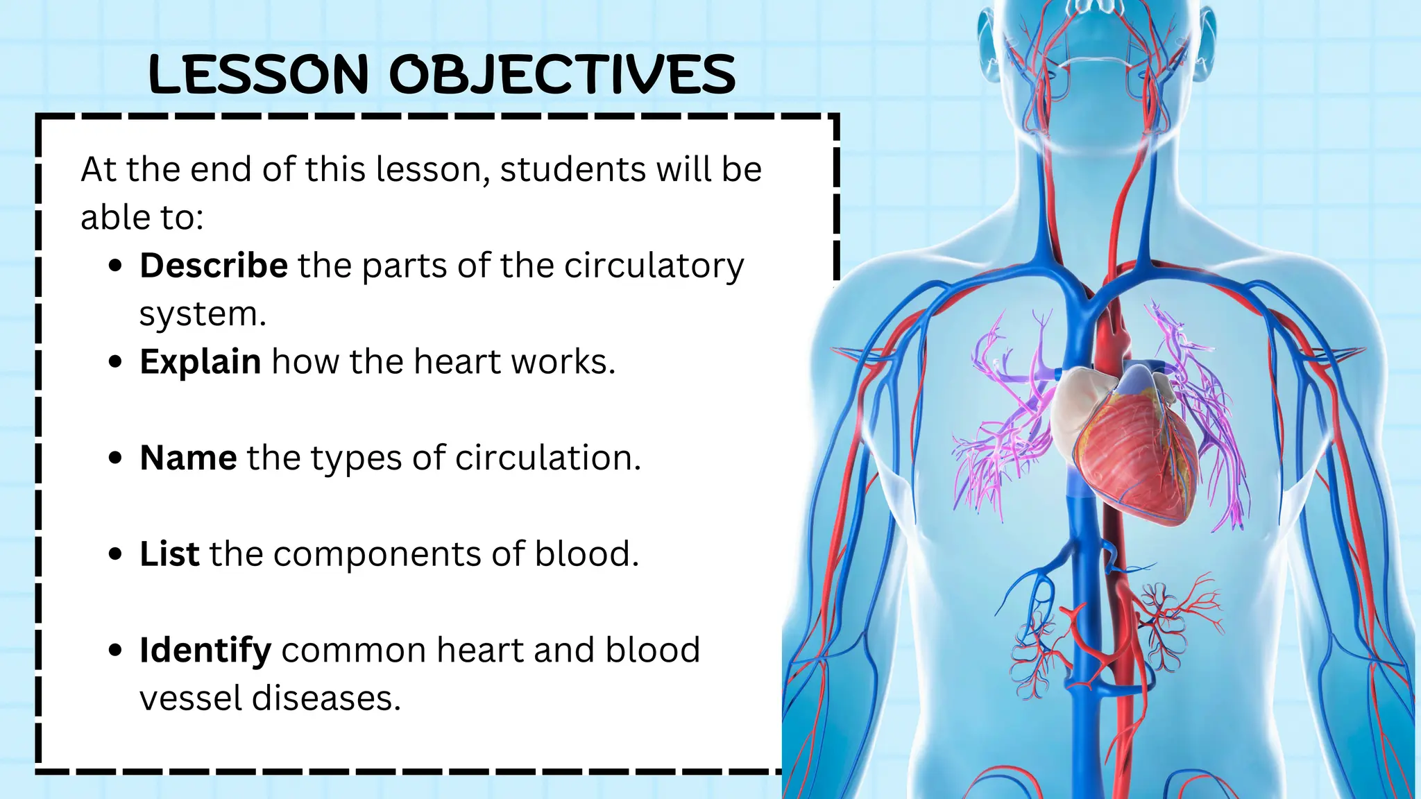
At the endof this lesson, students will be
able to:
Describe the parts of the circulatory
system.
Explain how the heart works.
Name the types of circulation.
List the components of blood.
Identify common heart and blood
vessel diseases.
LESSON OBJECTIVES
3.
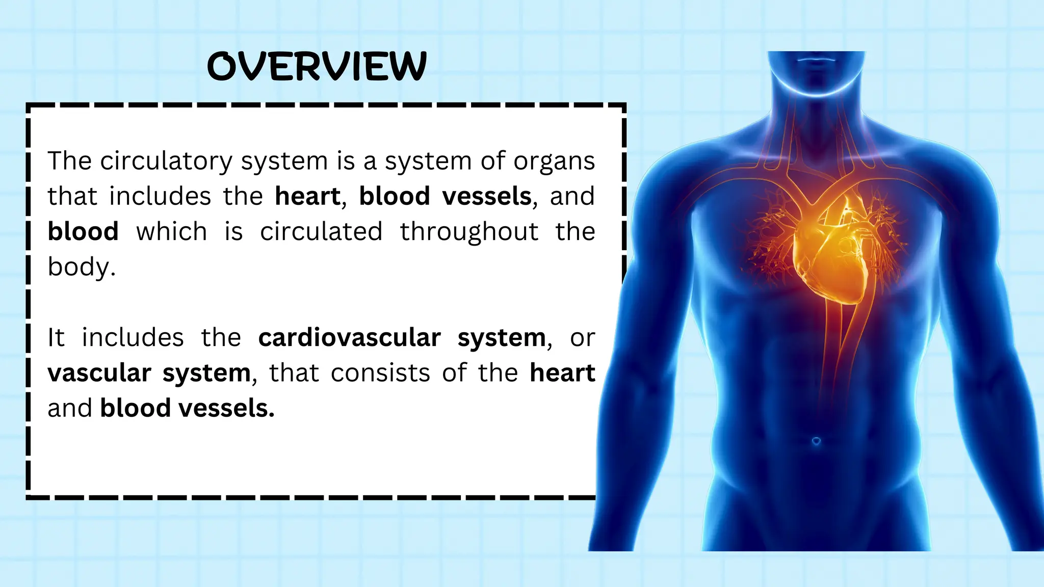
The circulatory systemis a system of organs
that includes the heart, blood vessels, and
blood which is circulated throughout the
body.
It includes the cardiovascular system, or
vascular system, that consists of the heart
and blood vessels.
OVERVIEW
4.
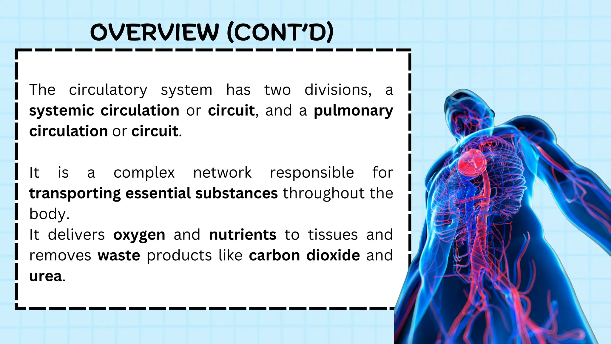
The circulatory systemhas two divisions, a
systemic circulation or circuit, and a pulmonary
circulation or circuit.
It is a complex network responsible for
transporting essential substances throughout the
body.
It delivers oxygen and nutrients to tissues and
removes waste products like carbon dioxide and
urea.
OVERVIEW (CONT’D)
5.
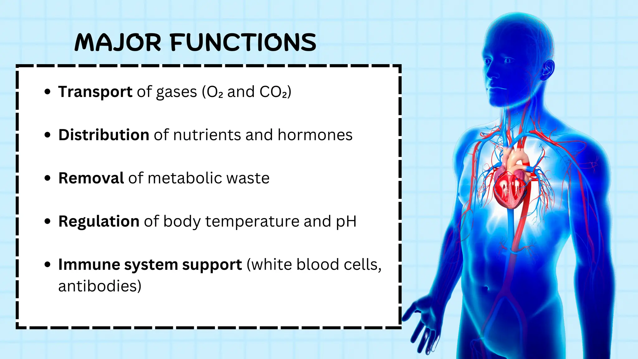
Transport of gases(O₂ and CO₂)
Distribution of nutrients and hormones
Removal of metabolic waste
Regulation of body temperature and pH
Immune system support (white blood cells,
antibodies)
MAJOR FUNCTIONS
6.
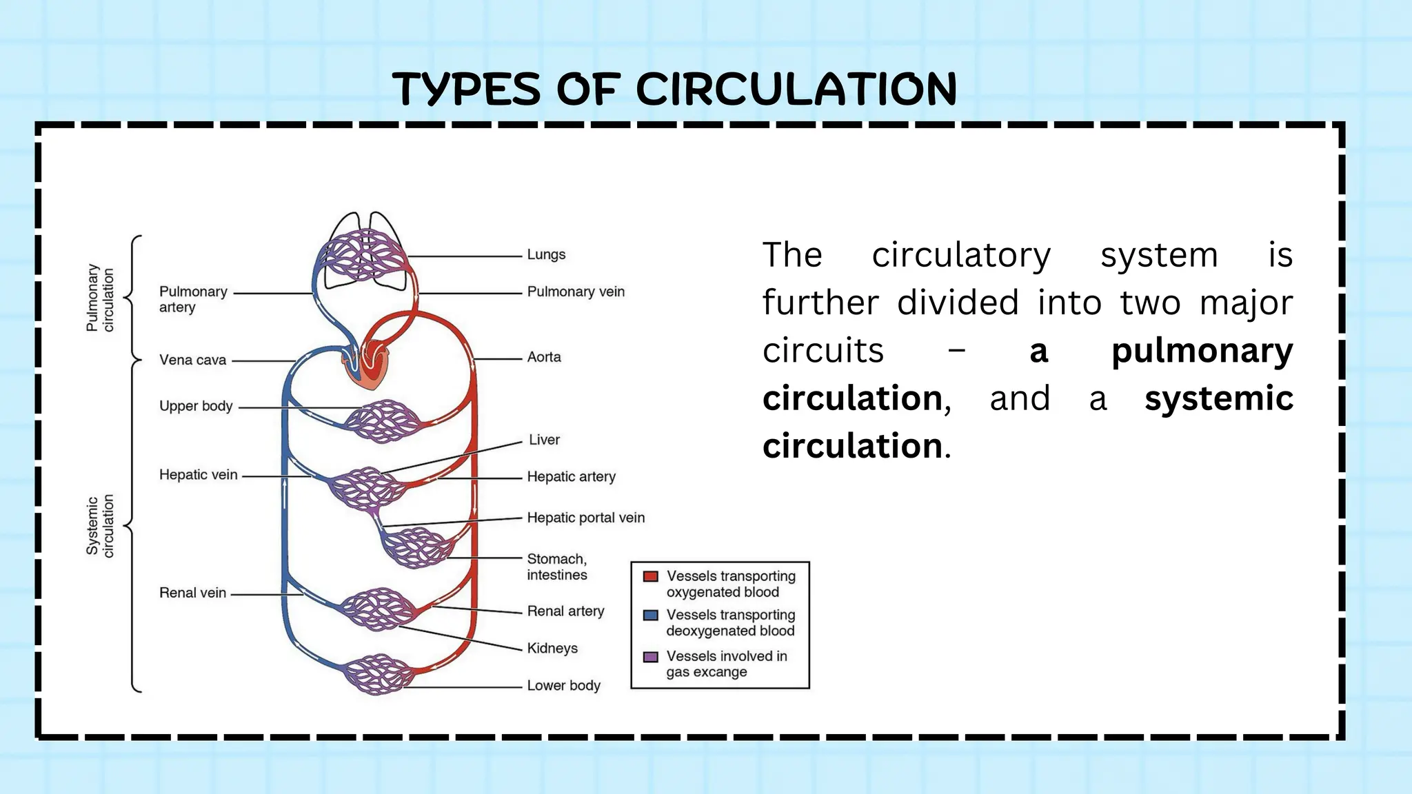
The circulatory systemis
further divided into two major
circuits – a pulmonary
circulation, and a systemic
circulation.
TYPES OF CIRCULATION
7.
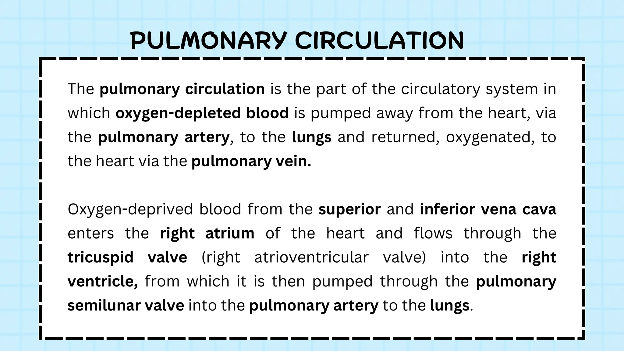
The pulmonary circulationis the part of the circulatory system in
which oxygen-depleted blood is pumped away from the heart, via
the pulmonary artery, to the lungs and returned, oxygenated, to
the heart via the pulmonary vein.
Oxygen-deprived blood from the superior and inferior vena cava
enters the right atrium of the heart and flows through the
tricuspid valve (right atrioventricular valve) into the right
ventricle, from which it is then pumped through the pulmonary
semilunar valve into the pulmonary artery to the lungs.
PULMONARY CIRCULATION
8.
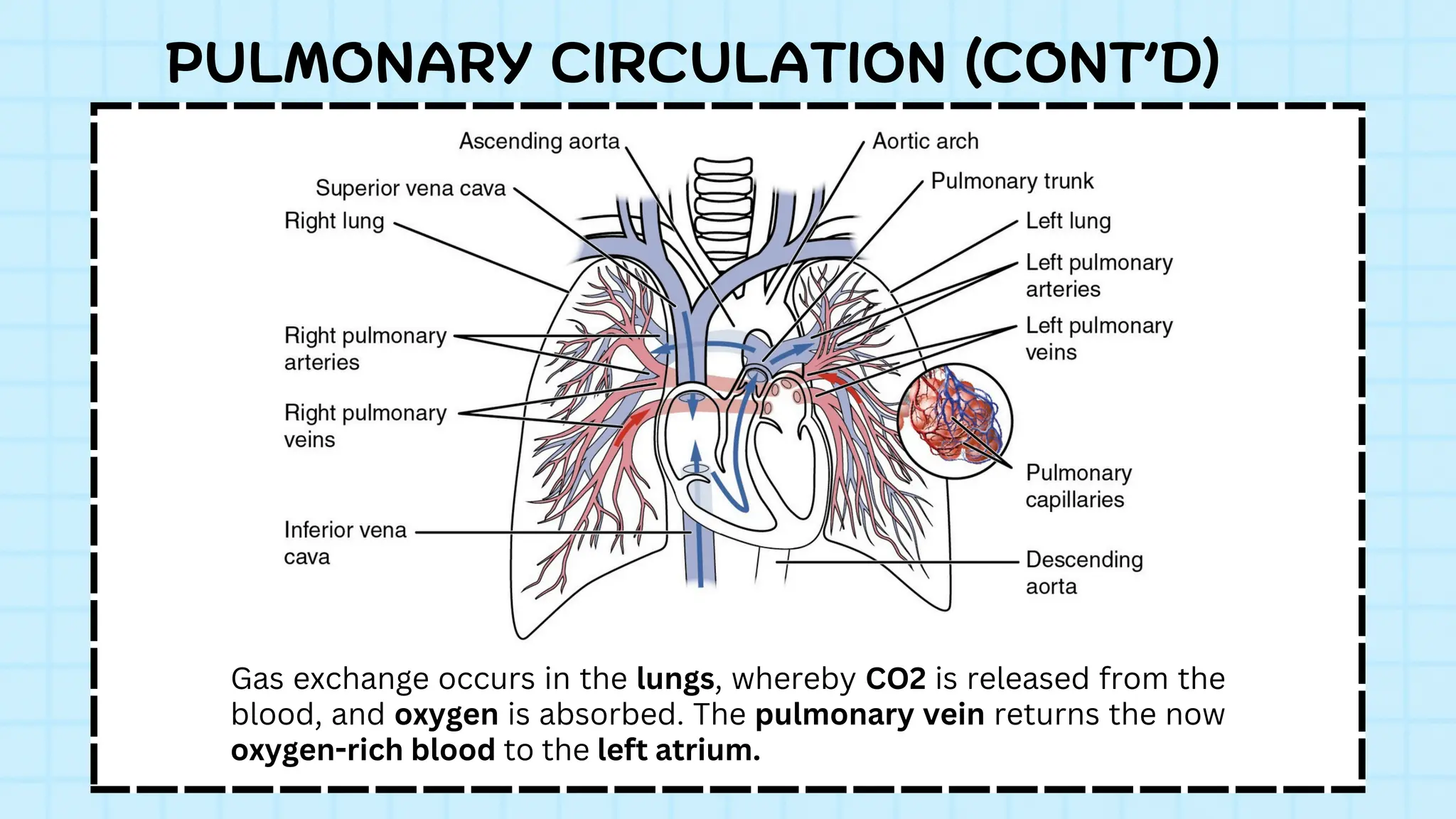
Gas exchange occursin the lungs, whereby CO2 is released from the
blood, and oxygen is absorbed. The pulmonary vein returns the now
oxygen-rich blood to the left atrium.
PULMONARY CIRCULATION (CONT’D)
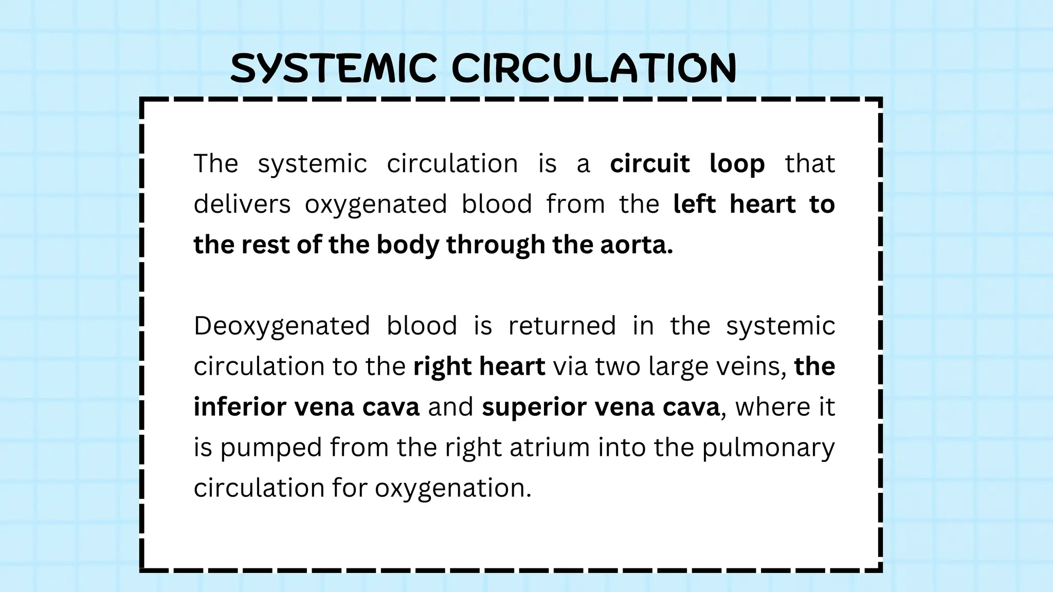
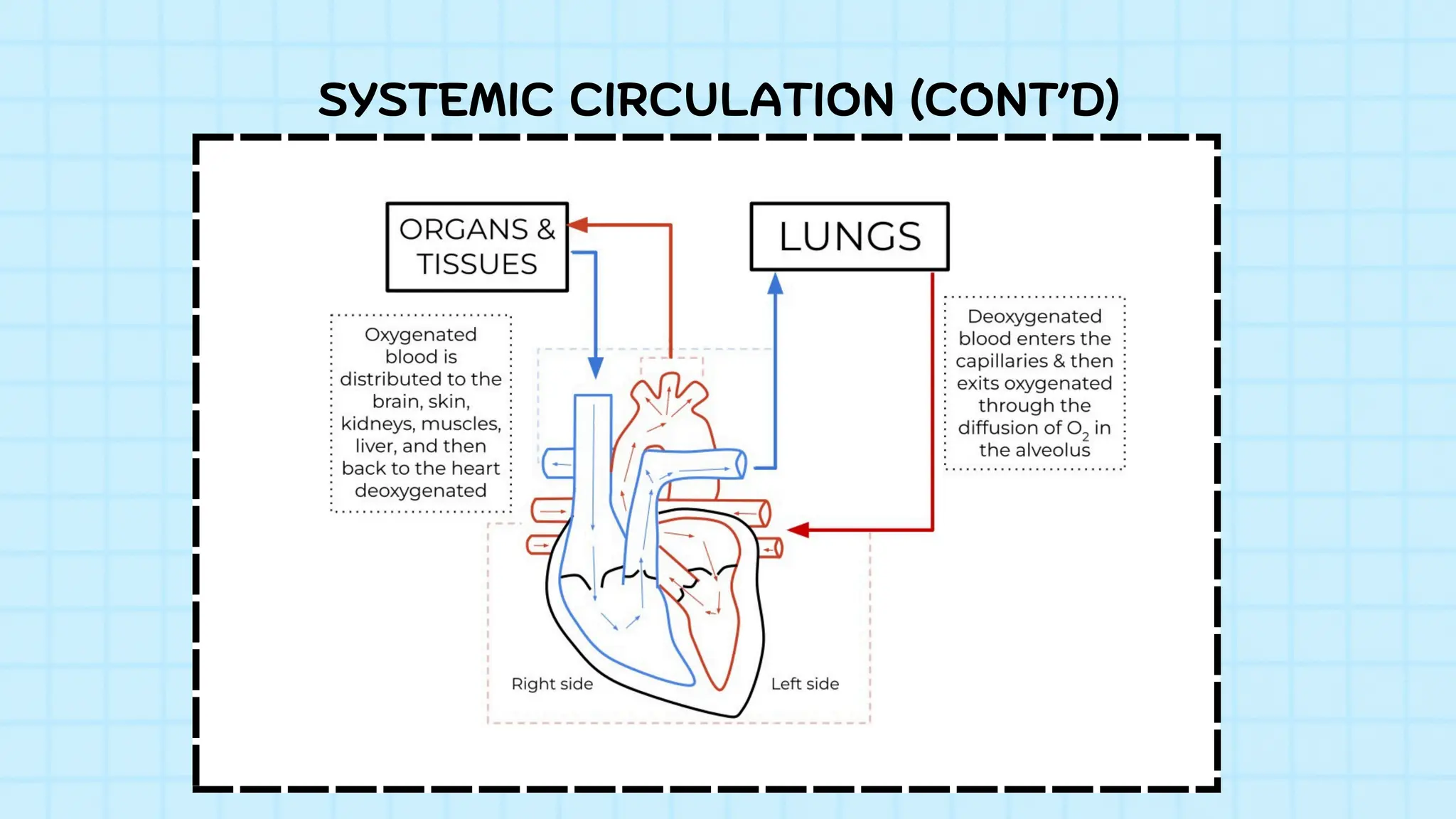
9.
The systemic circulationis a circuit loop that
delivers oxygenated blood from the left heart to
the rest of the body through the aorta.
Deoxygenated blood is returned in the systemic
circulation to the right heart via two large veins, the
inferior vena cava and superior vena cava, where it
is pumped from the right atrium into the pulmonary
circulation for oxygenation.
SYSTEMIC CIRCULATION
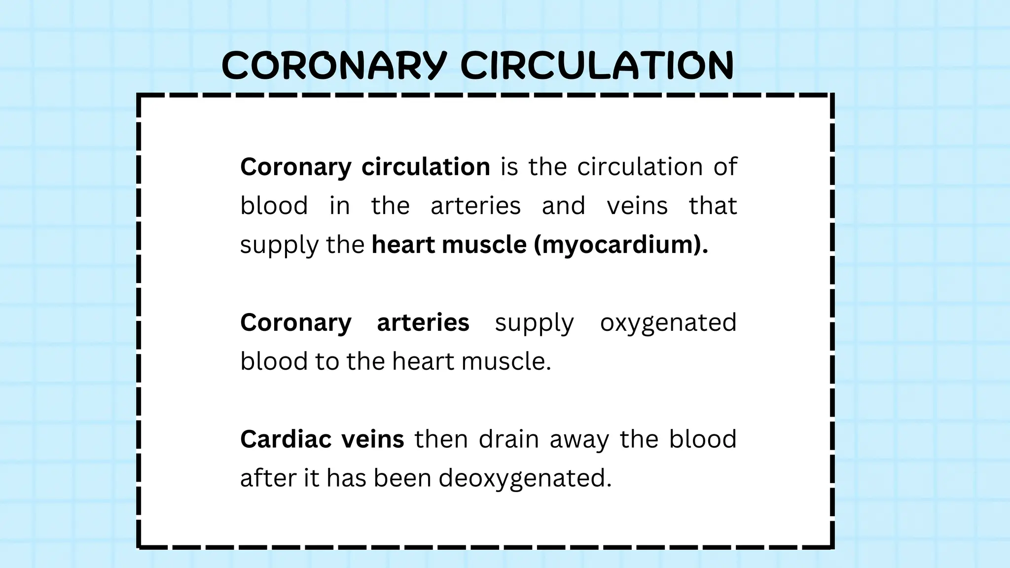
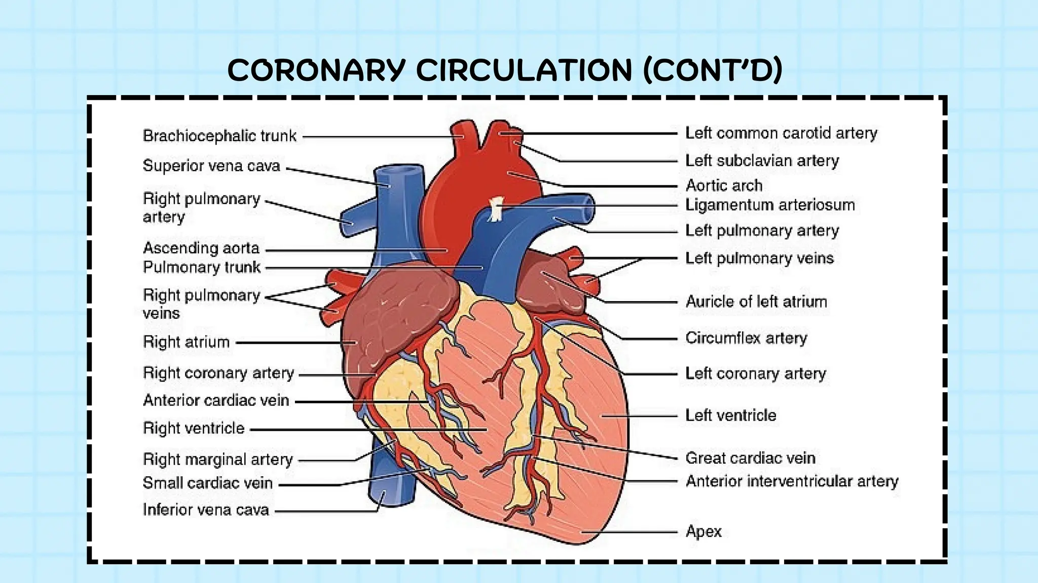
Coronary circulation isthe circulation of
blood in the arteries and veins that
supply the heart muscle (myocardium).
Coronary arteries supply oxygenated
blood to the heart muscle.
Cardiac veins then drain away the blood
after it has been deoxygenated.
CORONARY CIRCULATION
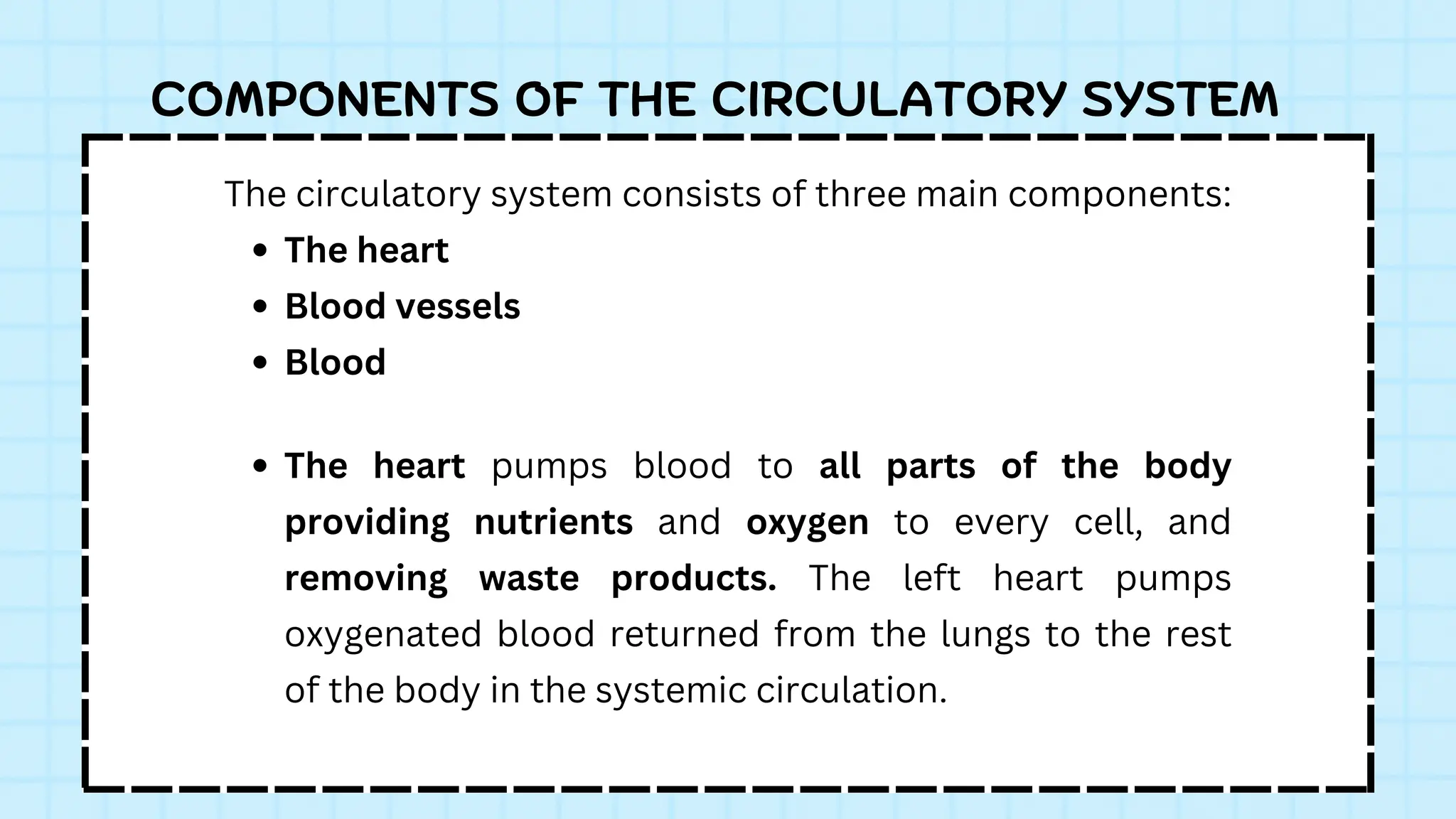
The circulatory systemconsists of three main components:
The heart
Blood vessels
Blood
COMPONENTS OF THE CIRCULATORY SYSTEM
The heart pumps blood to all parts of the body
providing nutrients and oxygen to every cell, and
removing waste products. The left heart pumps
oxygenated blood returned from the lungs to the rest
of the body in the systemic circulation.
14.
THE HEART
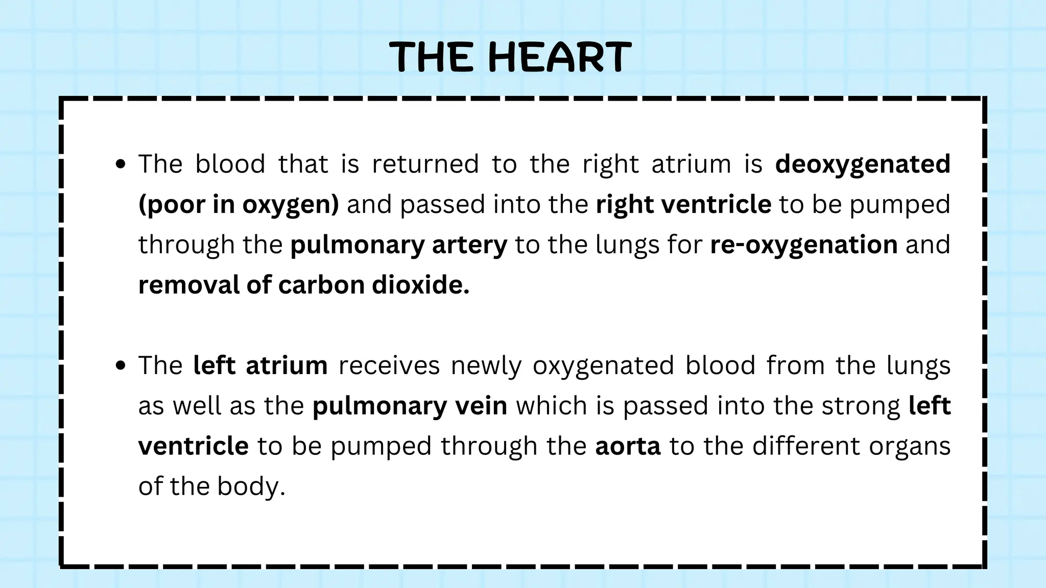
The bloodthat is returned to the right atrium is deoxygenated
(poor in oxygen) and passed into the right ventricle to be pumped
through the pulmonary artery to the lungs for re-oxygenation and
removal of carbon dioxide.
The left atrium receives newly oxygenated blood from the lungs
as well as the pulmonary vein which is passed into the strong left
ventricle to be pumped through the aorta to the different organs
of the body.
15.
COMPONENTS OF THECIRCULATORY SYSTEM (CONT’D)
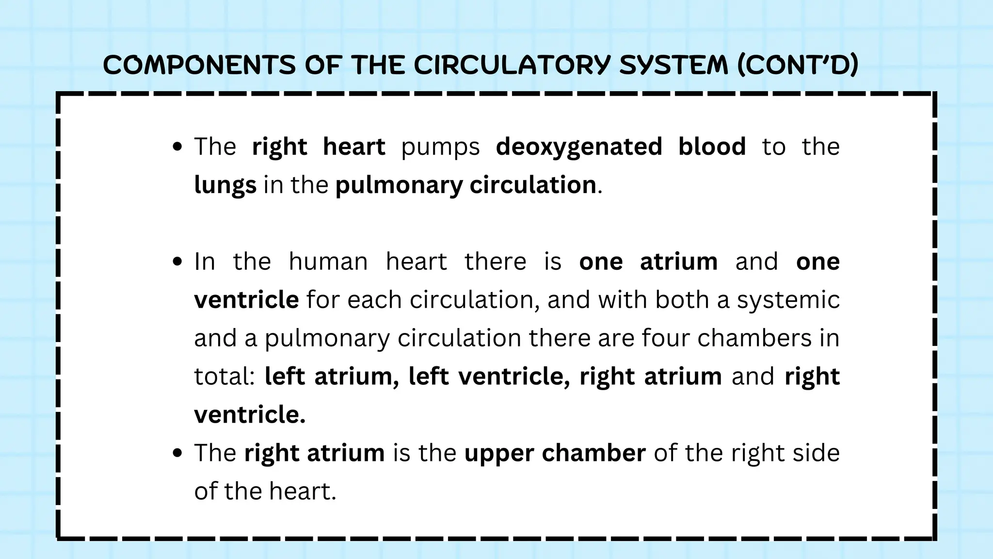
The right heart pumps deoxygenated blood to the
lungs in the pulmonary circulation.
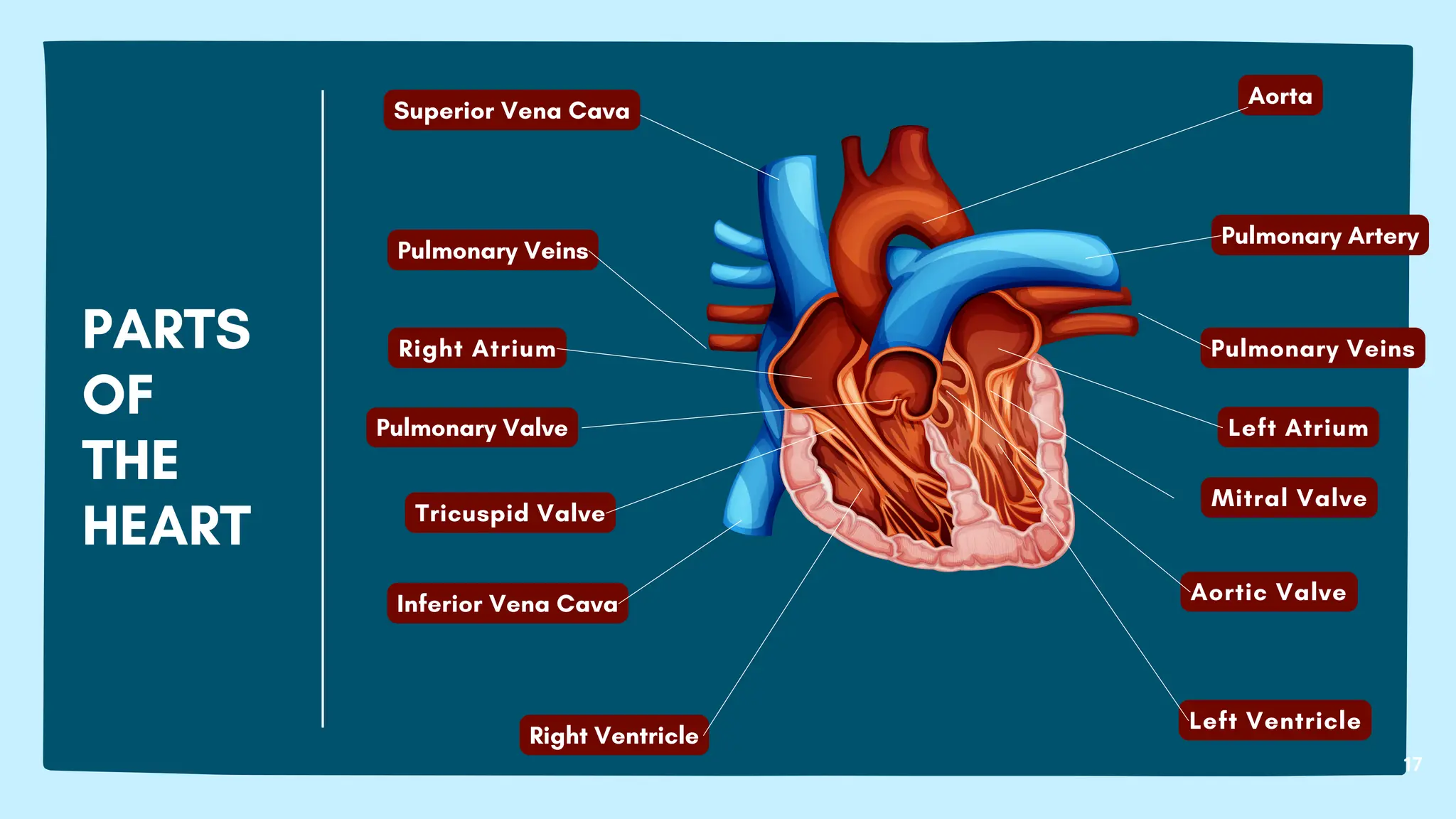
In the human heart there is one atrium and one
ventricle for each circulation, and with both a systemic
and a pulmonary circulation there are four chambers in
total: left atrium, left ventricle, right atrium and right
ventricle.
The right atrium is the upper chamber of the right side
of the heart.
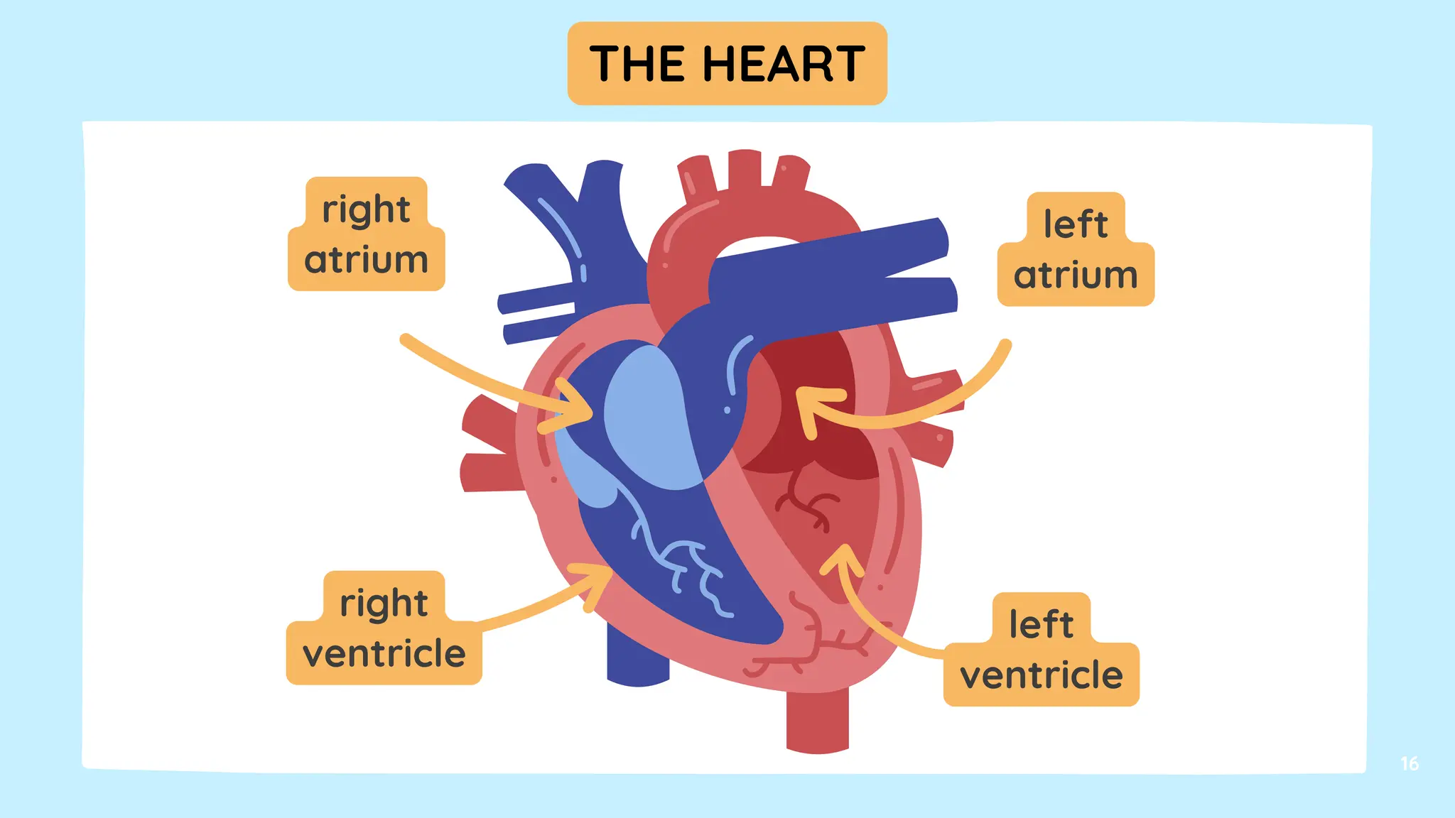
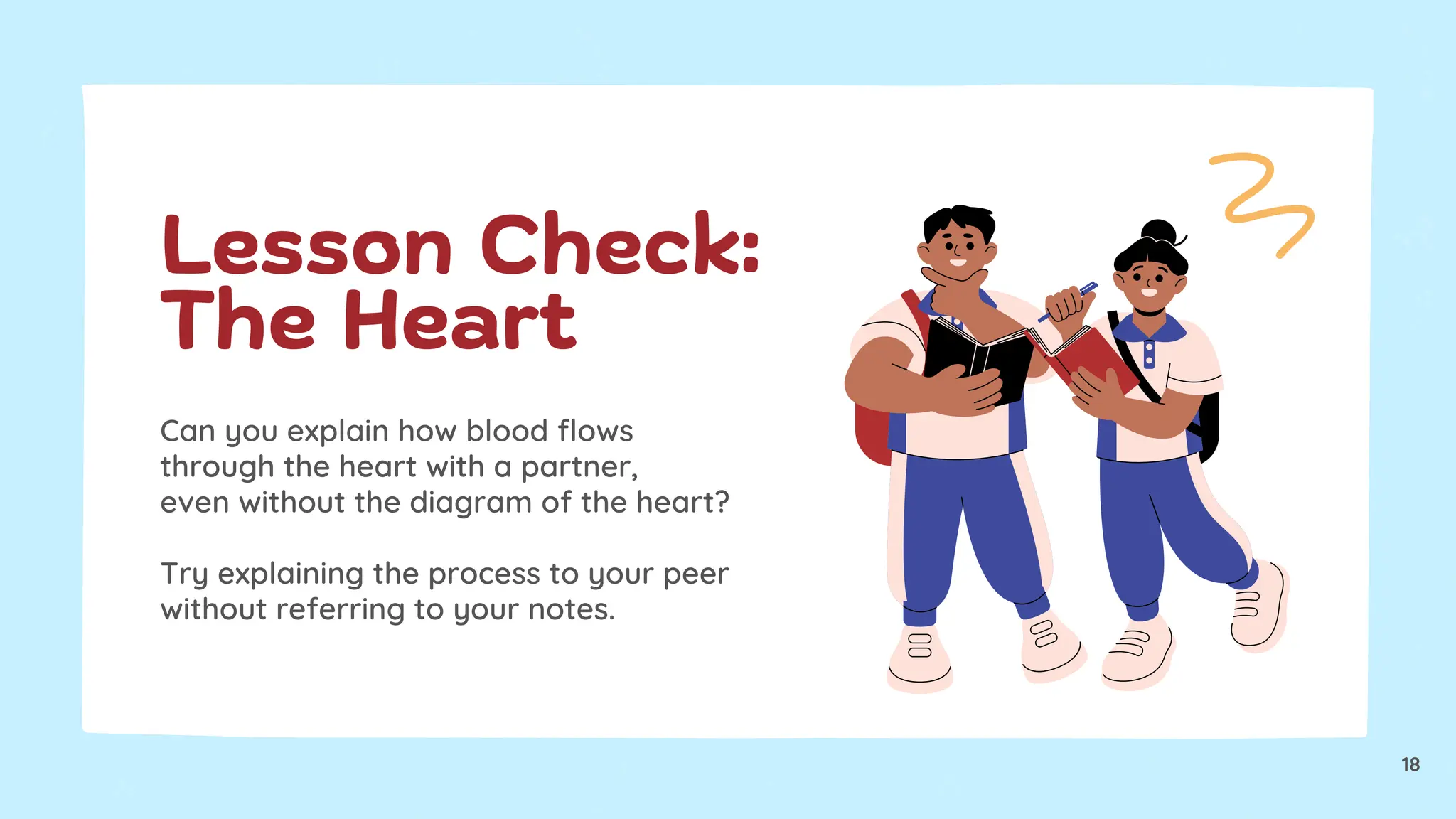
Can you explainhow blood flows
through the heart with a partner,
even without the diagram of the heart?
Try explaining the process to your peer
without referring to your notes.
Lesson Check:
The Heart
18
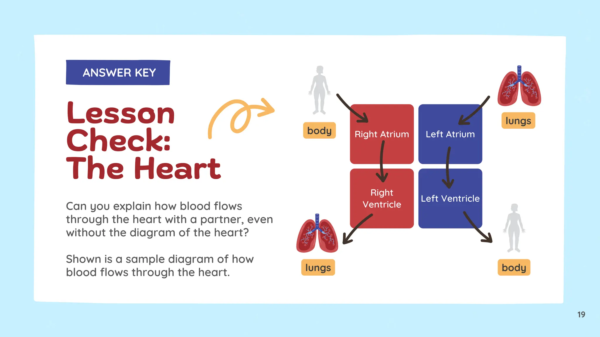
19.
Can you explainhow blood flows
through the heart with a partner, even
without the diagram of the heart?
Shown is a sample diagram of how
blood flows through the heart.
Lesson
Check:
The Heart
ANSWER KEY
Right Atrium Left Atrium
Right
Ventricle
Left Ventricle
body
lungs
lungs
body
19
20.
COMPONENTS OF THECIRCULATORY SYSTEM (CONT’D)
Blood vessels
The blood vessels of the circulatory system are the
arteries, veins, and capillaries.
The large arteries and veins that take blood to, and away
from the heart are known as the great vessels.
Arteries: Oxygenated blood enters the systemic circulation
when leaving the left ventricle, via the aortic semilunar
valve.
21.
COMPONENTS OF THECIRCULATORY SYSTEM (CONT’D)
The first part of the systemic circulation is the aorta, a massive and
thick-walled artery.
The aorta arches and gives branches supplying the upper part of
the body after passing through the aortic opening of the diaphragm
at the level of thoracic ten vertebra, it enters the abdomen.
Later, it descends down and supplies branches to abdomen, pelvis,
perineum and the lower limbs.
22.
COMPONENTS OF THECIRCULATORY SYSTEM (CONT’D)
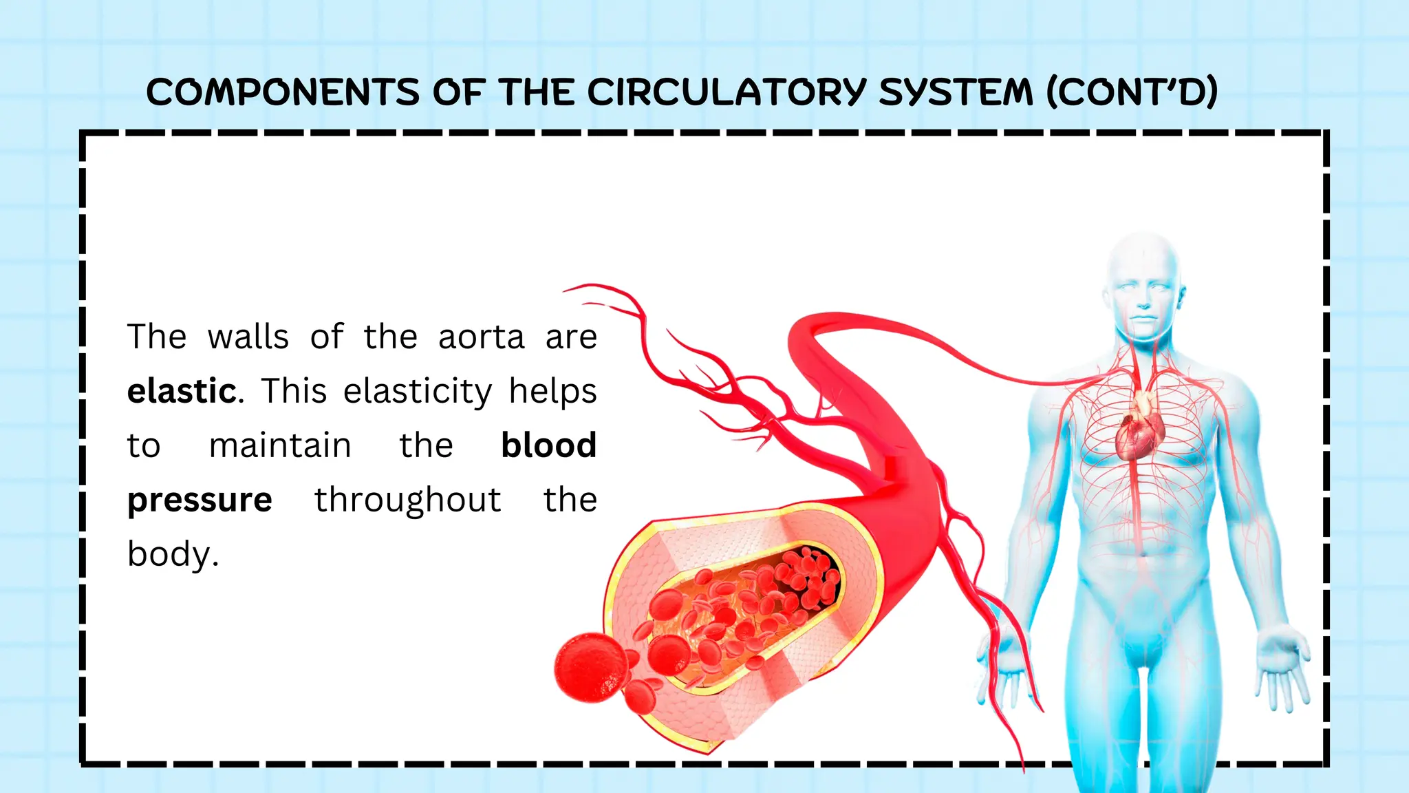
The walls of the aorta are
elastic. This elasticity helps
to maintain the blood
pressure throughout the
body.
23.
COMPONENTS OF THECIRCULATORY SYSTEM (CONT’D)
Veins
Capillaries merge into venules, which merge into veins.
The venous system feeds into the two major veins: the
superior vena cava – which mainly drains tissues above the
heart – and the inferior vena cava – which mainly drains
tissues below the heart.
These two large veins empty into the right atrium of the
heart.
24.
COMPONENTS OF THECIRCULATORY SYSTEM (CONT’D)
Capillaries
Arteries branch into small passages called arterioles and
then into the capillaries.
The capillaries merge to bring blood into the venous
system.
The total length of muscle capillaries in a 70 kg human is
estimated to be between 9,000 and 19,000 km.
25.
BLOOD
25
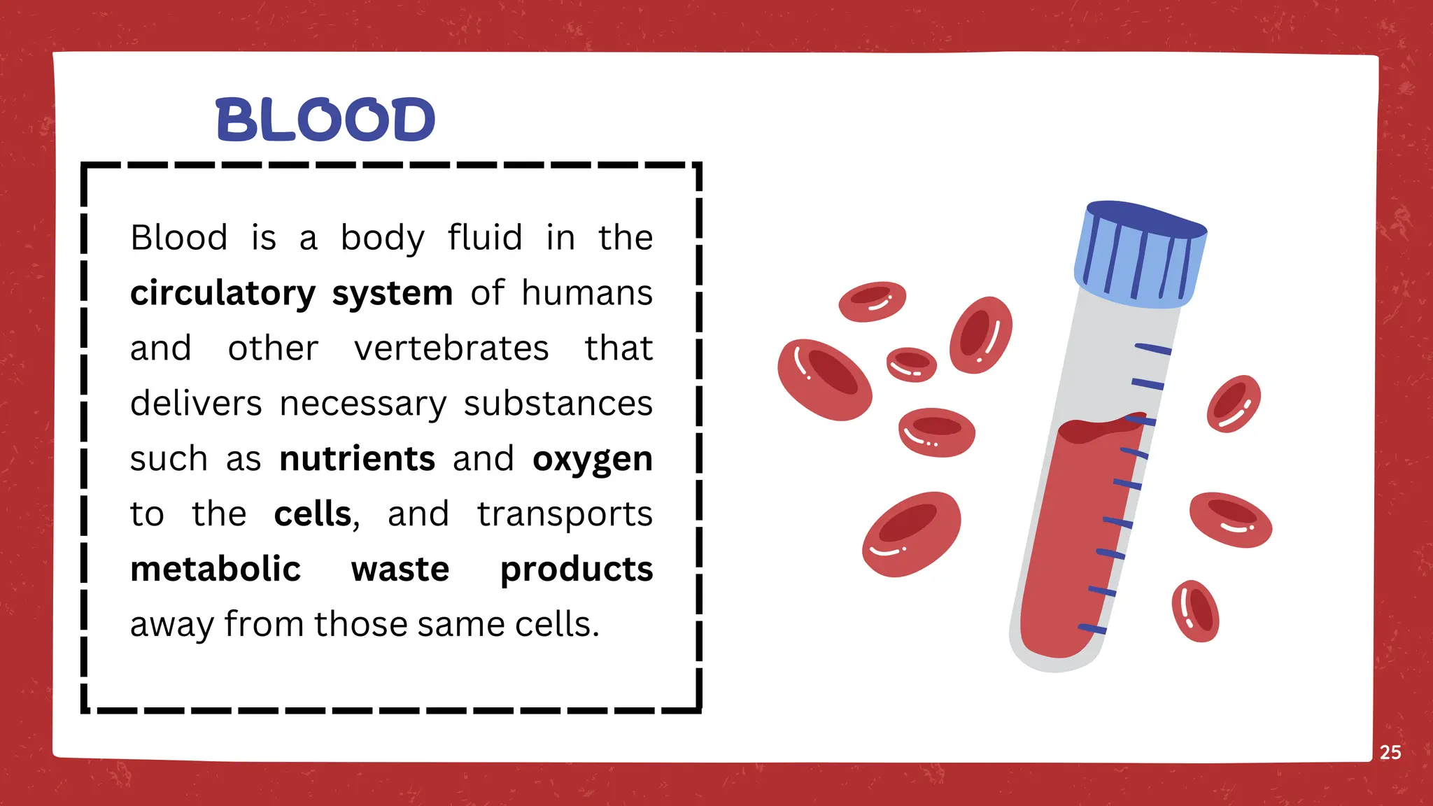
Blood is abody fluid in the
circulatory system of humans
and other vertebrates that
delivers necessary substances
such as nutrients and oxygen
to the cells, and transports
metabolic waste products
away from those same cells.
26.
BLOOD
26
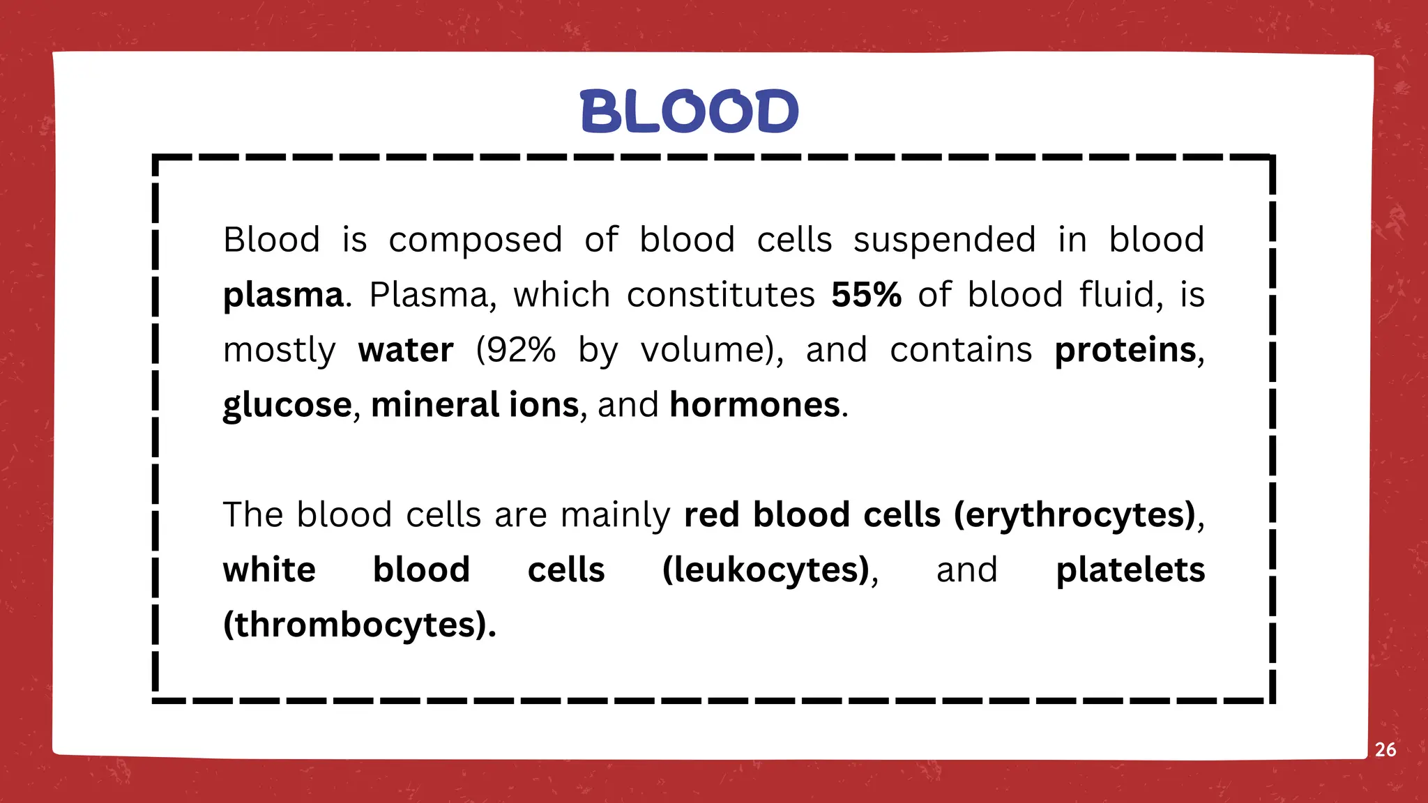
Blood is composedof blood cells suspended in blood
plasma. Plasma, which constitutes 55% of blood fluid, is
mostly water (92% by volume), and contains proteins,
glucose, mineral ions, and hormones.
The blood cells are mainly red blood cells (erythrocytes),
white blood cells (leukocytes), and platelets
(thrombocytes).
27.
BLOOD
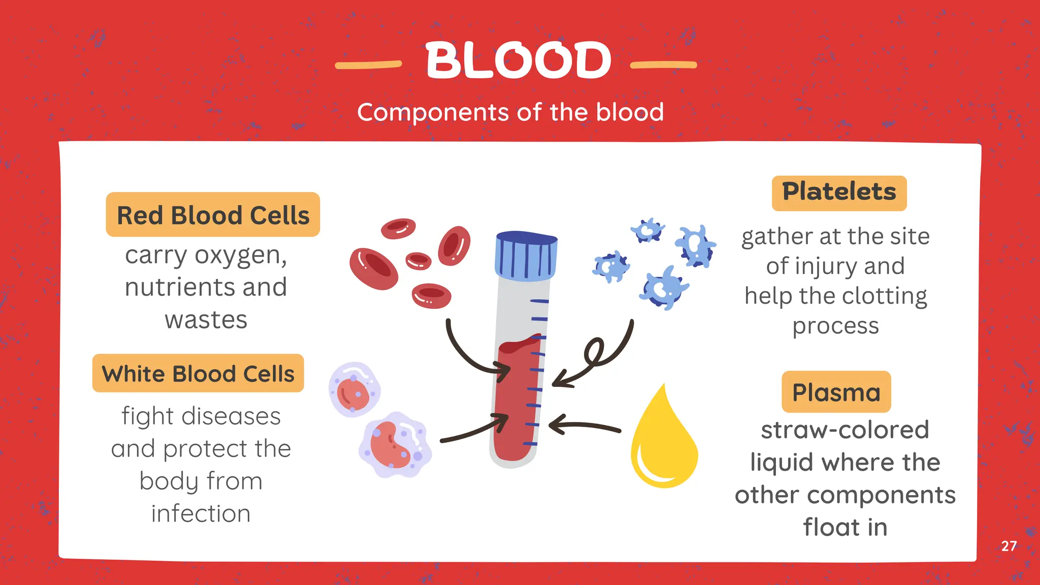
Components of theblood
carry oxygen,
nutrients and
wastes
fight diseases
and protect the
body from
infection
straw-colored
liquid where the
other components
float in
gather at the site
of injury and
help the clotting
process
Red Blood Cells
White Blood Cells
Platelets
Plasma
27
28.
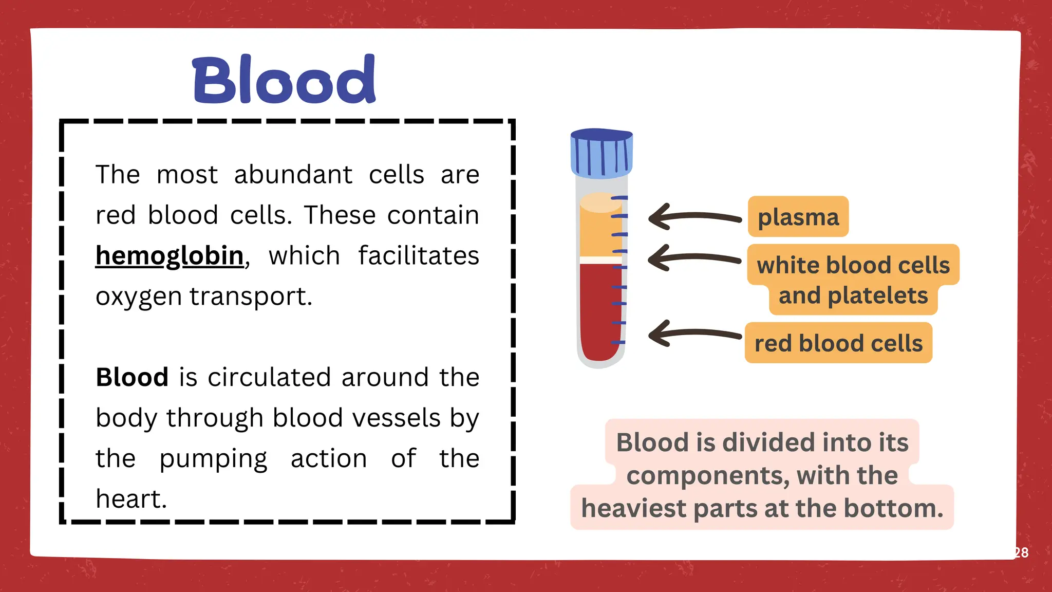
Blood is dividedinto its
components, with the
heaviest parts at the bottom.
plasma
white blood cells
and platelets
red blood cells
28
The most abundant cells are
red blood cells. These contain
hemoglobin, which facilitates
oxygen transport.
Blood is circulated around the
body through blood vessels by
the pumping action of the
heart.
Blood
29.
29
Blood performs manyimportant functions within the body,
including:
Supply of oxygen to tissues (bound to hemoglobin, which is
carried in red cells)
Supply of nutrients such as glucose, amino acids, and fatty
acids (dissolved in the blood or bound to plasma proteins
(e.g., blood lipids))
Removal of waste such as carbon dioxide, urea, and lactic
acid.
Function of the blood
30.
30
Immunological functions, includingcirculation of white
blood cells, and detection of foreign material by antibodies
Coagulation, the response to a broken blood vessel, the
conversion of blood from a liquid to a semisolid gel to stop
bleeding
Messenger functions, including the transport of hormones
and the signaling of tissue damage
Regulation of core body temperature
Hydraulic functions
Function of the blood
31.
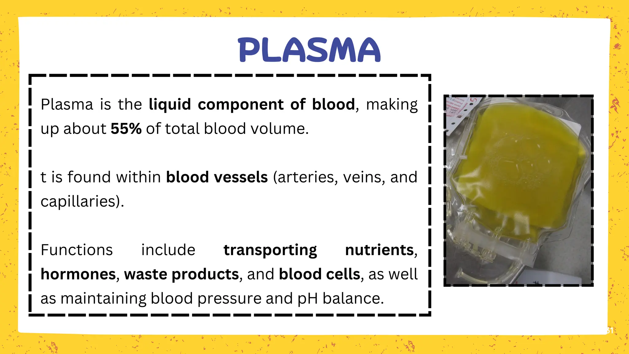
Plasma is theliquid component of blood, making
up about 55% of total blood volume.
t is found within blood vessels (arteries, veins, and
capillaries).
Functions include transporting nutrients,
hormones, waste products, and blood cells, as well
as maintaining blood pressure and pH balance.
31
PLASMA
32.
32
Interstitial fluid isthe fluid that surrounds and bathes the cells
in tissues.
It is found in the interstitial spaces (spaces between cells)
outside blood vessels.
Functions include facilitating the exchange of nutrients, gases,
and waste products between blood and cells.
Interstitial Fluid
33.
33
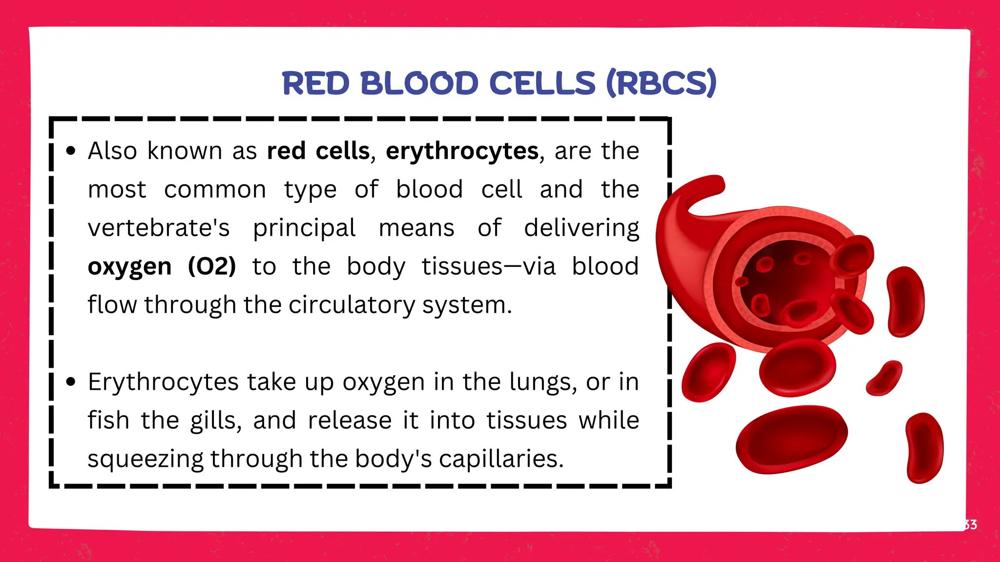
Also known asred cells, erythrocytes, are the
most common type of blood cell and the
vertebrate's principal means of delivering
oxygen (O2) to the body tissues—via blood
flow through the circulatory system.
Erythrocytes take up oxygen in the lungs, or in
fish the gills, and release it into tissues while
squeezing through the body's capillaries.
RED BLOOD CELLS (RBCS)
34.
34
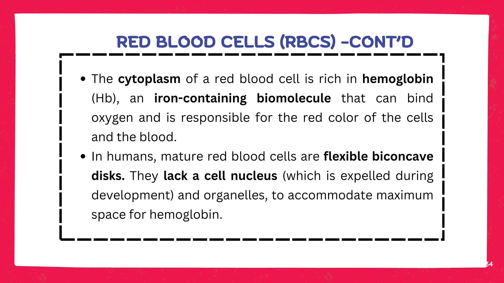
The cytoplasm ofa red blood cell is rich in hemoglobin
(Hb), an iron-containing biomolecule that can bind
oxygen and is responsible for the red color of the cells
and the blood.
In humans, mature red blood cells are flexible biconcave
disks. They lack a cell nucleus (which is expelled during
development) and organelles, to accommodate maximum
space for hemoglobin.
RED BLOOD CELLS (RBCS) -CONT’D
35.
35
Approximately 2.4 millionnew erythrocytes are produced
per second in human adults.
The cells develop in the bone marrow and circulate for
about 100–120 days in the body before their components
are recycled by macrophages.
Nearly half of the blood's volume (40% to 45%) is red
blood cells.
RED BLOOD CELLS (RBCS) -CONT’D
36.
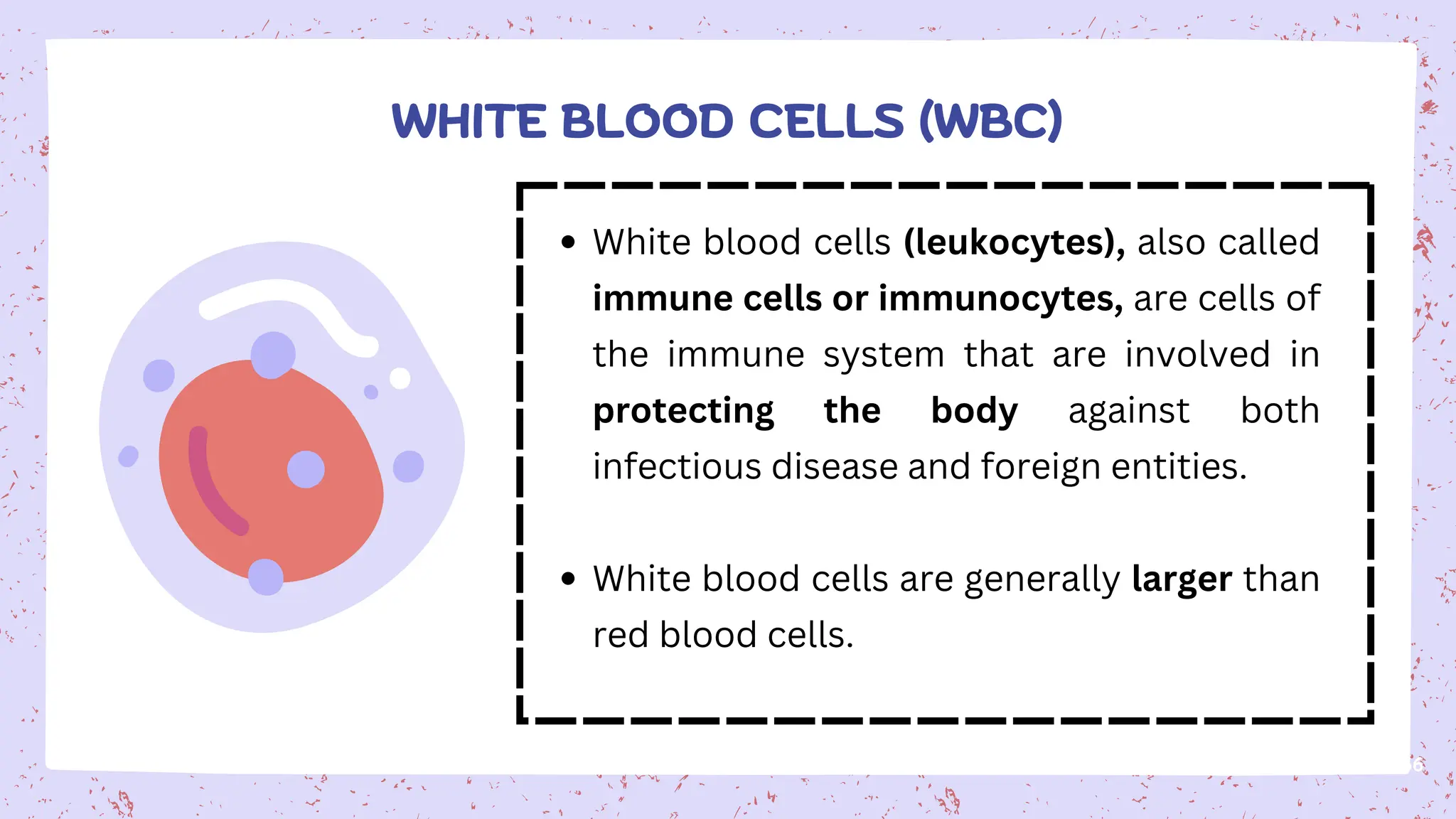
White blood cells(leukocytes), also called
immune cells or immunocytes, are cells of
the immune system that are involved in
protecting the body against both
infectious disease and foreign entities.
White blood cells are generally larger than
red blood cells.
36
WHITE BLOOD CELLS (WBC)
37.
37
All white bloodcells are produced and derived from
multipotent cells in the bone marrow known as
hematopoietic stem cells.
Leukocytes are found throughout the body, including the
blood and lymphatic system.
All white blood cells have nuclei, which distinguishes
them from the other blood cells.
WHITE BLOOD CELLS (WBC) - CONT’D
38.
38
White blood cellsare classified into Granulocytes and
Agranulocytes based on the presence of granules in the
cytoplasm.
Granulocytes (Contain granules; have lobed nuclei)
Neutrophils (55–70%): First responders to infection, perform
phagocytosis, short-lived (1–2 days).
Eosinophils (1–4%): Combat parasites, involved in allergic
reactions, regulate histamine activity.
Basophils (<1%): Release histamine and heparin, important in
inflammation and allergic responses.
TYPES OF WHITE BLOOD CELLS
39.
39
Agranulocytes (No visiblegranules; large nuclei)
Lymphocytes (20–40%): Key players in adaptive
immunity. B cells produce antibodies, T cells kill
infected cells, NK cells kill virus-infected and tumor
cells.
Monocytes (2–8%): Largest WBC, migrate to tissues
to become macrophages, perform phagocytosis and
antigen presentation.
TYPES OF WHITE BLOOD CELLS - CONT’D
40.
40
FUNCTIONS OF WHITEBLOOD CELLS
Defense against bacteria, viruses, fungi,
parasites.
Phagocytosis of foreign particles.
Antibody production by B lymphocytes.
Immune regulation by T cells.
Mediation of inflammation and allergy.
41.
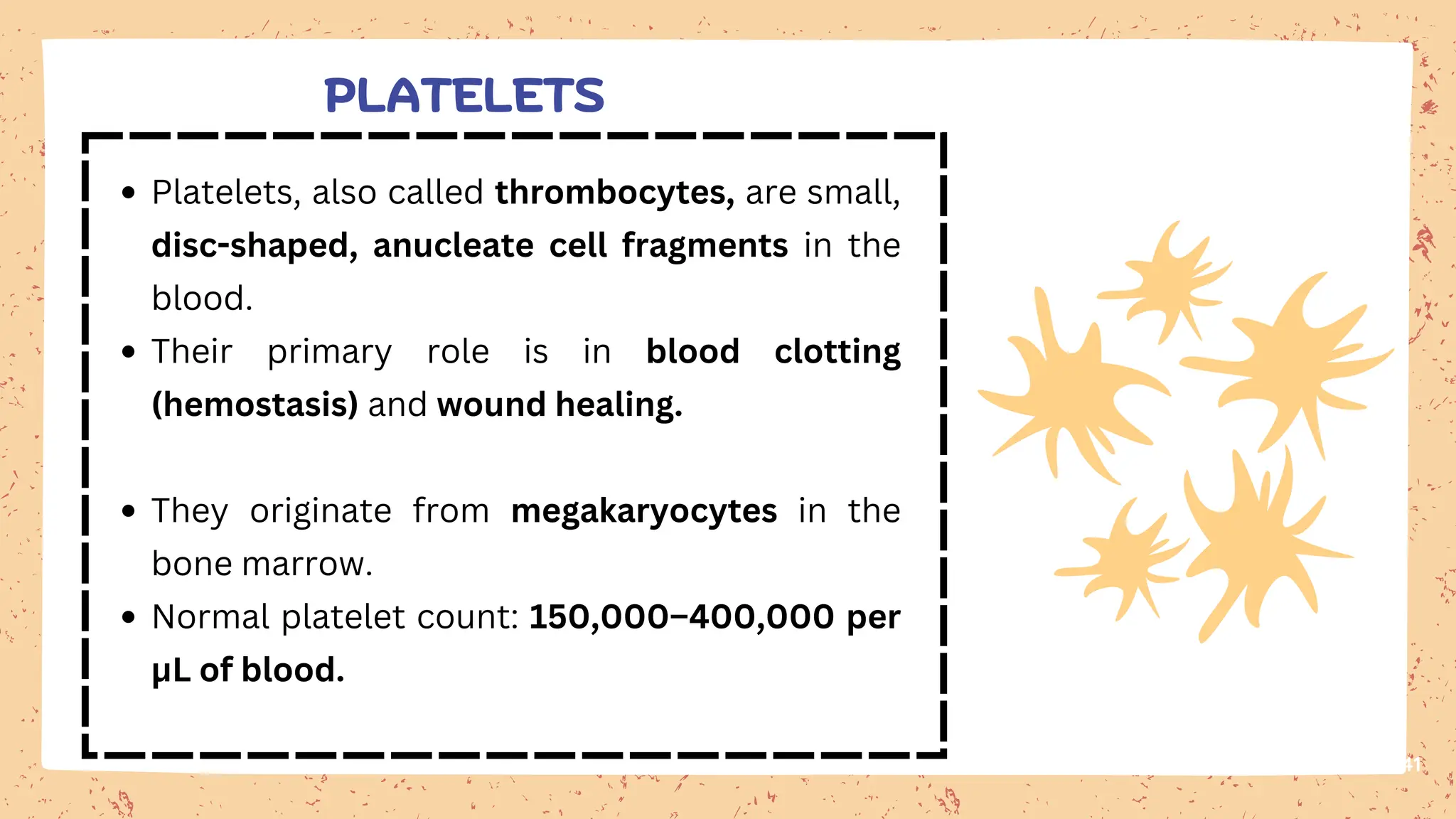
Platelets, also calledthrombocytes, are small,
disc-shaped, anucleate cell fragments in the
blood.
Their primary role is in blood clotting
(hemostasis) and wound healing.
They originate from megakaryocytes in the
bone marrow.
Normal platelet count: 150,000–400,000 per
μL of blood.
41
PLATELETS
42.
42
PLATELETS - CONT’D
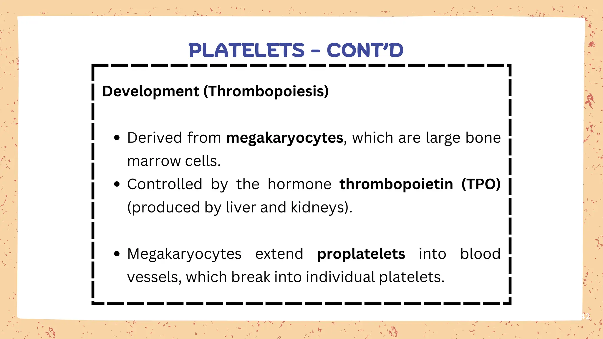
Development(Thrombopoiesis)
Derived from megakaryocytes, which are large bone
marrow cells.
Controlled by the hormone thrombopoietin (TPO)
(produced by liver and kidneys).
Megakaryocytes extend proplatelets into blood
vessels, which break into individual platelets.
43.
43
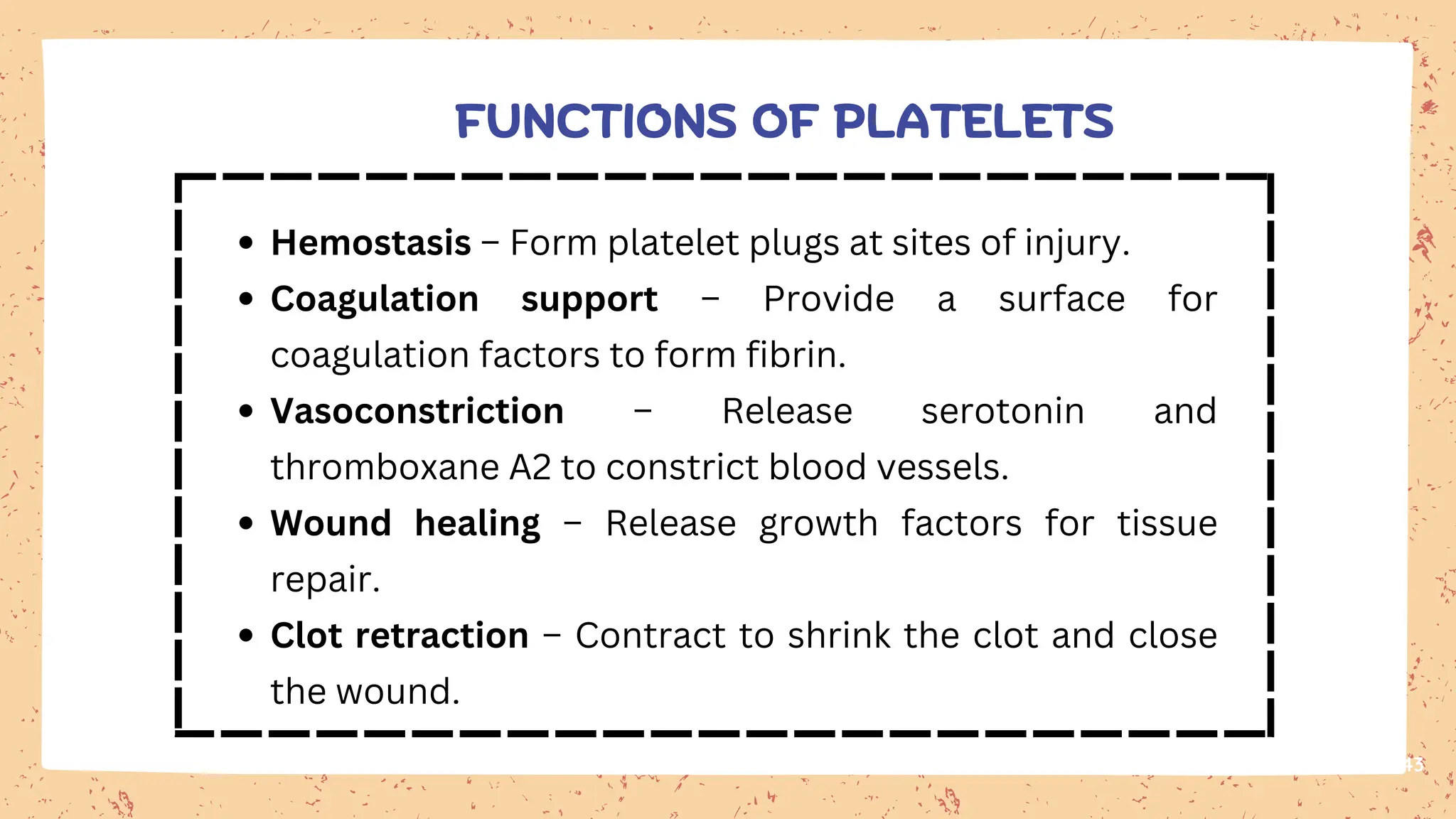
Hemostasis – Formplatelet plugs at sites of injury.
Coagulation support – Provide a surface for
coagulation factors to form fibrin.
Vasoconstriction – Release serotonin and
thromboxane A2 to constrict blood vessels.
Wound healing – Release growth factors for tissue
repair.
Clot retraction – Contract to shrink the clot and close
the wound.
FUNCTIONS OF PLATELETS