Features
• Circulatory systemsgenerally have three main features:
• Fluid (blood or hemolymph) that transports materials
• System of blood vessels
• A heart to pump the fluid through the vessels
Components
• Blood ismade up of four major components. What do
each of these do?
• Plasma: the liquid portion.
• Red blood cells.
• White cells.
• Platelets.
8.
Red blood cells
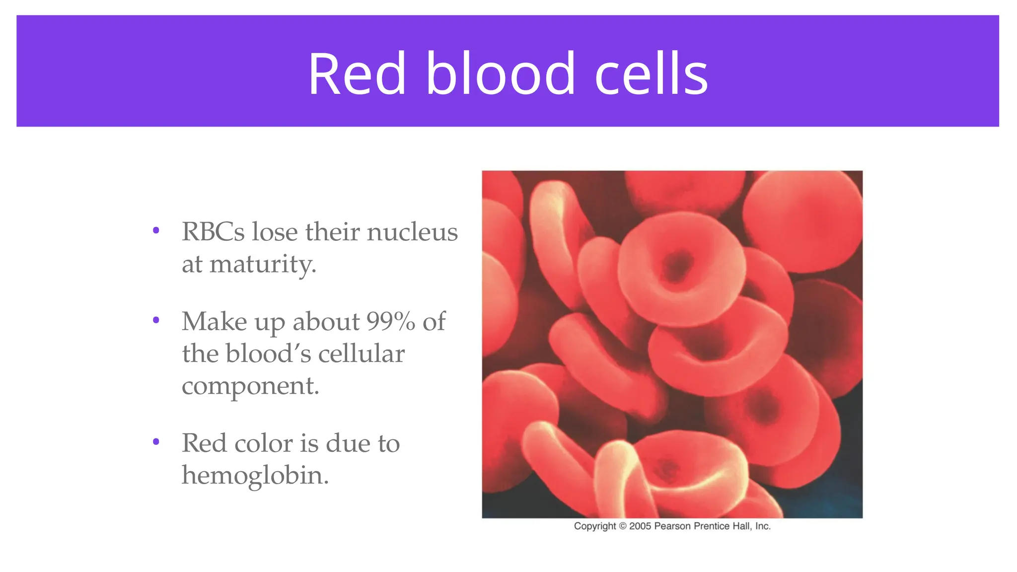
•RBCs lose their nucleus
at maturity.
• Make up about 99% of
the blood’s cellular
component.
• Red color is due to
hemoglobin.
9.
Hemoglobin
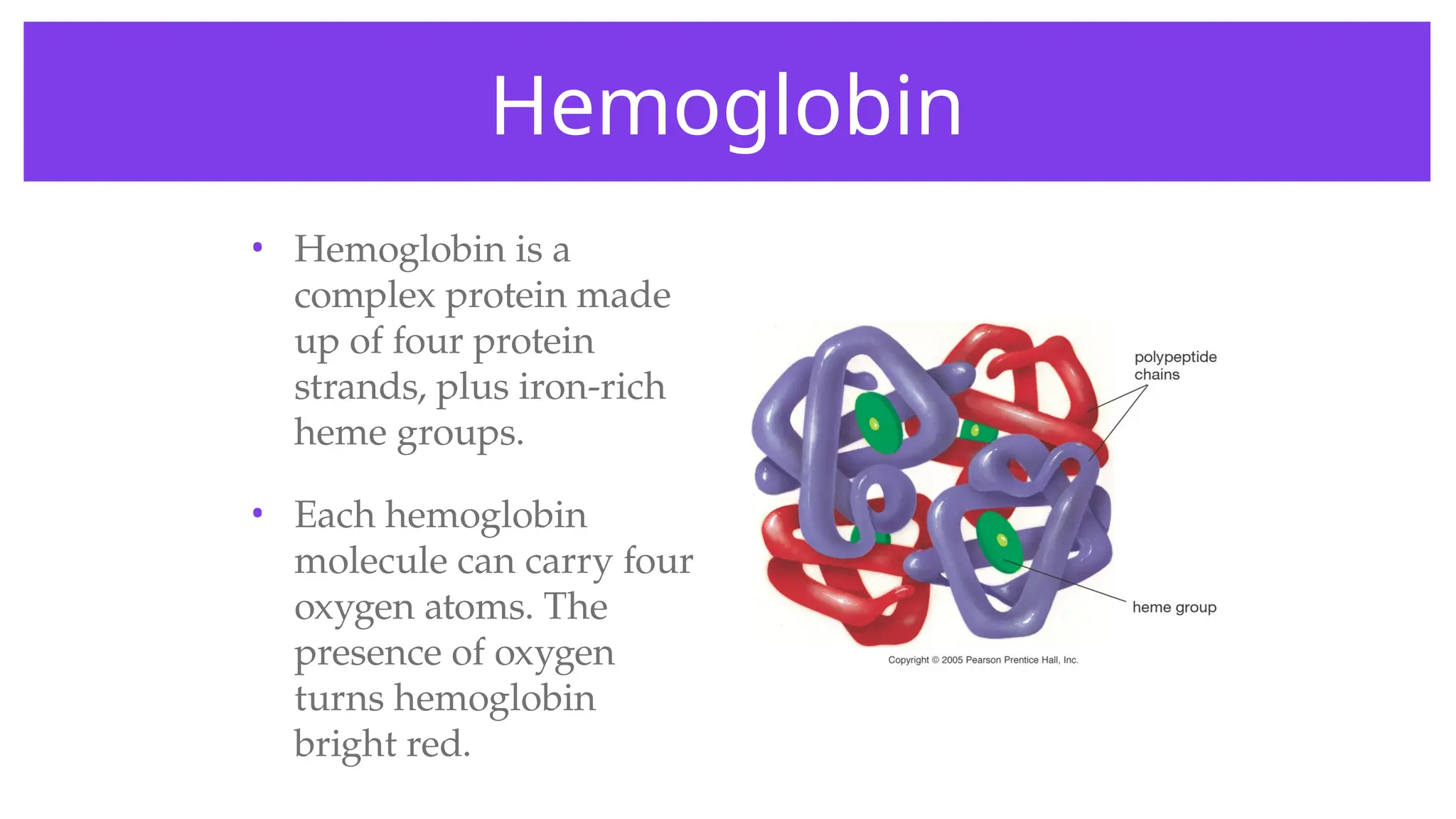
• Hemoglobin isa
complex protein made
up of four protein
strands, plus iron-rich
heme groups.
• Each hemoglobin
molecule can carry four
oxygen atoms. The
presence of oxygen
turns hemoglobin
bright red.
10.
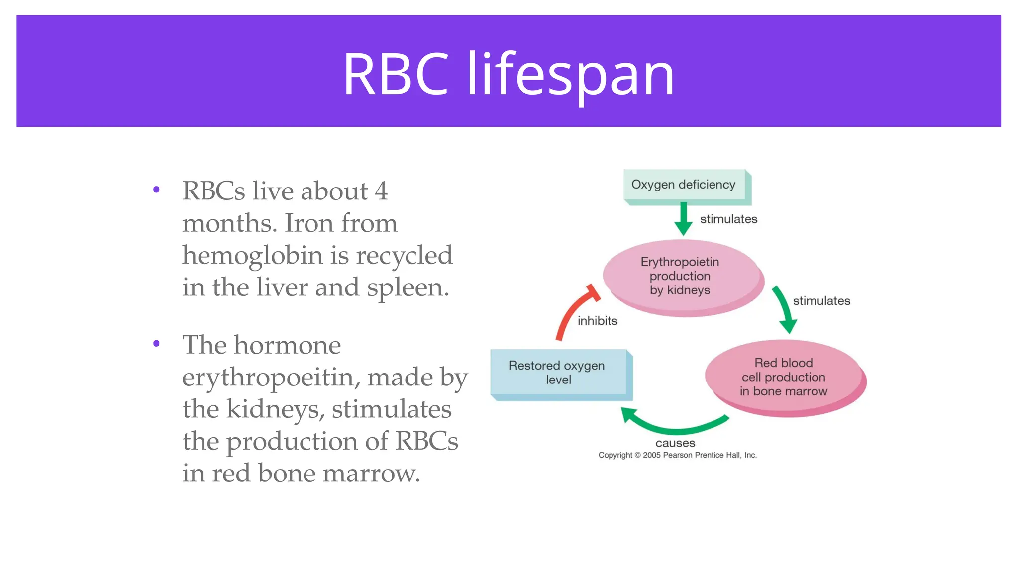
RBC lifespan
• RBCslive about 4
months. Iron from
hemoglobin is recycled
in the liver and spleen.
• The hormone
erythropoeitin, made by
the kidneys, stimulates
the production of RBCs
in red bone marrow.
11.
If your dietis poor in iron, what will
happen to your RBCs?
1 2 3
33% 33%
33%
1. You will make fewer
because there is less
iron to make
hemoglobin.
2. You will make more
to make up for the
lack of iron in
hemoglobin.
3. You will make just as
many.
12.
• One ofthe illegal drugs that some top Olympic athletes
have been caught using is erythropoetin. What would
this hormone do that would give athletes an edge in
competitions?
W
O
R
K
T
O
G
E
T
H
E
R
13.
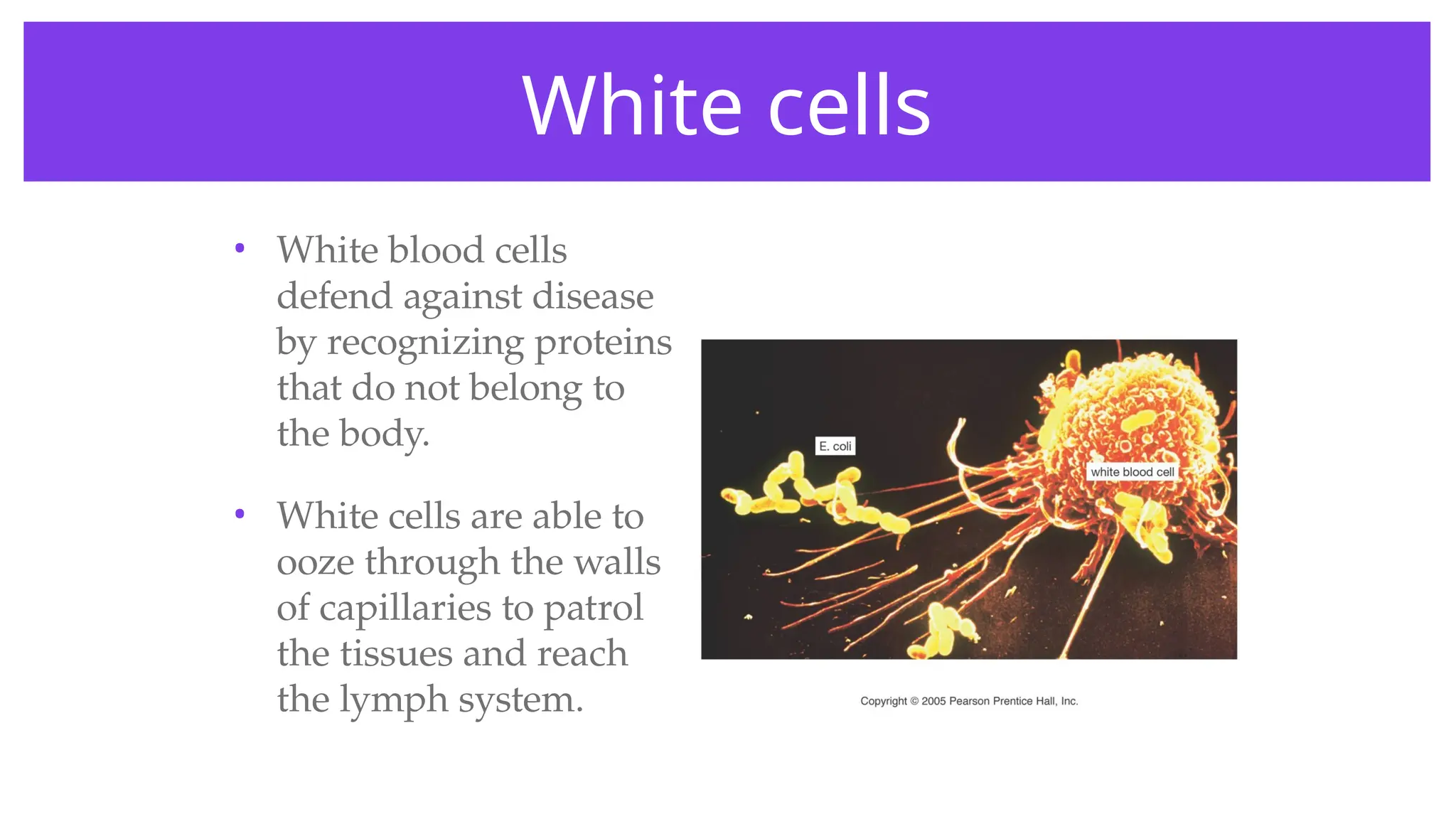
White cells
• Whiteblood cells
defend against disease
by recognizing proteins
that do not belong to
the body.
• White cells are able to
ooze through the walls
of capillaries to patrol
the tissues and reach
the lymph system.
14.
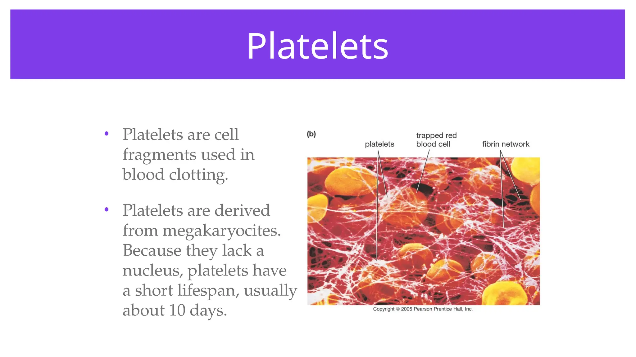
Platelets
• Platelets arecell
fragments used in
blood clotting.
• Platelets are derived
from megakaryocites.
Because they lack a
nucleus, platelets have
a short lifespan, usually
about 10 days.
15.
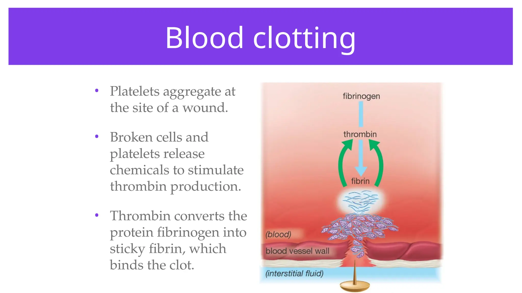
• Platelets aggregateat
the site of a wound.
• Broken cells and
platelets release
chemicals to stimulate
thrombin production.
• Thrombin converts the
protein fibrinogen into
sticky fibrin, which
binds the clot.
Blood clotting
16.
Which blood cellstransport
oxygen?
1 2 3 4
25% 25%
25%
25%
1. White cells
2. Red cells
3. Platelets
4. All blood cells
17.
• If aperson had a defect in the gene for fibrinogen, what
health problems could this cause?
W
O
R
K
T
O
G
E
T
H
E
R
Classes of bloodvessels
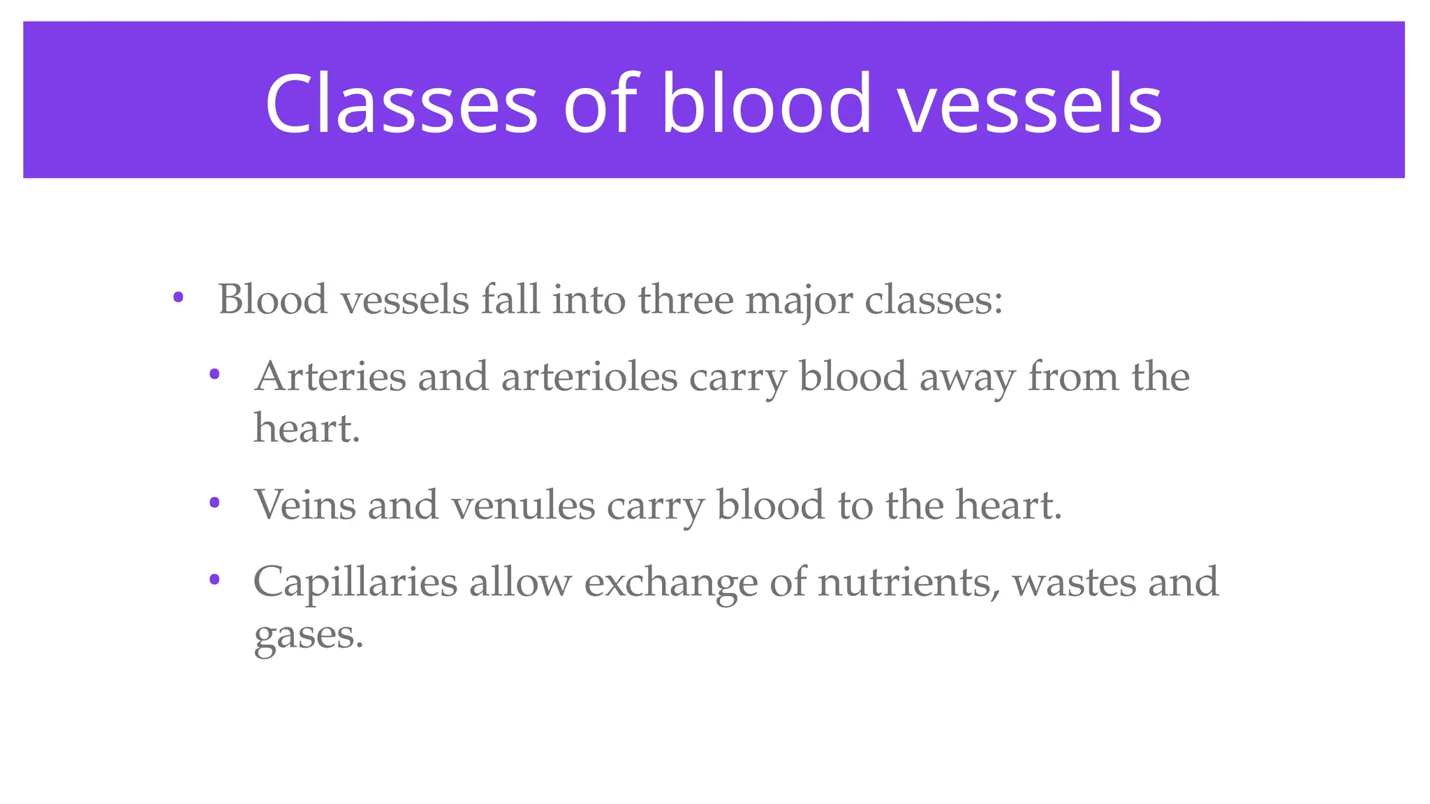
• Blood vessels fall into three major classes:
• Arteries and arterioles carry blood away from the
heart.
• Veins and venules carry blood to the heart.
• Capillaries allow exchange of nutrients, wastes and
gases.
20.
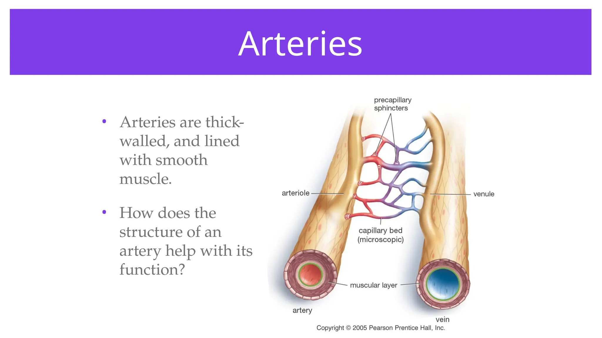
Arteries
• Arteries arethick-
walled, and lined
with smooth
muscle.
• How does the
structure of an
artery help with its
function?
21.
Arterioles
• Arterioles branchoff of arteries.
• Arterioles can constrict to direct and control
blood flow. They may, for example, increase or
decrease blood supply to the skin.
• How might arterioles be involved when:
• Your skin turns red when you are hot.
• A person’s face turns pale with fright.
22.
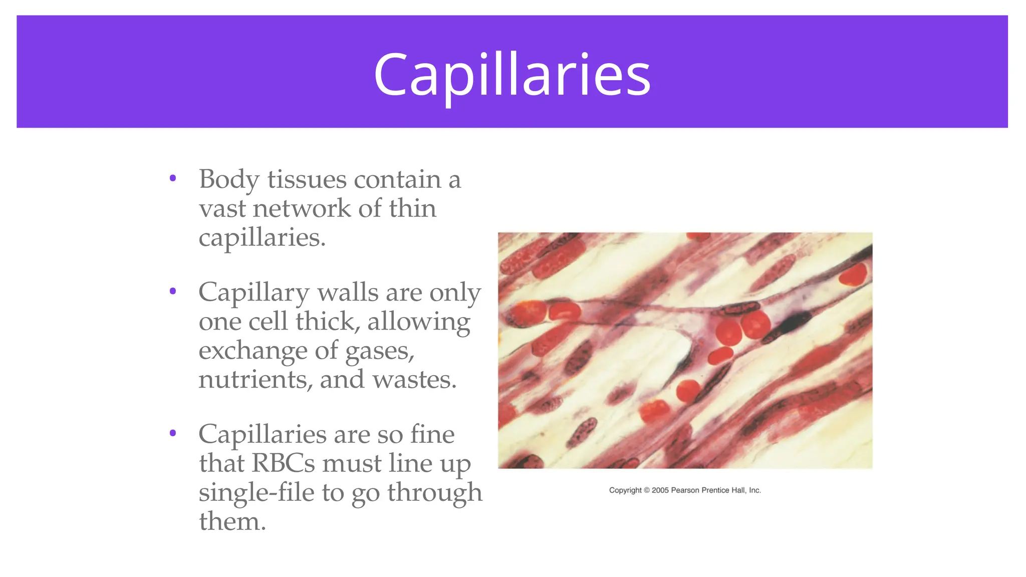
Capillaries
• Body tissuescontain a
vast network of thin
capillaries.
• Capillary walls are only
one cell thick, allowing
exchange of gases,
nutrients, and wastes.
• Capillaries are so fine
that RBCs must line up
single-file to go through
them.
23.
Venules
• Venules arethin-walled collectors of blood.
• Low pressure in the venules allows the capillary beds to
drain into them.
24.
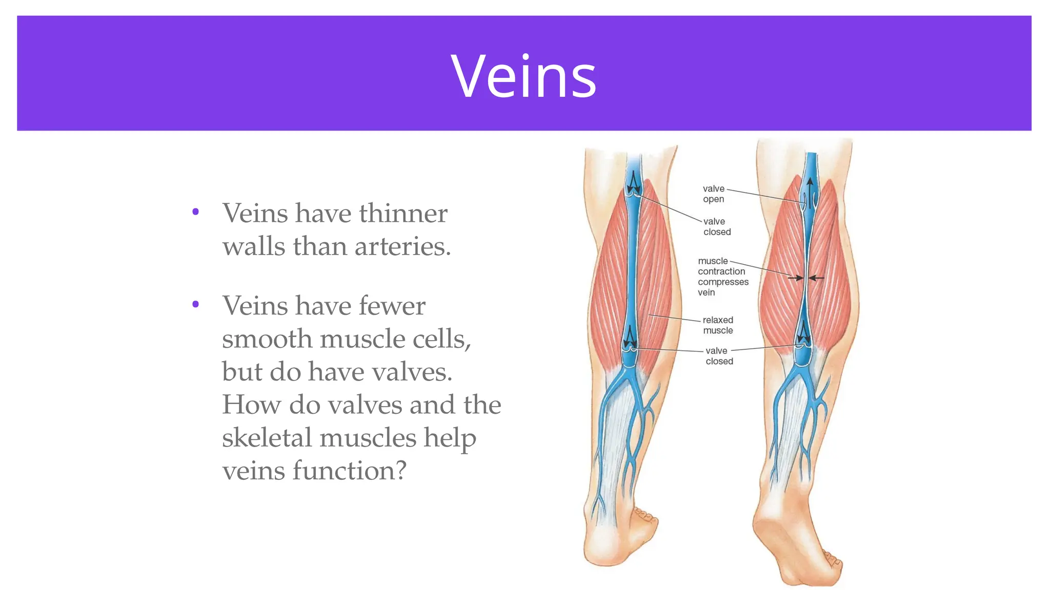
Veins
• Veins havethinner
walls than arteries.
• Veins have fewer
smooth muscle cells,
but do have valves.
How do valves and the
skeletal muscles help
veins function?
25.
• Besides theability to contract and move blood, why do
arteries need to be so thick and strong?
• Varicose veins are veins in the legs that are swollen,
stretched, and painful. What factors could lead to this
condition, and how can varicose veins be prevented?
W
O
R
K
T
O
G
E
T
H
E
R
26.
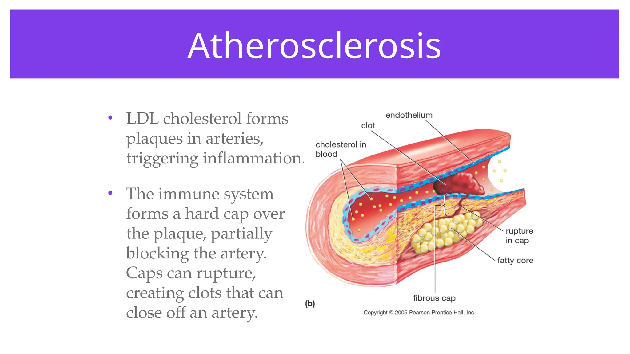
Atherosclerosis
• LDL cholesterolforms
plaques in arteries,
triggering inflammation.
• The immune system
forms a hard cap over
the plaque, partially
blocking the artery.
Caps can rupture,
creating clots that can
close off an artery.
27.
Preventing heart attacks
•Both genetic and environmental factors
contribute to atherosclerosis.
• Blood LDL cholesterol can be reduced by a low-
fat diet that emphasizes high-fiber foods,
antioxidants, and “good” fats
(monounsaturated fats, omega-3 oils), and
reduce trans-fats.
• Regular exercise also contributes significantly
to LDL cholesterol reduction.
28.
What is alwaystrue of arteries?
1 2 3 4
25% 25%
25%
25%
1. Always carry
oxygenated blood.
2. Always carry
deoxygenated blood.
3. Always carry blood
to the heart.
4. Always carry blood
away from the heart.
29.
Besides having toconstrict to move
blood, why are artery walls so thick and
strong?
1 2 3
33% 33%
33%
1. Arteries must move
oxygenated blood.
2. Arteries must
withstand very high
blood pressure when
the heart contracts.
3. Arteries must move
blood out to all parts
of the body.
30.
Why are capillarywalls so thin?
1 2 3
33% 33%
33%
1. Because capillaries
are thin and narrow
2. To allow exchange of
gases and nutrients.
3. To force RBCs to
move through in
single file.
31.
• Some peoplewho are at high risk for heart attacks may
be advised by their doctors to take low doses of aspirin
daily. What effects does aspirin have that would help
prevent heart attacks?
W
O
R
K
T
O
G
E
T
H
E
R
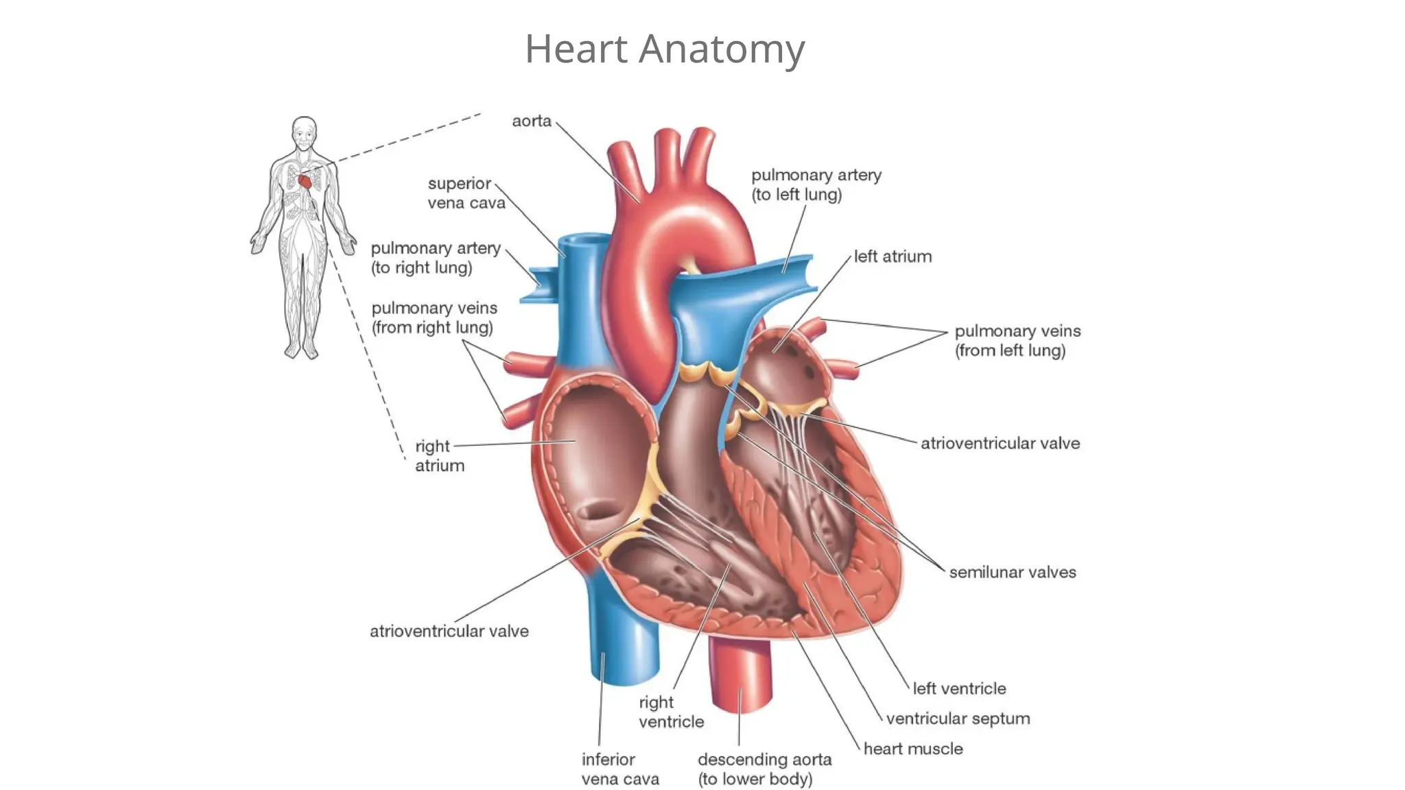
The Vertebrate Heart
•Vertebrate hearts are separated into two
types of chambers
• Atria (singular: atrium): receive blood
from body or lungs. Contractions of the
atria send blood through a valve to the
ventricles.
• Ventricles: receive blood from atria,
contract to send blood to body or lungs.
34.
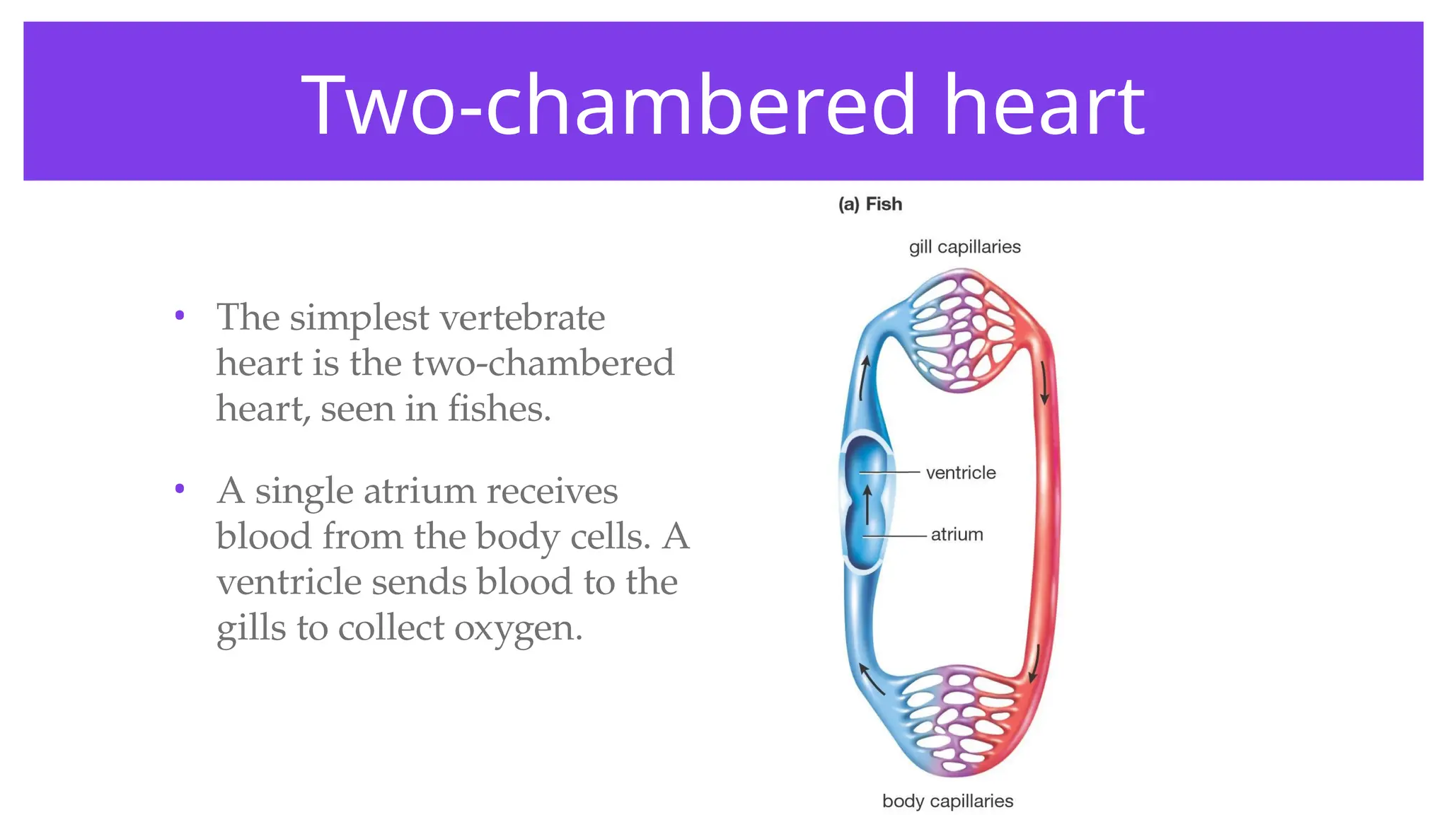
Two-chambered heart
• Thesimplest vertebrate
heart is the two-chambered
heart, seen in fishes.
• A single atrium receives
blood from the body cells. A
ventricle sends blood to the
gills to collect oxygen.
35.
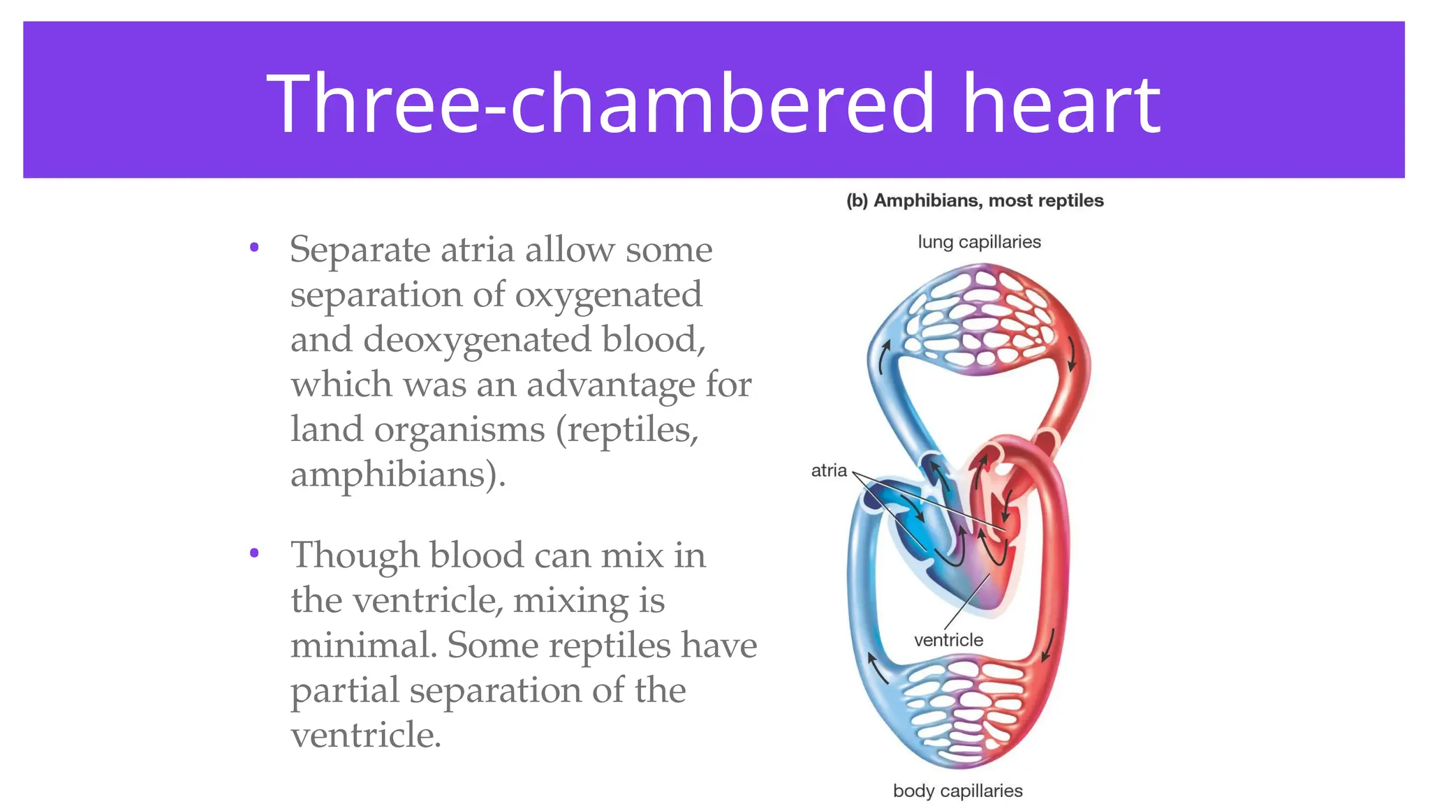
Three-chambered heart
• Separateatria allow some
separation of oxygenated
and deoxygenated blood,
which was an advantage for
land organisms (reptiles,
amphibians).
• Though blood can mix in
the ventricle, mixing is
minimal. Some reptiles have
partial separation of the
ventricle.
36.
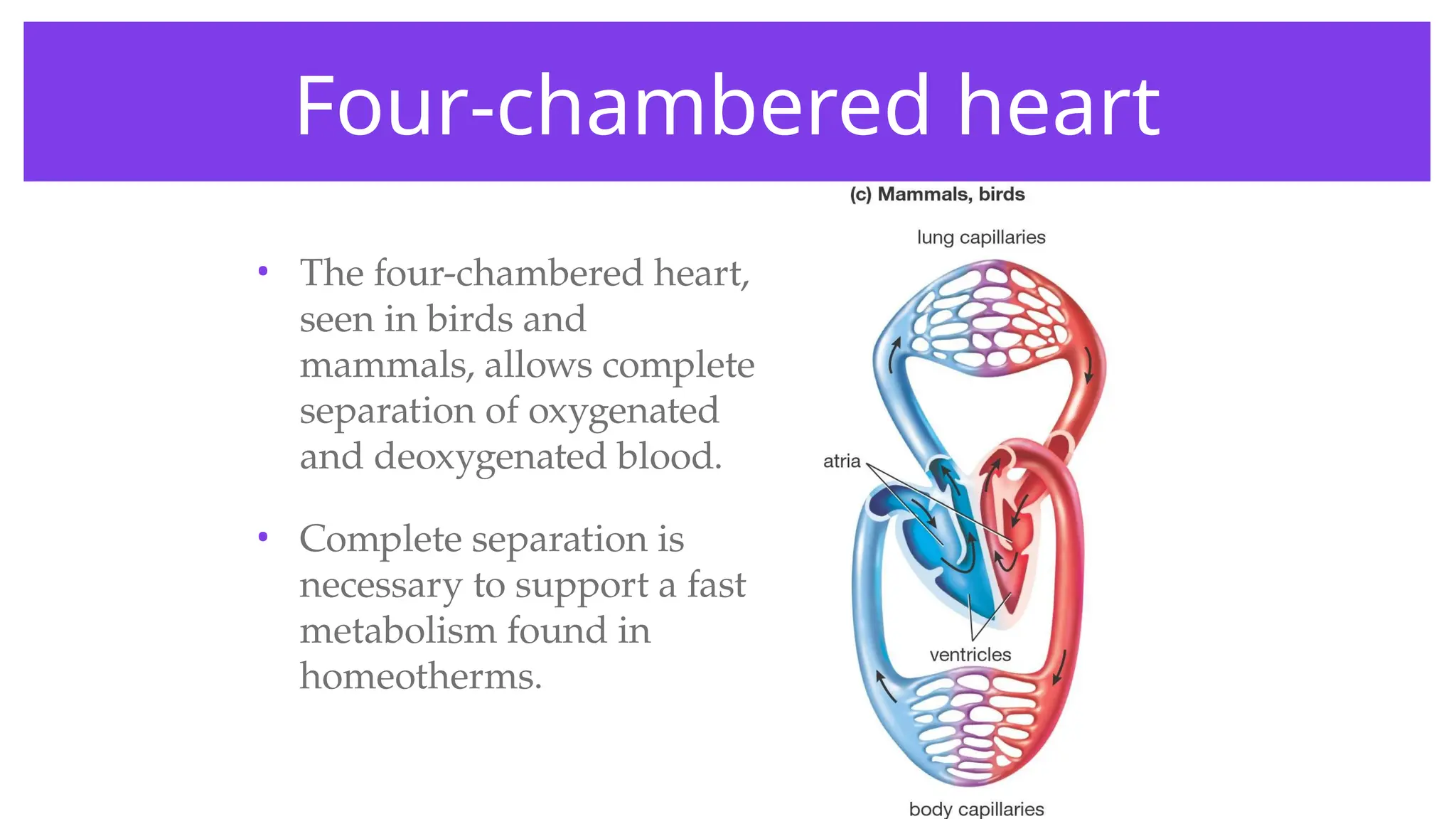
Four-chambered heart
• Thefour-chambered heart,
seen in birds and
mammals, allows complete
separation of oxygenated
and deoxygenated blood.
• Complete separation is
necessary to support a fast
metabolism found in
homeotherms.
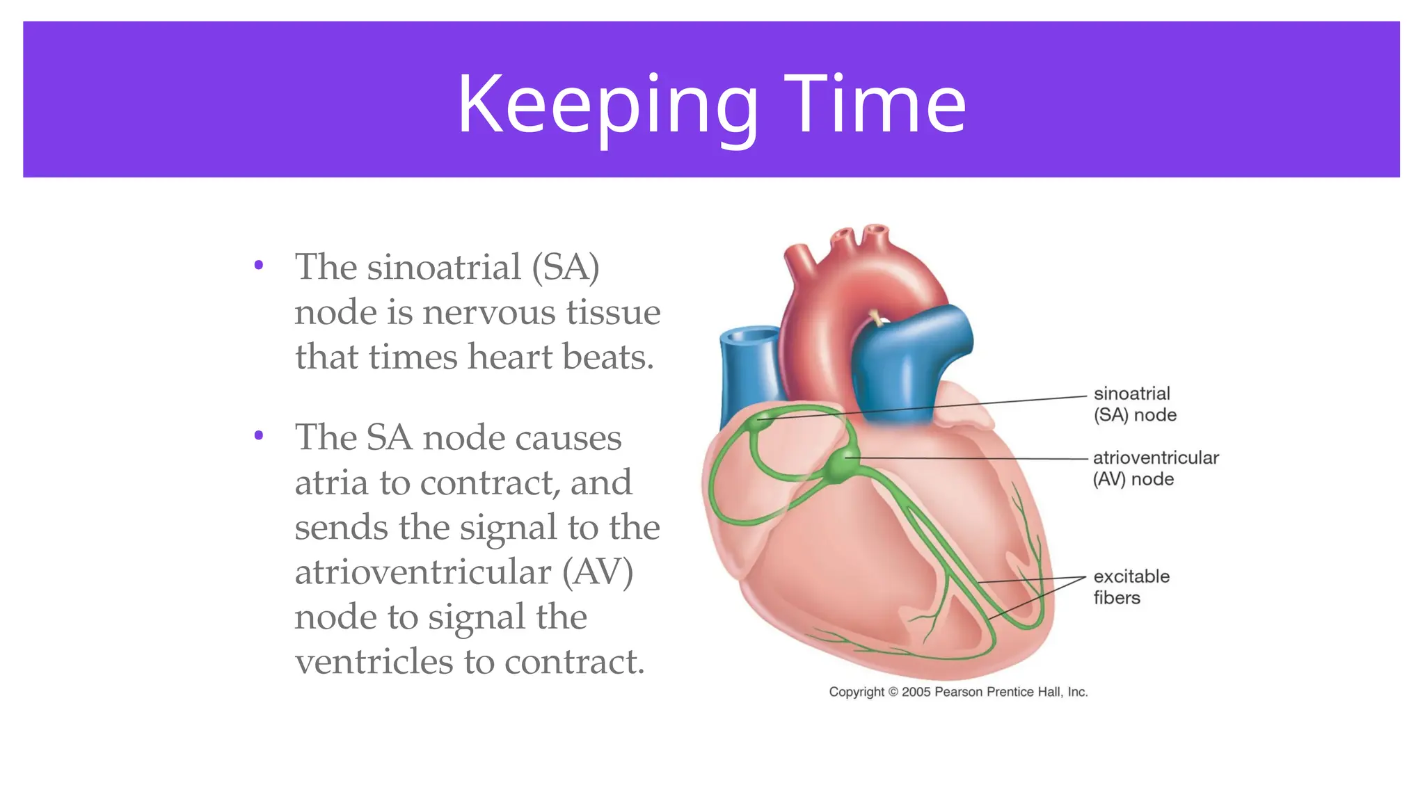
Keeping Time
• Thesinoatrial (SA)
node is nervous tissue
that times heart beats.
• The SA node causes
atria to contract, and
sends the signal to the
atrioventricular (AV)
node to signal the
ventricles to contract.
40.
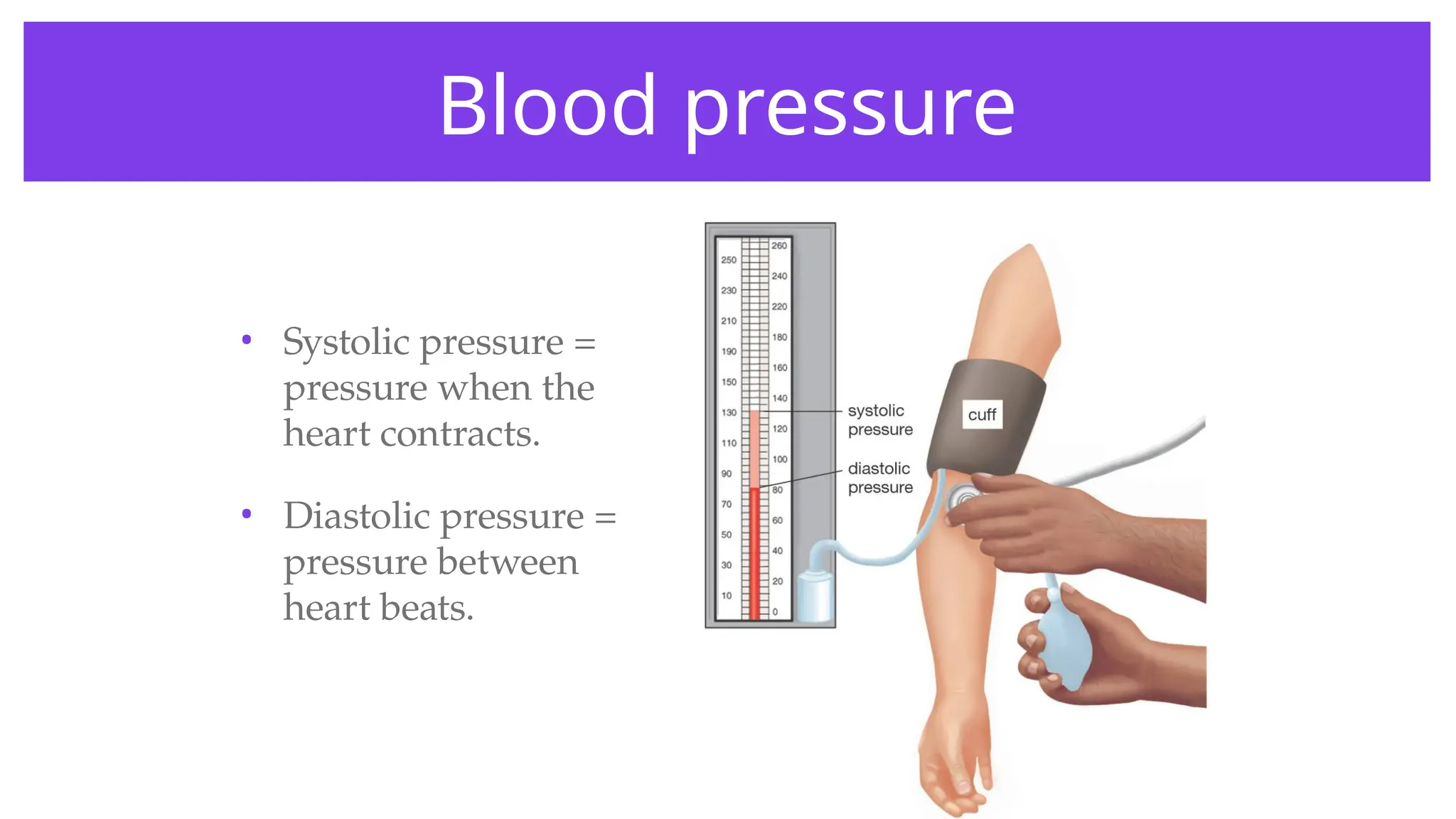
Blood pressure
• Systolicpressure =
pressure when the
heart contracts.
• Diastolic pressure =
pressure between
heart beats.
41.
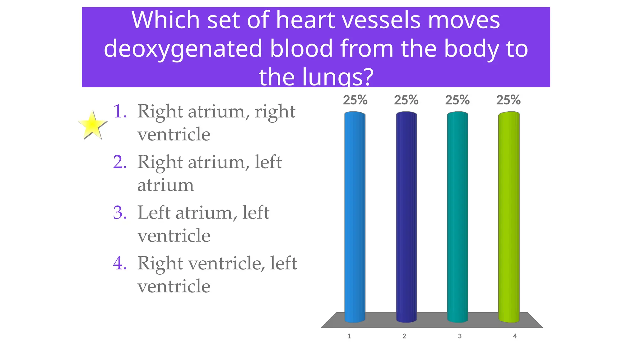
Which set ofheart vessels moves
deoxygenated blood from the body to
the lungs?
1 2 3 4
25% 25%
25%
25%
1. Right atrium, right
ventricle
2. Right atrium, left
atrium
3. Left atrium, left
ventricle
4. Right ventricle, left
ventricle
42.
If your bloodpressure is 90/70, the
70 represents:
1 2 3 4
25% 25%
25%
25%
1. Systolic pressure –
heart contracts
2. Systolic pressure –
heart is relaxed
3. Diastolic pressure –
heart contracts
4. Diastolic pressure –
heart is relaxed
43.
An electric pacemakercan be connected
to the heart to replace a faulty:
1 2 3 4
25% 25%
25%
25%
1. AV node
2. Bicuspid valve
3. SA node
4. Tricuspid valve
44.
• Hypertension (highblood pressure) puts people at risk
for heart disease. What long-term effects would an
increase in blood pressure have on the heart?
• What other organ system is involved in hypertension?
W
O
R
K
T
O
G
E
T
H
E
R
45.
Types of circulatorysystems
• Animals that have a circulatory system have one of two
kinds:
• Open: fluid is circulated through an open body
chamber.
• Closed: fluid is circulated through blood vessels.
46.
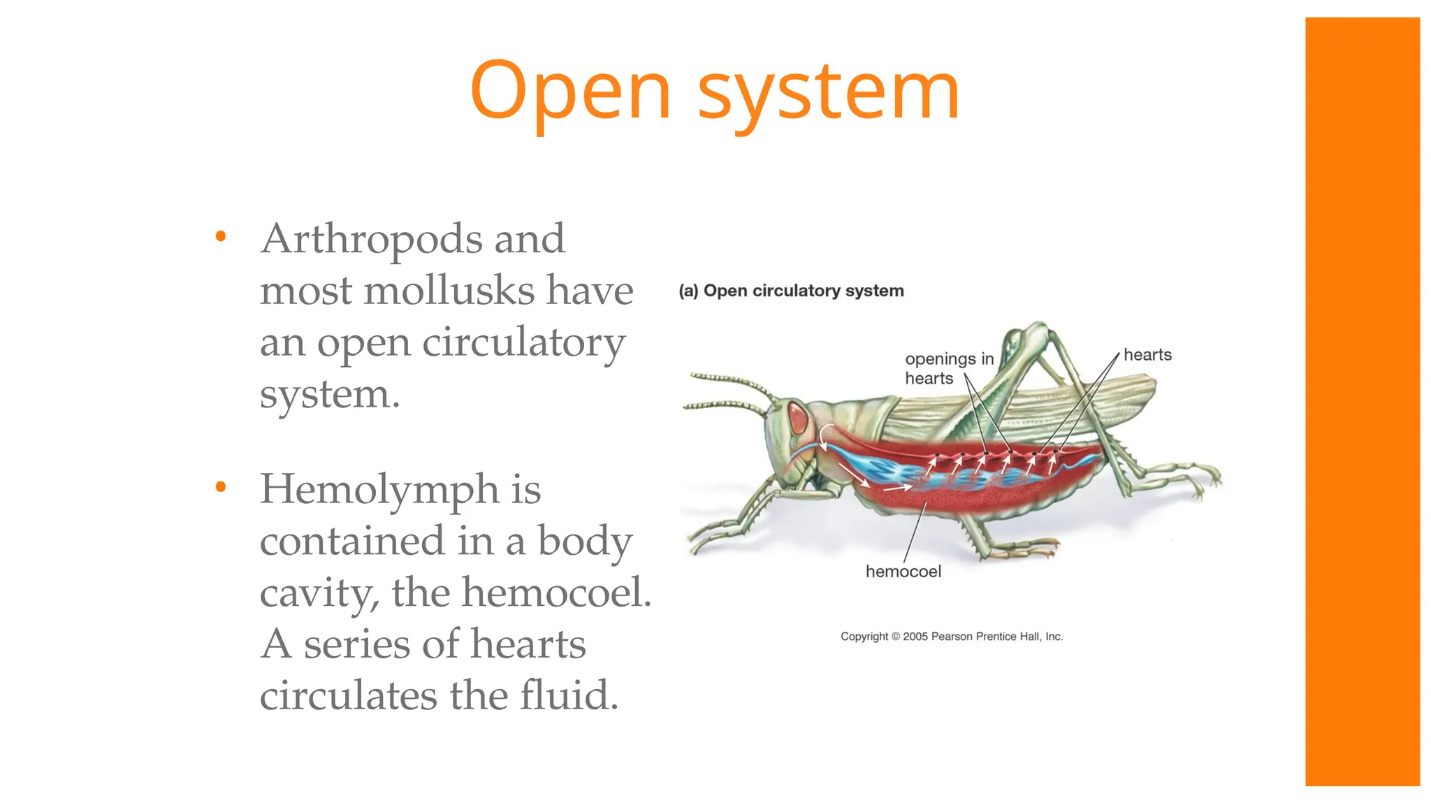
Open system
• Arthropodsand
most mollusks have
an open circulatory
system.
• Hemolymph is
contained in a body
cavity, the hemocoel.
A series of hearts
circulates the fluid.
47.
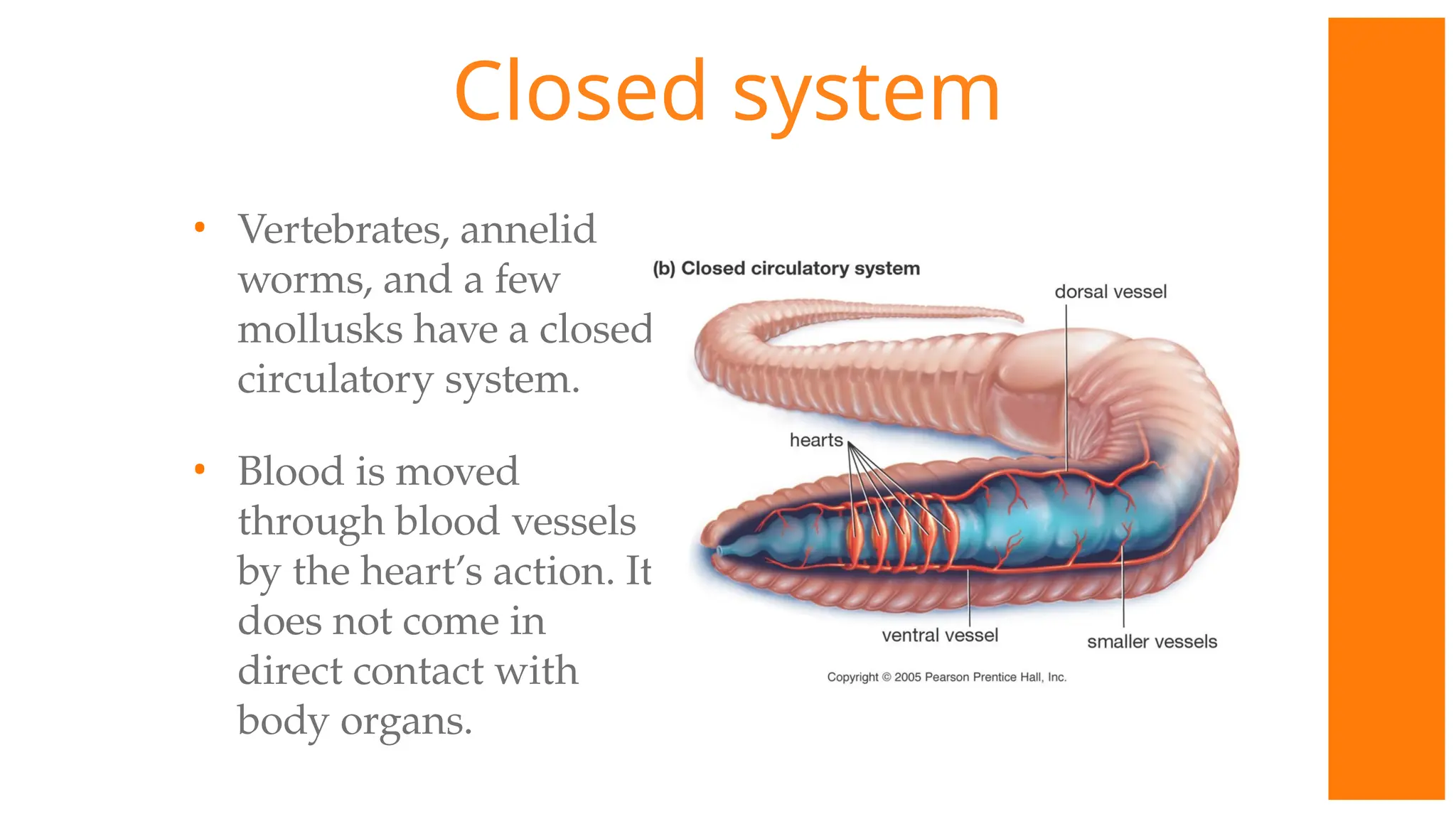
Closed system
• Vertebrates,annelid
worms, and a few
mollusks have a closed
circulatory system.
• Blood is moved
through blood vessels
by the heart’s action. It
does not come in
direct contact with
body organs.
48.
Why does anopen circulatory
system limit body size?
1 2 3 4
25% 25%
25%
25%
1. Hearts are too small
for growth.
2. Too little blood to
support a larger
animal.
3. Less efficient in
moving oxygen to
body tissues.
4. Hemocoel must be
shed for growth.
49.
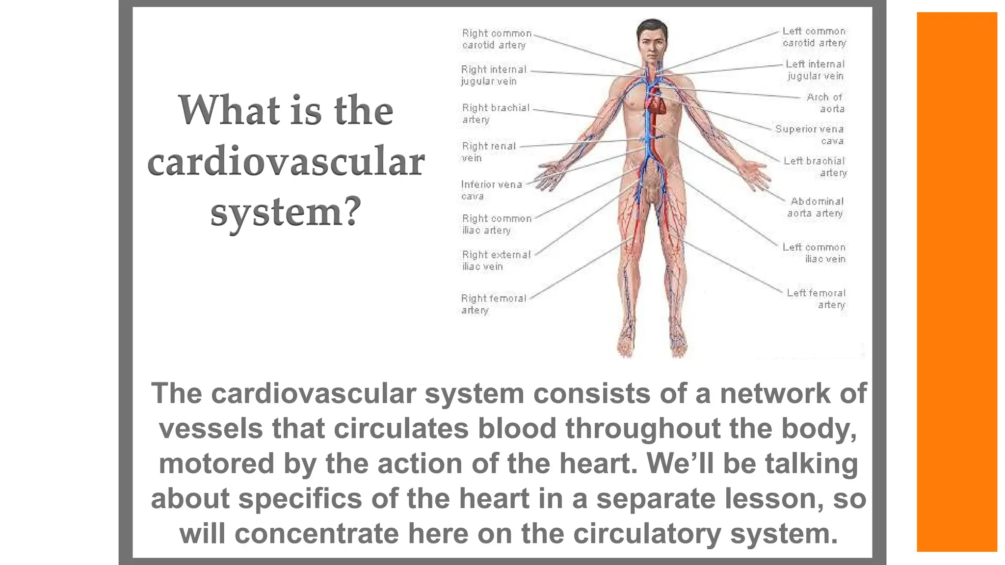
The cardiovascular systemconsists of a network of
vessels that circulates blood throughout the body,
motored by the action of the heart. We’ll be talking
about specifics of the heart in a separate lesson, so
will concentrate here on the circulatory system.
50.
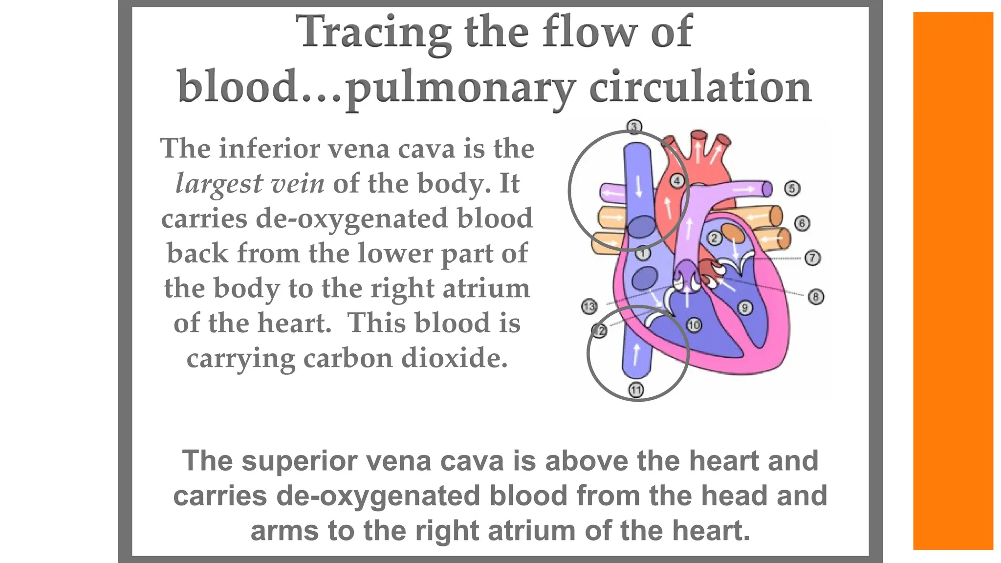
The inferior venacava is the
largest vein of the body. It
carries de-oxygenated blood
back from the lower part of
the body to the right atrium
of the heart. This blood is
carrying carbon dioxide.
The superior vena cava is above the heart and
carries de-oxygenated blood from the head and
arms to the right atrium of the heart.
51.
From the right
atrium,the blood
flows through the
tricuspid valve to
the right ventricle
and then onto the
lungs through the
pulmonary valve
and pulmonary
artery.
52.
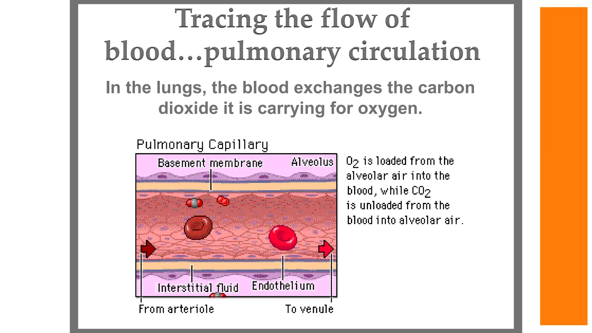
In the lungs,the blood exchanges the carbon
dioxide it is carrying for oxygen.
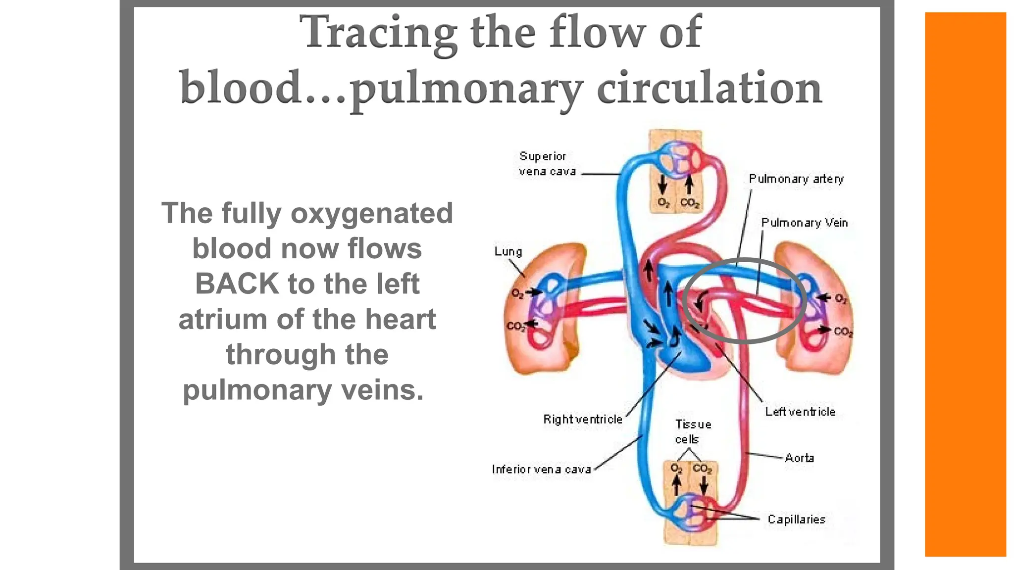
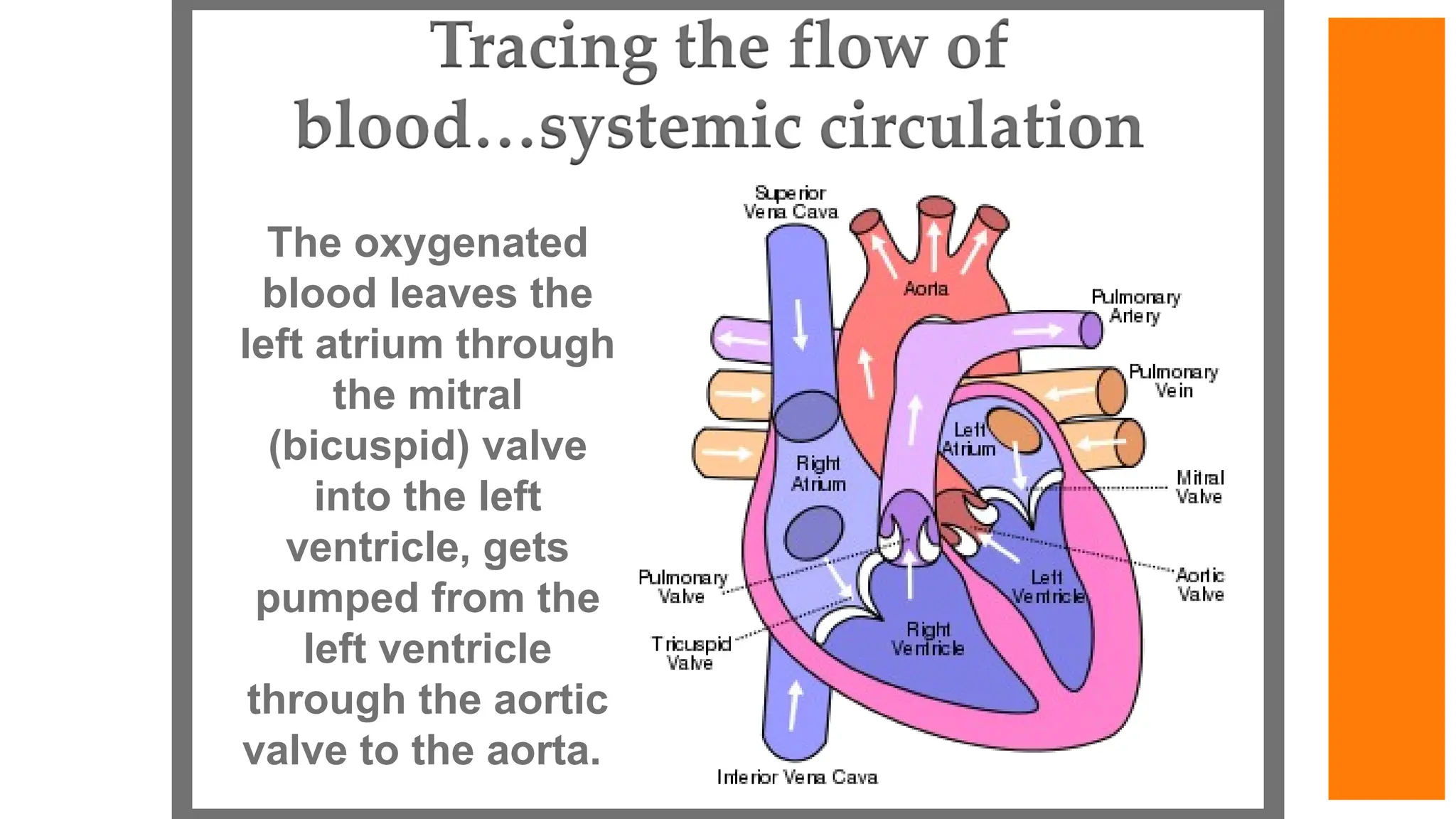
The oxygenated
blood leavesthe
left atrium through
the mitral
(bicuspid) valve
into the left
ventricle, gets
pumped from the
left ventricle
through the aortic
valve to the aorta.
55.
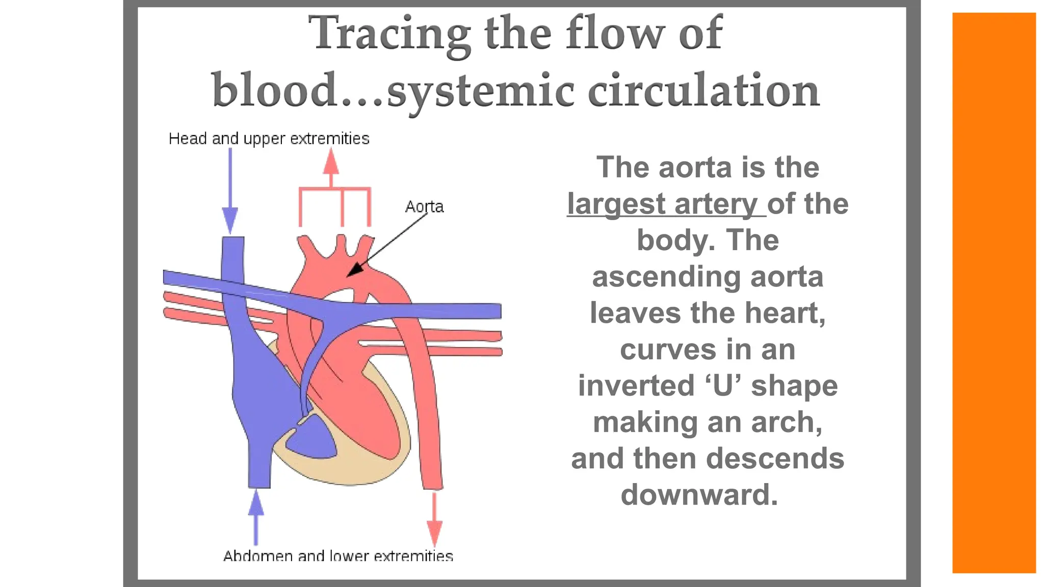
The aorta isthe
largest artery of the
body. The
ascending aorta
leaves the heart,
curves in an
inverted ‘U’ shape
making an arch,
and then descends
downward.
56.
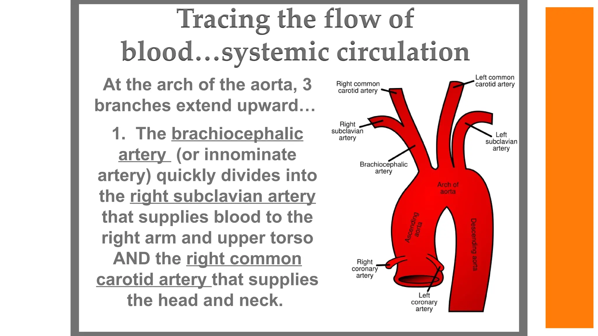
At the archof the aorta, 3
branches extend upward…
1. The brachiocephalic
artery (or innominate
artery) quickly divides into
the right subclavian artery
that supplies blood to the
right arm and upper torso
AND the right common
carotid artery that supplies
the head and neck.
57.
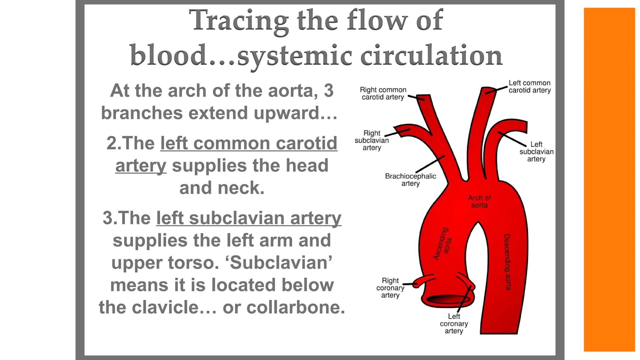
At the archof the aorta, 3
branches extend upward…
2.The left common carotid
artery supplies the head
and neck.
3.The left subclavian artery
supplies the left arm and
upper torso. ‘Subclavian’
means it is located below
the clavicle… or collarbone.
58.
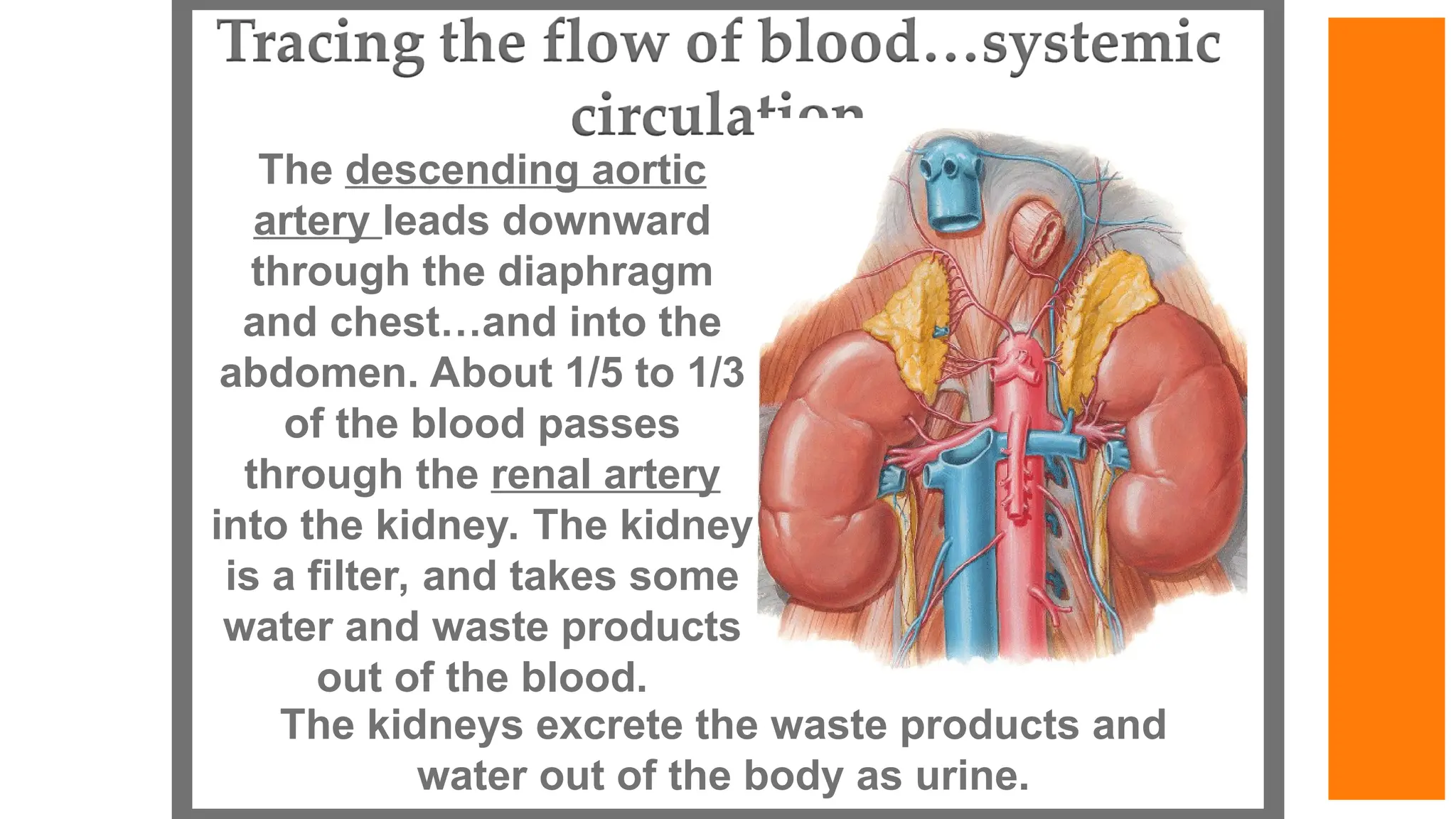
The descending aortic
arteryleads downward
through the diaphragm
and chest…and into the
abdomen. About 1/5 to 1/3
of the blood passes
through the renal artery
into the kidney. The kidney
is a filter, and takes some
water and waste products
out of the blood.
The kidneys excrete the waste products and
water out of the body as urine.
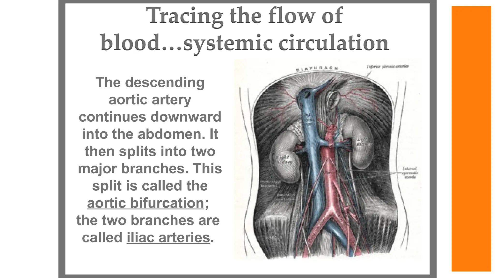
59.
The descending
aortic artery
continuesdownward
into the abdomen. It
then splits into two
major branches. This
split is called the
aortic bifurcation;
the two branches are
called iliac arteries.
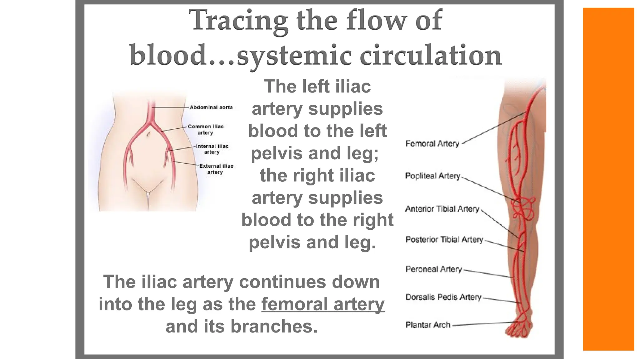
60.
The left iliac
arterysupplies
blood to the left
pelvis and leg;
the right iliac
artery supplies
blood to the right
pelvis and leg.
The iliac artery continues down
into the leg as the femoral artery
and its branches.
61.
Arteries are elastictubes that carry
blood in pulsating waves. The blood
exerts pressure against the walls of
the arteries as it passes through. The
peak pressure occurs during the
heart’s contraction, and is called
systolic pressure. The minimum
pressure occurs between
contractions when the heart expands
and refills, and is called diastolic
pressure. This pressure variation
within the artery produces a pulse.
All arteries have a pulse.
62.
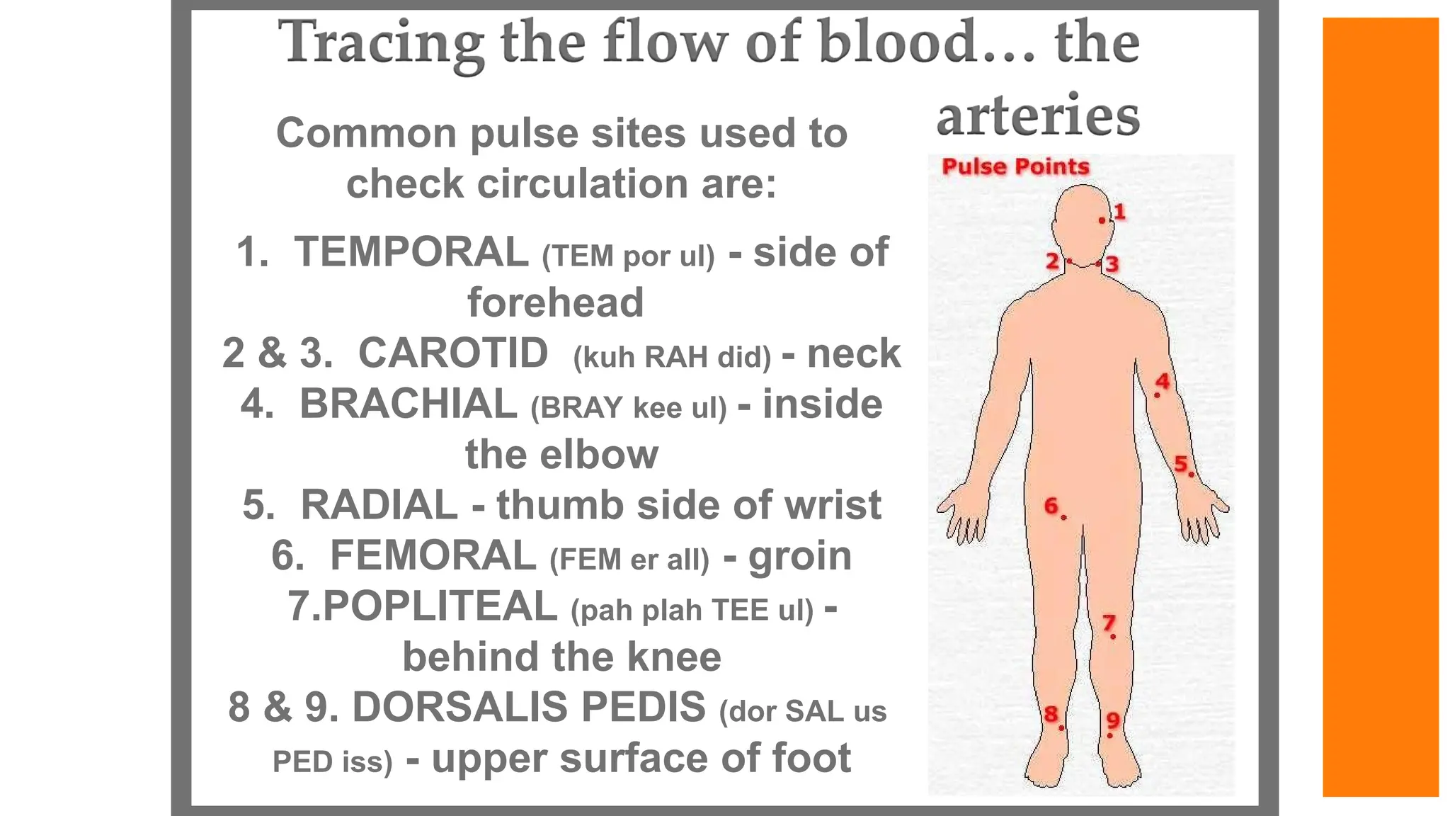
Common pulse sitesused to
check circulation are:
1. TEMPORAL (TEM por ul) - side of
forehead
2 & 3. CAROTID (kuh RAH did) - neck
4. BRACHIAL (BRAY kee ul) - inside
the elbow
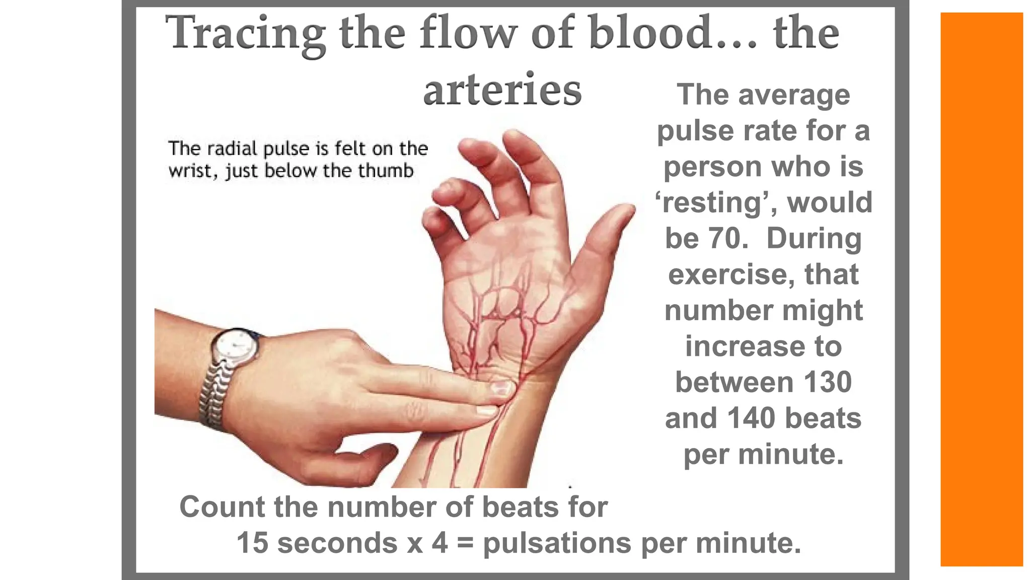
5. RADIAL - thumb side of wrist
6. FEMORAL (FEM er all) - groin
7.POPLITEAL (pah plah TEE ul) -
behind the knee
8 & 9. DORSALIS PEDIS (dor SAL us
PED iss) - upper surface of foot
63.
Count the numberof beats for
15 seconds x 4 = pulsations per minute.
The average
pulse rate for a
person who is
‘resting’, would
be 70. During
exercise, that
number might
increase to
between 130
and 140 beats
per minute.
64.
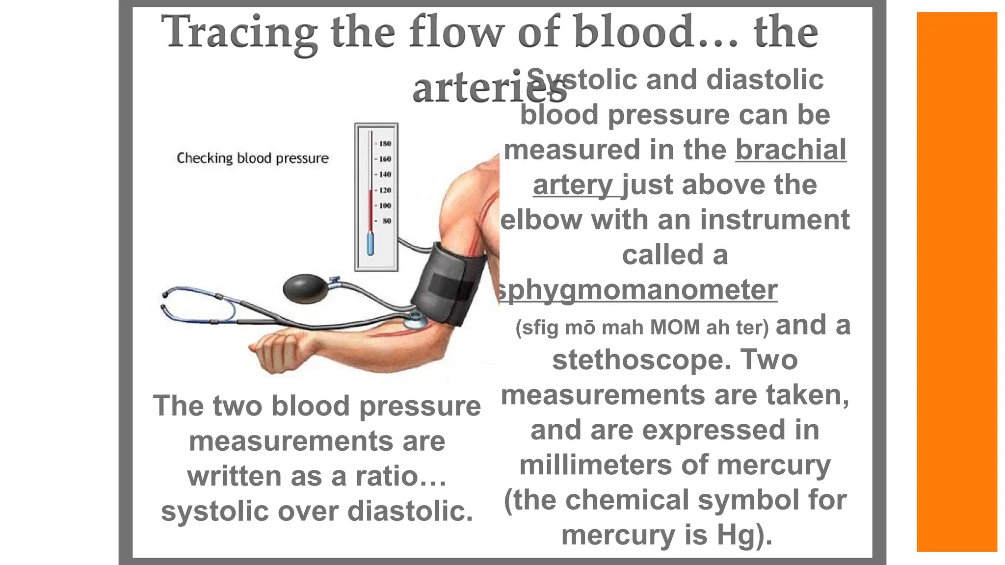
Systolic and diastolic
bloodpressure can be
measured in the brachial
artery just above the
elbow with an instrument
called a
sphygmomanometer
(sfig mō mah MOM ah ter) and a
stethoscope. Two
measurements are taken,
and are expressed in
millimeters of mercury
(the chemical symbol for
mercury is Hg).
The two blood pressure
measurements are
written as a ratio…
systolic over diastolic.
65.
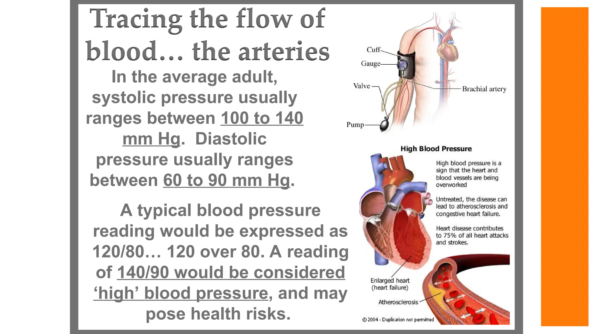
In the averageadult,
systolic pressure usually
ranges between 100 to 140
mm Hg. Diastolic
pressure usually ranges
between 60 to 90 mm Hg.
A typical blood pressure
reading would be expressed as
120/80… 120 over 80. A reading
of 140/90 would be considered
‘high’ blood pressure, and may
pose health risks.
66.
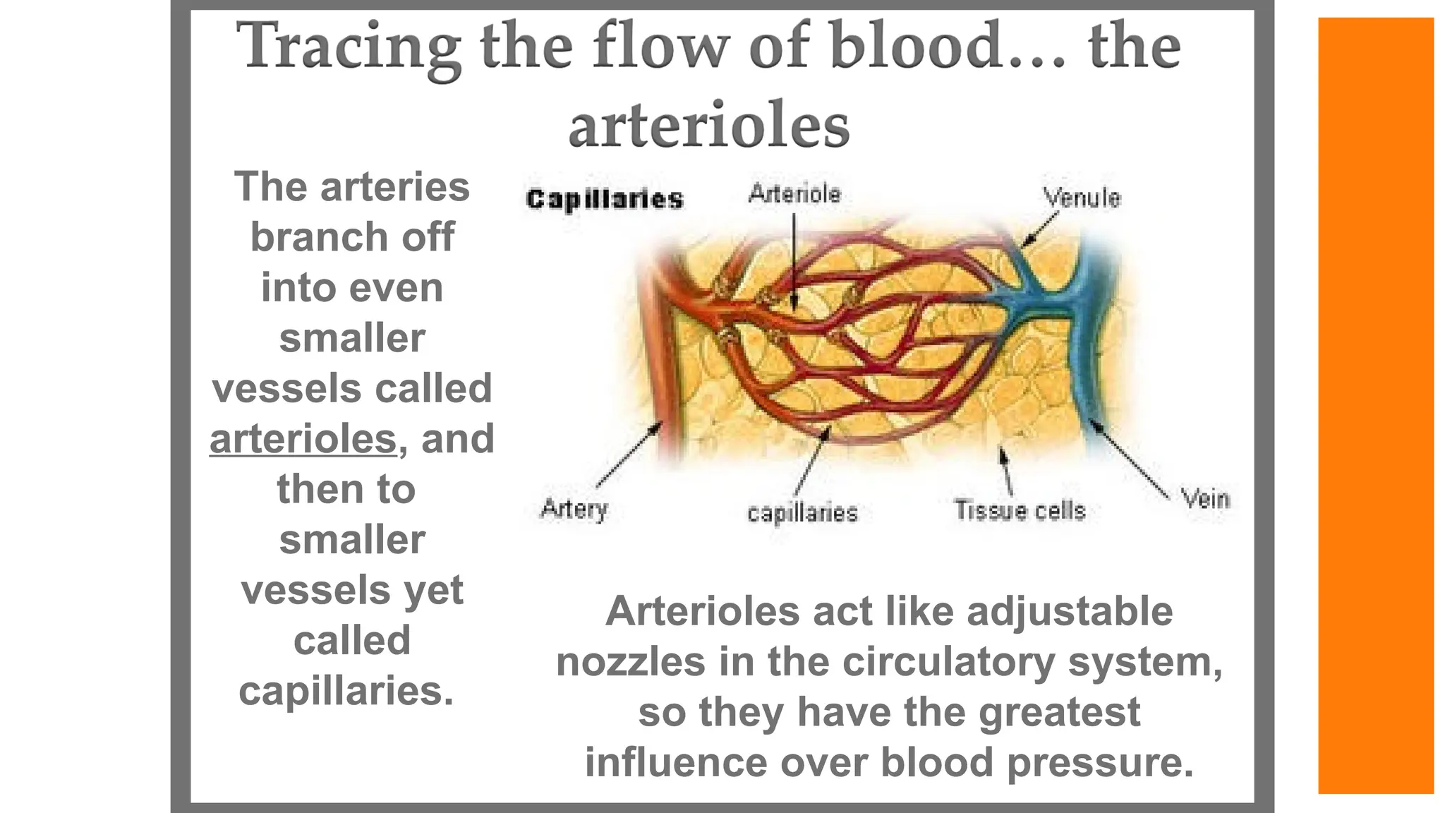
Arterioles act likeadjustable
nozzles in the circulatory system,
so they have the greatest
influence over blood pressure.
The arteries
branch off
into even
smaller
vessels called
arterioles, and
then to
smaller
vessels yet
called
capillaries.
67.
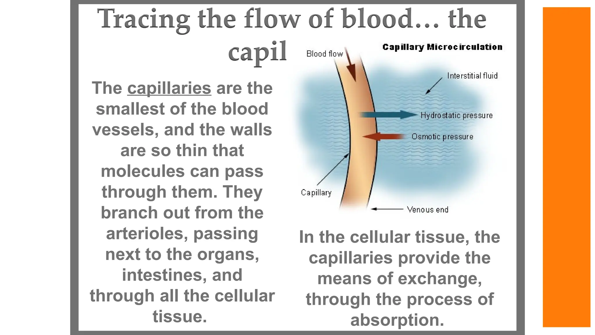
The capillaries arethe
smallest of the blood
vessels, and the walls
are so thin that
molecules can pass
through them. They
branch out from the
arterioles, passing
next to the organs,
intestines, and
through all the cellular
tissue.
In the cellular tissue, the
capillaries provide the
means of exchange,
through the process of
absorption.
68.
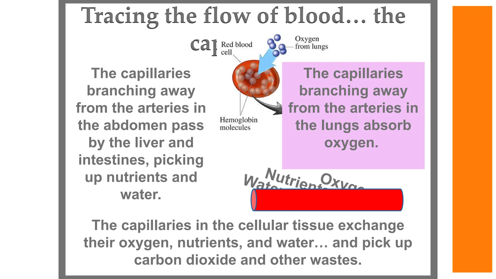
The capillaries
branching away
fromthe arteries in
the abdomen pass
by the liver and
intestines, picking
up nutrients and
water.
The capillaries in the cellular tissue exchange
their oxygen, nutrients, and water… and pick up
carbon dioxide and other wastes.
The capillaries
branching away
from the arteries in
the lungs absorb
oxygen.
Nutrients
Water
Oxygen
69.
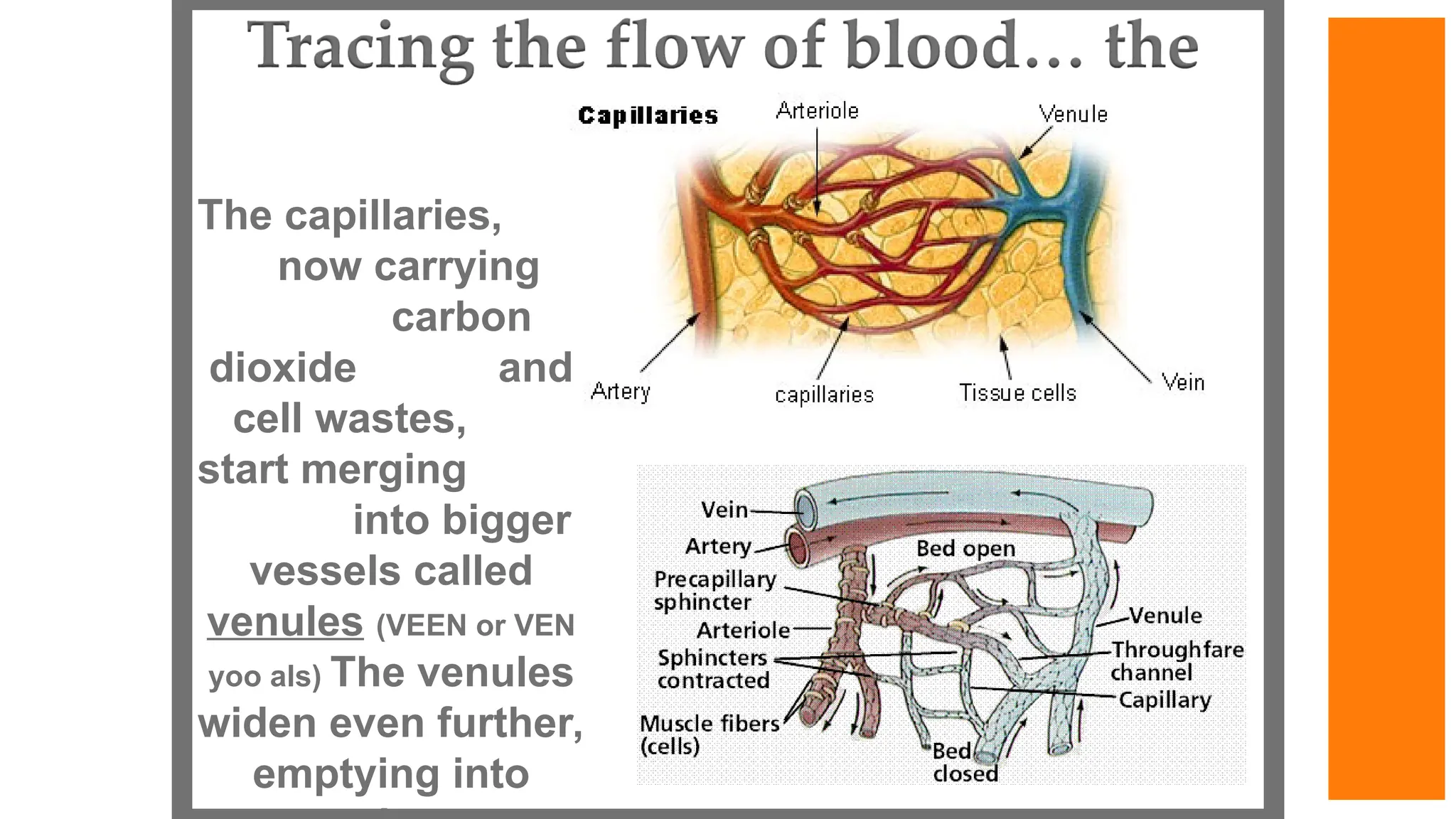
The capillaries,
now carrying
carbon
dioxideand
cell wastes,
start merging
into bigger
vessels called
venules (VEEN or VEN
yoo als) The venules
widen even further,
emptying into
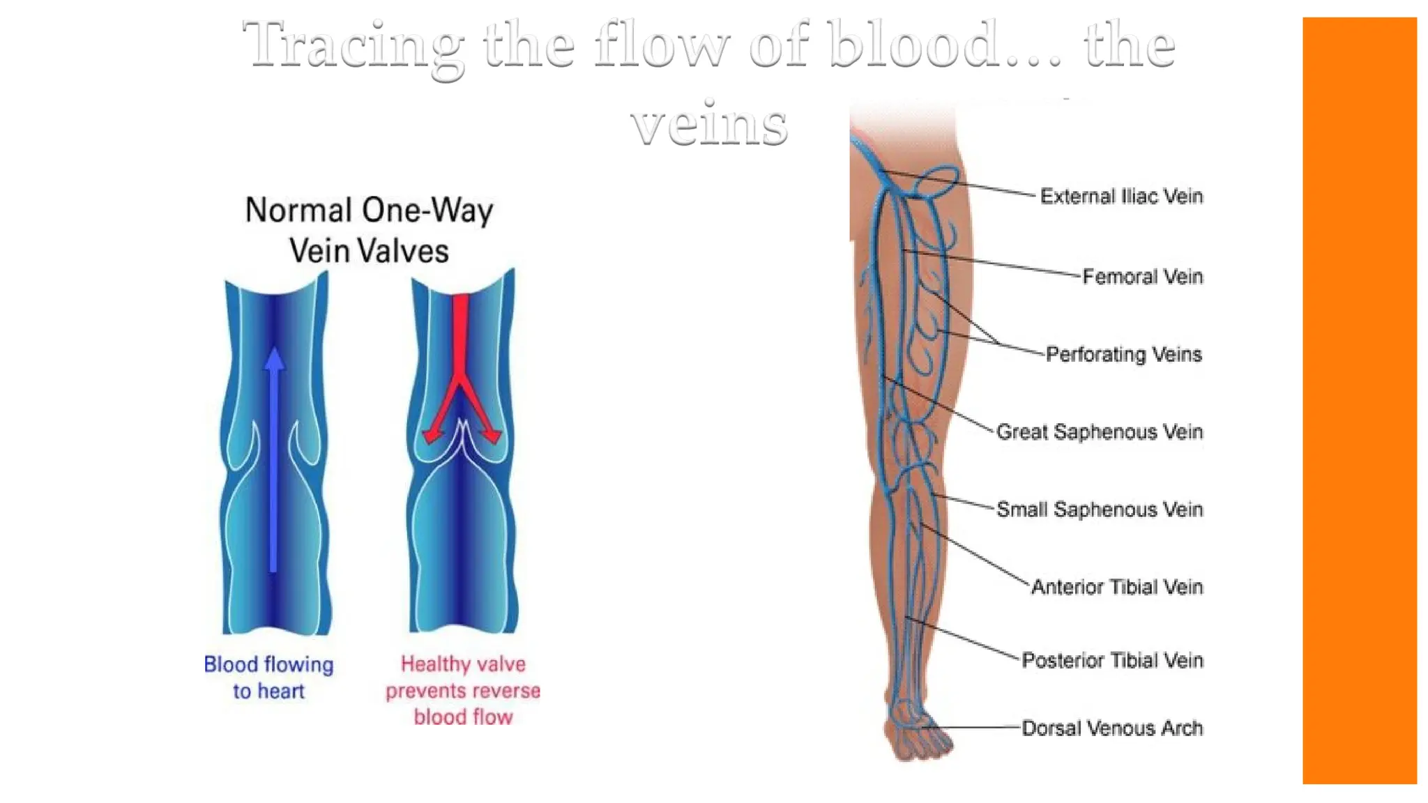
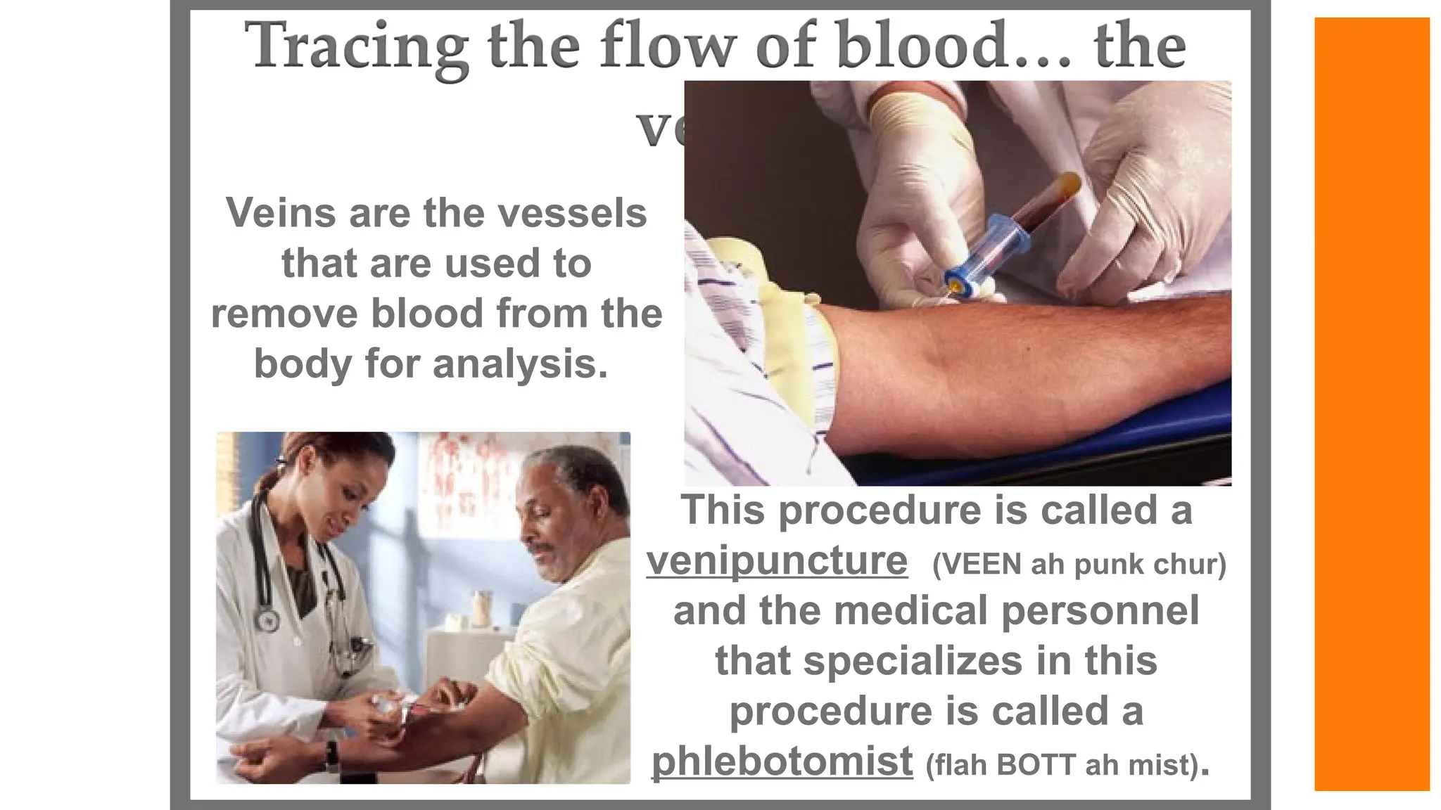
Veins are thevessels
that are used to
remove blood from the
body for analysis.
This procedure is called a
venipuncture (VEEN ah punk chur)
and the medical personnel
that specializes in this
procedure is called a
phlebotomist (flah BOTT ah mist).
72.
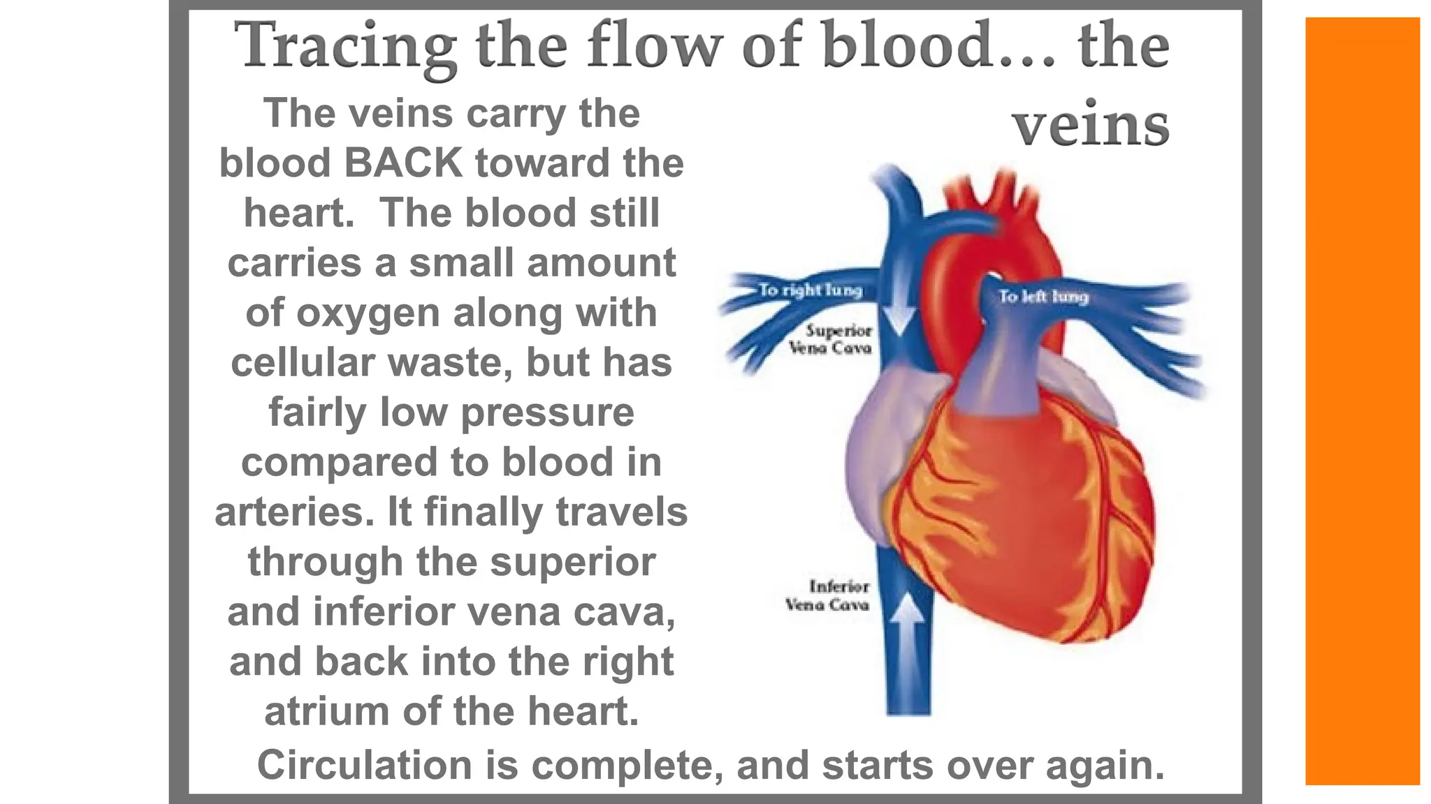
The veins carrythe
blood BACK toward the
heart. The blood still
carries a small amount
of oxygen along with
cellular waste, but has
fairly low pressure
compared to blood in
arteries. It finally travels
through the superior
and inferior vena cava,
and back into the right
atrium of the heart.
Circulation is complete, and starts over again.