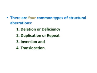
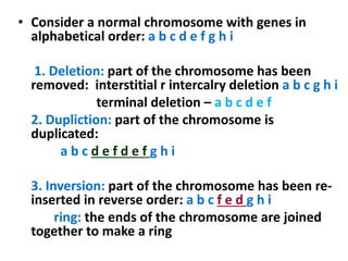
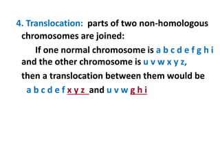
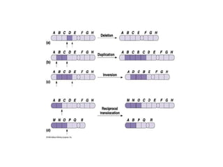
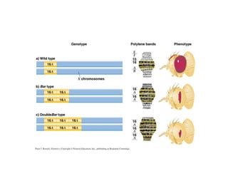
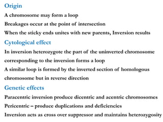
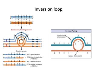
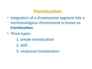
Chromosomal Aberrations
• The somatic (2n) and gametic (n) chromosome
numbers of a species ordinarily remain constant.
• This is due to the extremely precise mitotic and
meiotic cell division.
• Somatic cells of a diploid species contain two copies
of each chromosome, which are called homologous
chromosome.
• Their gametes, therefore contain only one copy of
each chromosome, that is they contain one
chromosome complement or genome.
• Each chromosome of a genome contains a definite
numbers and kinds of genes, which are arranged in
a definite sequence.
Chromosomal Aberrations
• Sometime due to mutation or spontaneously
(without any known causal factors), variation
in chromosomal number or structure do arise
in nature. - Chromosomal aberrations.
• Chromosomal aberration may be grouped
into two broad classes:
1. Structural and
2. Numerical
Structural Chromosomal Aberrations
• Chromosome structure variations result from
chromosome breakage.
• Broken chromosomes tend to re-join; if there is
more than one break, rejoining occurs at random
and not necessarily with the correct ends.
• The result is structural changes in the
chromosomes.
• Chromosome breakage is caused by X-rays,
various chemicals, and can also occur
spontaneously.
• There are four common types of structural
aberrations:
1. Deletion or Deficiency
2. Duplication or Repeat
3. Inversion and
4. Translocation.
• Consider a normal chromosome with genes in
alphabetical order: a b c d e f g h i
1. Deletion: part of the chromosome has been
removed: interstitial r intercalry deletion a b c g h i
terminal deletion – a b c d e f
2. Dupliction: part of the chromosome is
duplicated:
a b c d e f d e f g h i
3. Inversion: part of the chromosome has been re-
inserted in reverse order: a b c f e d g h i
ring: the ends of the chromosome are joined
together to make a ring
4. Translocation: parts of two non-homologous
chromosomes are joined:
If one normal chromosome is a b c d e f g h i
and the other chromosome is u v w x y z,
then a translocation between them would be
a b c d e f x y z and u v w g h i
Deletion or deficiency
Loss of a chromosome segment is known as deletion or
deficiency
It can be terminal deletion or interstitial or intercalary
deletion.
A single break near the end of the chromosome would be
expected to result in terminal deficiency.
If two breaks occur, a section may be deleted and an
intercalary deficiency created.
Terminal deficiencies might seem less complicated.
But majority of deficiencies detected are intercalary type
within the chromosome.
Deletion was the first structural aberration detected by
Bridges in 1917 from his genetic studies on
X chromosome of Drosophila.
• Deletion generally produce striking genetic and
physiological effects.
• When homozygous, most deletions are lethal, because
most genes are necessary for life and a homozygous
deletion would have zero copies of some genes.
• When heterozygous, the genes on the normal
homologue are hemizygous: there is only 1 copy of
those genes.
• Crossing over is absent in deleted region of a
chromosome since this region is present in only one
copy in deletion heterozygotes.
• In Drosophila, several deficiencies induced the mutants
like Blond, Pale, Beaded, Carved, Notch, Minute etc.
Cytological Effect
In deletion heterozygotes, deletion loop occurs during
pairing of homologus chromosomes
The portion of the normal chromosomes homologus to
the deficient segment bulges out
Genetic Effect
When a segment of a chromosome is absent, some
genes are also absent
If these lost genes are physiologically important,
deletion leads to death of the organism
Deletion in Prokaryotes
Deletions are found in prokaryotes as well, e.g., E.coli, T4
phage and Lambda phage.
In E.coli, deletions of up to 1 % of the bacterial
chromosome are known.
In lambda phage, however 20% of the genome may be
missing in some of the deletions.
Deletion in Human
Chromosome deletions are usually lethal even as
heterozygotes, resulting in zygotic loss, stillbirths, or
infant death.
Sometimes, infants with small chromosome deficiencies
however, survive long enough to permit the abnormal
phenotype they express.
Cri-du-chat (Cat cry syndrome):
The name of the syndrome came from a catlike mewing cry
from small weak infants with the disorder.
Other characteristics are microcephaly (small head), broad
face and saddle nose, physical and mental retardation.
Cri-du-chat patients die in infancy or early childhood.
The chromosome deficiency is in the short arm of
chromosome 5 .
.
Myelocytic leukemia
Another human disorder that is associated with
a chromosome abnormality is chronic
myelocytic leukemia.
A deletion of chromosome 22 was described by
P.C.Nowell and Hungerford and was called
“Philadelphia” (Ph’) chromosome after the
city in which the discovery was made
Duplication
The presence of an additional chromosome
segment, as compared to that normally present
in a nucleus is known as Duplication.
• In a diploid organism, presence of a
chromosome segment in more than two copies
per nucleus is called duplication.
• Four types of duplication:
1. Tandem duplication
2. Reverse tandem duplication
3. Displaced duplication
4. Translocation duplication
• The extra chromosome segment may be located
immediately after the normal segment in precisely
the same orientation forms the tandem
• When the gene sequence in the extra segment of a
tandem in the reverse order i.e, inverted , it is
known as reverse tandem duplication
• In some cases, the extra segment may be located in
the same chromosome but away from the normal
segment – termed as displaced duplication
• The additional chromosome segment is located in a
non-homologous chromosome is translocation
duplication.
Cytological Effect
During meiotic pairing of heterozygotes, the
chromosome with duplicated segment forms a loop
Genetic Effect
The duplications are not lethal
The unusual dosage of genes can be investigated
Duplications are useful in evolution of new characters
without loss of original traits
Relocations of chromosomal materials due to
duplication results in an altered phenotype : Position
effect
Origin
• Origin of duplication involves chromosome breakage
and reunion of chromosome segment with its
homologous chromosome.
• As a result, one of the two homologous involved in the
production of a duplication ends up with a deficiency,
while the other has a duplication for the concerned
segment.
• Another phenomenon, known as unequal crossing
over, also leads to exactly the same consequences for
small chromosome segments.
• For e.g., duplication of the band 16A of X chromosome
of Drosophila produces Bar eye.
• This duplication is believed to originate due to
unequal crossing over between the two normal X
chromosomes of female.
Position Effect
 Altered phenotype due to relocation of
chromosomal material
 A fly homozygous for Bar eye has four 16A
segments, two in each chromosome
 A fly heterozygous for ultra Bar has also four
16A segments- one in normal and three in duplic.
chromo
 Flies homozygous for ultra Bar produced smaller
size eyes
Inversion
• When a segment of chromosome is oriented in the reverse
direction, such segment said to be inverted and the
phenomenon is termed as inversion.
• The existence of inversion was first detected by Strutevant
and Plunkett in 1926.
• Inversion occur when parts of chromosomes become
detached , turn through 1800 and are reinserted in such a way
that the genes are in reversed order.
• Inversion may be classified into two types:
– Pericentric - include the centromere
– Paracentric - do not include the centromere
• An inversion consists of two breaks in one
chromosome.
• The area between the breaks is inverted
(turned around), and then reinserted and the
breaks then unite to the rest of the
chromosome.
• If the inverted area includes the centromere it
is called a pericentric inversion.
• If it does not, it is called a paracentric
inversion.
Inversions in natural populations
• In natural populations, pericentric inversions
are much less frequent than paracentric
inversions.
• In many sp, however, pericentric inversions are
relatively common, e.g., in some grasshoppers.
• Paracentric inversions appear to be very
frequent in natural populations of Drosophila.
Origin
A chromosome may form a loop
Breakages occur at the point of intersection
When the sticky ends unites with new parents, Inversion results
Cytological effect
In inversion heterozygote the part of the uninverted chromosome
corresponding to the inversion forms a loop
A similar loop is formed by the inverted section of homologous
chromosome but in reverse direction
Genetic effects
Paracentric inversion produce dicentric and acentric chromosomes
Pericentric – produce duplications and deficiencies
Inversion acts as cross over suppressor and maintains heterozygosity
Inversion loop
Translocation
• Integration of a chromosome segment into a
nonhomologous chromosome is known as
translocation.
• Three types:
1. simple translocation
2. shift
3. reciprocal translocation.
• Simple translocation: In this case, terminal
segment of a chromosome is integrated at one
end of a non-homologous region. Simple
translocations are rather rare.
• Shift: In shift, an intercalary segment of a
chromosome is integrated within a non-
homologous chromosome. Such
translocations are known in the populations of
Drosophila, Neurospora etc.
• Reciprocal translocation: It is produced when
two non-homologous chromosomes exchange
segments – i.e., segments reciprocally
transferred.
• Translocation of this type is most common
Cytological effect
• In the translocation heterozygote, pairing of
homologous chromosomal segments is
effected by cross-shaped configuration
• This cross opens out into a ring as chiasma
terminalizes
• The meiotic products are of three kinds :
normal, balanced and unbalanced
A … B C D 1
A… BCK 2
D… MNO 3
K…MNO 4
Cross shape pairing in tranlocation
heterozygote
Genetic effect
• Translocation alter the linkage relationships
of genes
• Heterozygotic translocation causes semi
sterility
• Phenotypic expression of gene may be
modified

6. Chromosomal aberrations - Structural

  • 1.
  • 2.
    Chromosomal Aberrations • Thesomatic (2n) and gametic (n) chromosome numbers of a species ordinarily remain constant. • This is due to the extremely precise mitotic and meiotic cell division. • Somatic cells of a diploid species contain two copies of each chromosome, which are called homologous chromosome. • Their gametes, therefore contain only one copy of each chromosome, that is they contain one chromosome complement or genome. • Each chromosome of a genome contains a definite numbers and kinds of genes, which are arranged in a definite sequence.
  • 3.
    Chromosomal Aberrations • Sometimedue to mutation or spontaneously (without any known causal factors), variation in chromosomal number or structure do arise in nature. - Chromosomal aberrations. • Chromosomal aberration may be grouped into two broad classes: 1. Structural and 2. Numerical
  • 4.
    Structural Chromosomal Aberrations •Chromosome structure variations result from chromosome breakage. • Broken chromosomes tend to re-join; if there is more than one break, rejoining occurs at random and not necessarily with the correct ends. • The result is structural changes in the chromosomes. • Chromosome breakage is caused by X-rays, various chemicals, and can also occur spontaneously.
  • 5.
    • There arefour common types of structural aberrations: 1. Deletion or Deficiency 2. Duplication or Repeat 3. Inversion and 4. Translocation.
  • 6.
    • Consider anormal chromosome with genes in alphabetical order: a b c d e f g h i 1. Deletion: part of the chromosome has been removed: interstitial r intercalry deletion a b c g h i terminal deletion – a b c d e f 2. Dupliction: part of the chromosome is duplicated: a b c d e f d e f g h i 3. Inversion: part of the chromosome has been re- inserted in reverse order: a b c f e d g h i ring: the ends of the chromosome are joined together to make a ring
  • 7.
    4. Translocation: partsof two non-homologous chromosomes are joined: If one normal chromosome is a b c d e f g h i and the other chromosome is u v w x y z, then a translocation between them would be a b c d e f x y z and u v w g h i
  • 9.
    Deletion or deficiency Lossof a chromosome segment is known as deletion or deficiency It can be terminal deletion or interstitial or intercalary deletion. A single break near the end of the chromosome would be expected to result in terminal deficiency. If two breaks occur, a section may be deleted and an intercalary deficiency created. Terminal deficiencies might seem less complicated. But majority of deficiencies detected are intercalary type within the chromosome. Deletion was the first structural aberration detected by Bridges in 1917 from his genetic studies on X chromosome of Drosophila.
  • 10.
    • Deletion generallyproduce striking genetic and physiological effects. • When homozygous, most deletions are lethal, because most genes are necessary for life and a homozygous deletion would have zero copies of some genes. • When heterozygous, the genes on the normal homologue are hemizygous: there is only 1 copy of those genes. • Crossing over is absent in deleted region of a chromosome since this region is present in only one copy in deletion heterozygotes. • In Drosophila, several deficiencies induced the mutants like Blond, Pale, Beaded, Carved, Notch, Minute etc.
  • 11.
    Cytological Effect In deletionheterozygotes, deletion loop occurs during pairing of homologus chromosomes The portion of the normal chromosomes homologus to the deficient segment bulges out Genetic Effect When a segment of a chromosome is absent, some genes are also absent If these lost genes are physiologically important, deletion leads to death of the organism
  • 12.
    Deletion in Prokaryotes Deletionsare found in prokaryotes as well, e.g., E.coli, T4 phage and Lambda phage. In E.coli, deletions of up to 1 % of the bacterial chromosome are known. In lambda phage, however 20% of the genome may be missing in some of the deletions. Deletion in Human Chromosome deletions are usually lethal even as heterozygotes, resulting in zygotic loss, stillbirths, or infant death. Sometimes, infants with small chromosome deficiencies however, survive long enough to permit the abnormal phenotype they express.
  • 13.
    Cri-du-chat (Cat crysyndrome): The name of the syndrome came from a catlike mewing cry from small weak infants with the disorder. Other characteristics are microcephaly (small head), broad face and saddle nose, physical and mental retardation. Cri-du-chat patients die in infancy or early childhood. The chromosome deficiency is in the short arm of chromosome 5 . .
  • 14.
    Myelocytic leukemia Another humandisorder that is associated with a chromosome abnormality is chronic myelocytic leukemia. A deletion of chromosome 22 was described by P.C.Nowell and Hungerford and was called “Philadelphia” (Ph’) chromosome after the city in which the discovery was made
  • 15.
    Duplication The presence ofan additional chromosome segment, as compared to that normally present in a nucleus is known as Duplication. • In a diploid organism, presence of a chromosome segment in more than two copies per nucleus is called duplication. • Four types of duplication: 1. Tandem duplication 2. Reverse tandem duplication 3. Displaced duplication 4. Translocation duplication
  • 16.
    • The extrachromosome segment may be located immediately after the normal segment in precisely the same orientation forms the tandem • When the gene sequence in the extra segment of a tandem in the reverse order i.e, inverted , it is known as reverse tandem duplication • In some cases, the extra segment may be located in the same chromosome but away from the normal segment – termed as displaced duplication • The additional chromosome segment is located in a non-homologous chromosome is translocation duplication.
  • 17.
    Cytological Effect During meioticpairing of heterozygotes, the chromosome with duplicated segment forms a loop Genetic Effect The duplications are not lethal The unusual dosage of genes can be investigated Duplications are useful in evolution of new characters without loss of original traits Relocations of chromosomal materials due to duplication results in an altered phenotype : Position effect
  • 18.
    Origin • Origin ofduplication involves chromosome breakage and reunion of chromosome segment with its homologous chromosome. • As a result, one of the two homologous involved in the production of a duplication ends up with a deficiency, while the other has a duplication for the concerned segment. • Another phenomenon, known as unequal crossing over, also leads to exactly the same consequences for small chromosome segments. • For e.g., duplication of the band 16A of X chromosome of Drosophila produces Bar eye. • This duplication is believed to originate due to unequal crossing over between the two normal X chromosomes of female.
  • 19.
    Position Effect  Alteredphenotype due to relocation of chromosomal material  A fly homozygous for Bar eye has four 16A segments, two in each chromosome  A fly heterozygous for ultra Bar has also four 16A segments- one in normal and three in duplic. chromo  Flies homozygous for ultra Bar produced smaller size eyes
  • 22.
    Inversion • When asegment of chromosome is oriented in the reverse direction, such segment said to be inverted and the phenomenon is termed as inversion. • The existence of inversion was first detected by Strutevant and Plunkett in 1926. • Inversion occur when parts of chromosomes become detached , turn through 1800 and are reinserted in such a way that the genes are in reversed order.
  • 23.
    • Inversion maybe classified into two types: – Pericentric - include the centromere – Paracentric - do not include the centromere
  • 24.
    • An inversionconsists of two breaks in one chromosome. • The area between the breaks is inverted (turned around), and then reinserted and the breaks then unite to the rest of the chromosome. • If the inverted area includes the centromere it is called a pericentric inversion. • If it does not, it is called a paracentric inversion.
  • 25.
    Inversions in naturalpopulations • In natural populations, pericentric inversions are much less frequent than paracentric inversions. • In many sp, however, pericentric inversions are relatively common, e.g., in some grasshoppers. • Paracentric inversions appear to be very frequent in natural populations of Drosophila.
  • 26.
    Origin A chromosome mayform a loop Breakages occur at the point of intersection When the sticky ends unites with new parents, Inversion results Cytological effect In inversion heterozygote the part of the uninverted chromosome corresponding to the inversion forms a loop A similar loop is formed by the inverted section of homologous chromosome but in reverse direction Genetic effects Paracentric inversion produce dicentric and acentric chromosomes Pericentric – produce duplications and deficiencies Inversion acts as cross over suppressor and maintains heterozygosity
  • 27.
  • 28.
    Translocation • Integration ofa chromosome segment into a nonhomologous chromosome is known as translocation. • Three types: 1. simple translocation 2. shift 3. reciprocal translocation.
  • 29.
    • Simple translocation:In this case, terminal segment of a chromosome is integrated at one end of a non-homologous region. Simple translocations are rather rare. • Shift: In shift, an intercalary segment of a chromosome is integrated within a non- homologous chromosome. Such translocations are known in the populations of Drosophila, Neurospora etc. • Reciprocal translocation: It is produced when two non-homologous chromosomes exchange segments – i.e., segments reciprocally transferred. • Translocation of this type is most common
  • 30.
    Cytological effect • Inthe translocation heterozygote, pairing of homologous chromosomal segments is effected by cross-shaped configuration • This cross opens out into a ring as chiasma terminalizes • The meiotic products are of three kinds : normal, balanced and unbalanced
  • 31.
    A … BC D 1 A… BCK 2 D… MNO 3 K…MNO 4
  • 32.
    Cross shape pairingin tranlocation heterozygote
  • 33.
    Genetic effect • Translocationalter the linkage relationships of genes • Heterozygotic translocation causes semi sterility • Phenotypic expression of gene may be modified