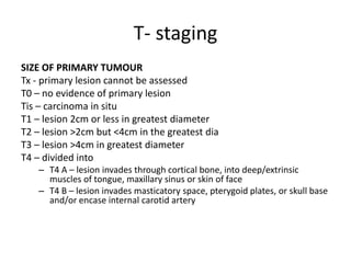
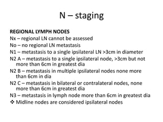
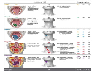
TNM STAGING
DR DAVIS NADAKKAVUKARAN
M.D.S.
ORAL AND MAXILLOFACIAL SUEGEON
MALABAR DENTAL COLLEGE MANOOR
• TNM staging system was first reported by Pierre
Dernoix in 1940’s
• The international union against cancer eventually
adapted the system and compiled first edition of
the TNM staging system in 1968 for 23 body sites
• Simply an anatomical staging system that
describes the anatomic extend of the primary
tumor as well as the involvement of regional
lymph nodes and distant metastasis
• Tumor size and extend of spread are
considered to be the best indicators of the
patient’s prognosis
• This system has three basic clinical features:
– Size of primary lesion
– Presence, number, size, and spread to the local
lymph nodes
– Presence or absence of distant metastasis
T- staging
SIZE OF PRIMARY TUMOUR
Tx - primary lesion cannot be assessed
T0 – no evidence of primary lesion
Tis – carcinoma in situ
T1 – lesion 2cm or less in greatest diameter
T2 – lesion >2cm but <4cm in the greatest dia
T3 – lesion >4cm in greatest diameter
T4 – divided into
– T4 A – lesion invades through cortical bone, into deep/extrinsic
muscles of tongue, maxillary sinus or skin of face
– T4 B – lesion invades masticatory space, pterygoid plates, or skull base
and/or encase internal carotid artery
N – staging
REGIONAL LYMPH NODES
Nx – regional LN cannot be assessed
No – no regional LN metastasis
N1 – metastasis to a single ipsilateral LN >3cm in diameter
N2 A – metastasis to a single ipsilateral node, >3cm but not
more than 6cm in greatest dia
N2 B – metastasis in multiple ipsilateral nodes none more
than 6cm in dia
N2 C – metastasis in bilateral or contralateral nodes, none
more than 6cm in greatest dia
N3 – metastasis in lymph node more than 6cm in greatest dia
 Midline nodes are considered ipsilateral nodes
M- staging
DISTANT METASTASIS
Mx – distant metastasis cannot be assessed
M0 – no distant metastasis
M1 – distant metastasis present
MANAGMENT
• Treatment depends on the stage of cancer at
diagnosis:
Early stage oral cancer: stage I and II - single
modality treatment
Locally advanced and operable cancers : stage III
and IV A – treated with combined modality
treatment
Early stage:
• Radiotherapy : brachytherapy or external
beam radiotherapy
– In brachytherapy, radiation is administered
interstitially through catheters surgically placed
across the lesion
– spares normal tissue such as bone , salivary
gland, and spinal cord
– ideal lesion should be less than 3 cm, superficial
invasion not close to the bone
• Surgical technique of early lesion:
1. Margins : all lesions should be excised with a
margin of atleast 1cm in all dimension.
2. Modality used : excision can be done using
the cautery, laser or knife
• Approaches to oral cavity:
1. Peroral – restricted to anteriorly placed lesion
2. Lipsplit – to raise a cheek flap for posteriorly
based gingivobuccal complex lesions and for
performing marginal mandibulectomy
3. Pull through approach : tongue and floor of
mouth lesion
4. Mandibulotomy - tongue and floor of mouth
close to mandible
• Marginal mandibulectomy :
– involves removal of rim of mandible
– Lesion reaching close to mandible
– Lesion superficially eroding bone
– A margin of .5 to 1cm of the mandible should be
preserved
Locally
advanced
Operable
III/IV A
Surgery followed
by radiotherapy
with or without
chemotherapy
Inoperable
IV B/ IV C
Palliative CT/RT
Palliative RT/CT
Symptomatic care
Management of neck lymph nodes
Node
negative
Early
cancer
T1, T2
Elective neck dissection: observe
Elective neck dissection preferred if :
T2 cancer
Poor follow up
Poor prognostic variables
Thick tumor >4mm
If removal of primary tumor need to
raise a check flap
Locally
advanced
T3, T4
Treatment of neck
mandatory
High chance of
metastasis
Surgery for neck
•Selective neck
dissection(I to III)
•Modified neck
dissection(I to V)
•Never perform
Node positive
Modified neck
dissection
Radical neck
dissection
Stage III and IV
High risk lesions:
T4 lesion
Multiple node positive
Perinodal extension
Cut margins positive
Concurrent chemotherapy
Cisplatin-100mg/m2 3weekly or
30-40mg/m2 weekly
Low risk lesions
Radiotherapy alone
57/60 gy 30 fractions
/6 week
5 tnm staging
5 tnm staging

5 tnm staging

  • 1.
    TNM STAGING DR DAVISNADAKKAVUKARAN M.D.S. ORAL AND MAXILLOFACIAL SUEGEON MALABAR DENTAL COLLEGE MANOOR
  • 2.
    • TNM stagingsystem was first reported by Pierre Dernoix in 1940’s • The international union against cancer eventually adapted the system and compiled first edition of the TNM staging system in 1968 for 23 body sites • Simply an anatomical staging system that describes the anatomic extend of the primary tumor as well as the involvement of regional lymph nodes and distant metastasis
  • 3.
    • Tumor sizeand extend of spread are considered to be the best indicators of the patient’s prognosis • This system has three basic clinical features: – Size of primary lesion – Presence, number, size, and spread to the local lymph nodes – Presence or absence of distant metastasis
  • 4.
    T- staging SIZE OFPRIMARY TUMOUR Tx - primary lesion cannot be assessed T0 – no evidence of primary lesion Tis – carcinoma in situ T1 – lesion 2cm or less in greatest diameter T2 – lesion >2cm but <4cm in the greatest dia T3 – lesion >4cm in greatest diameter T4 – divided into – T4 A – lesion invades through cortical bone, into deep/extrinsic muscles of tongue, maxillary sinus or skin of face – T4 B – lesion invades masticatory space, pterygoid plates, or skull base and/or encase internal carotid artery
  • 6.
    N – staging REGIONALLYMPH NODES Nx – regional LN cannot be assessed No – no regional LN metastasis N1 – metastasis to a single ipsilateral LN >3cm in diameter N2 A – metastasis to a single ipsilateral node, >3cm but not more than 6cm in greatest dia N2 B – metastasis in multiple ipsilateral nodes none more than 6cm in dia N2 C – metastasis in bilateral or contralateral nodes, none more than 6cm in greatest dia N3 – metastasis in lymph node more than 6cm in greatest dia  Midline nodes are considered ipsilateral nodes
  • 8.
    M- staging DISTANT METASTASIS Mx– distant metastasis cannot be assessed M0 – no distant metastasis M1 – distant metastasis present
  • 10.
    MANAGMENT • Treatment dependson the stage of cancer at diagnosis: Early stage oral cancer: stage I and II - single modality treatment Locally advanced and operable cancers : stage III and IV A – treated with combined modality treatment
  • 11.
    Early stage: • Radiotherapy: brachytherapy or external beam radiotherapy – In brachytherapy, radiation is administered interstitially through catheters surgically placed across the lesion – spares normal tissue such as bone , salivary gland, and spinal cord – ideal lesion should be less than 3 cm, superficial invasion not close to the bone
  • 12.
    • Surgical techniqueof early lesion: 1. Margins : all lesions should be excised with a margin of atleast 1cm in all dimension. 2. Modality used : excision can be done using the cautery, laser or knife
  • 13.
    • Approaches tooral cavity: 1. Peroral – restricted to anteriorly placed lesion 2. Lipsplit – to raise a cheek flap for posteriorly based gingivobuccal complex lesions and for performing marginal mandibulectomy 3. Pull through approach : tongue and floor of mouth lesion 4. Mandibulotomy - tongue and floor of mouth close to mandible
  • 14.
    • Marginal mandibulectomy: – involves removal of rim of mandible – Lesion reaching close to mandible – Lesion superficially eroding bone – A margin of .5 to 1cm of the mandible should be preserved
  • 15.
    Locally advanced Operable III/IV A Surgery followed byradiotherapy with or without chemotherapy Inoperable IV B/ IV C Palliative CT/RT Palliative RT/CT Symptomatic care
  • 16.
    Management of necklymph nodes
  • 17.
    Node negative Early cancer T1, T2 Elective neckdissection: observe Elective neck dissection preferred if : T2 cancer Poor follow up Poor prognostic variables Thick tumor >4mm If removal of primary tumor need to raise a check flap Locally advanced T3, T4 Treatment of neck mandatory High chance of metastasis Surgery for neck •Selective neck dissection(I to III) •Modified neck dissection(I to V) •Never perform
  • 18.
  • 19.
    Stage III andIV High risk lesions: T4 lesion Multiple node positive Perinodal extension Cut margins positive Concurrent chemotherapy Cisplatin-100mg/m2 3weekly or 30-40mg/m2 weekly Low risk lesions Radiotherapy alone 57/60 gy 30 fractions /6 week