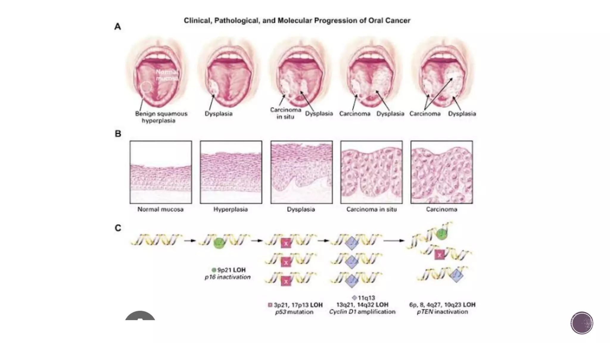
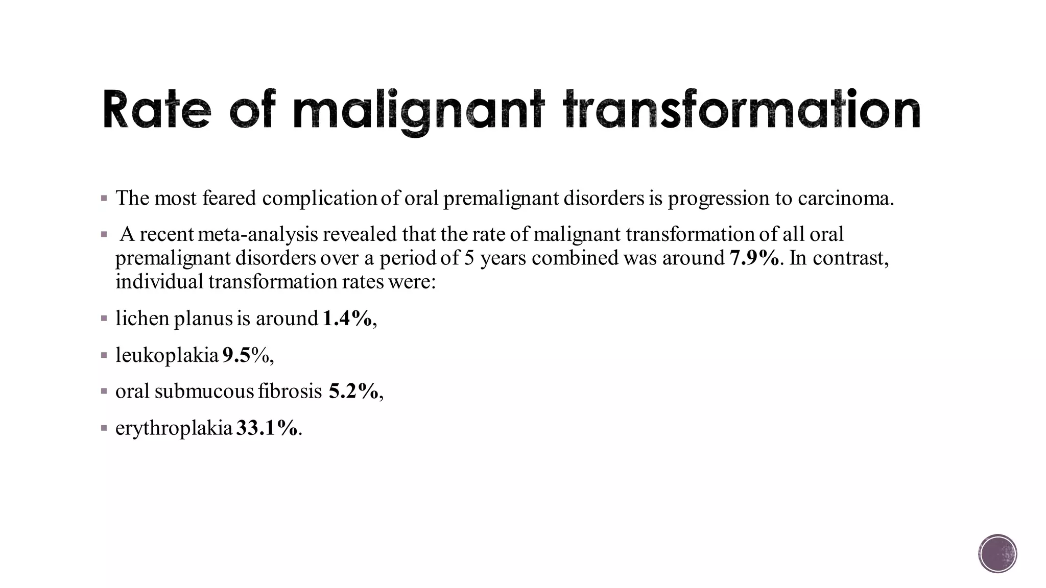
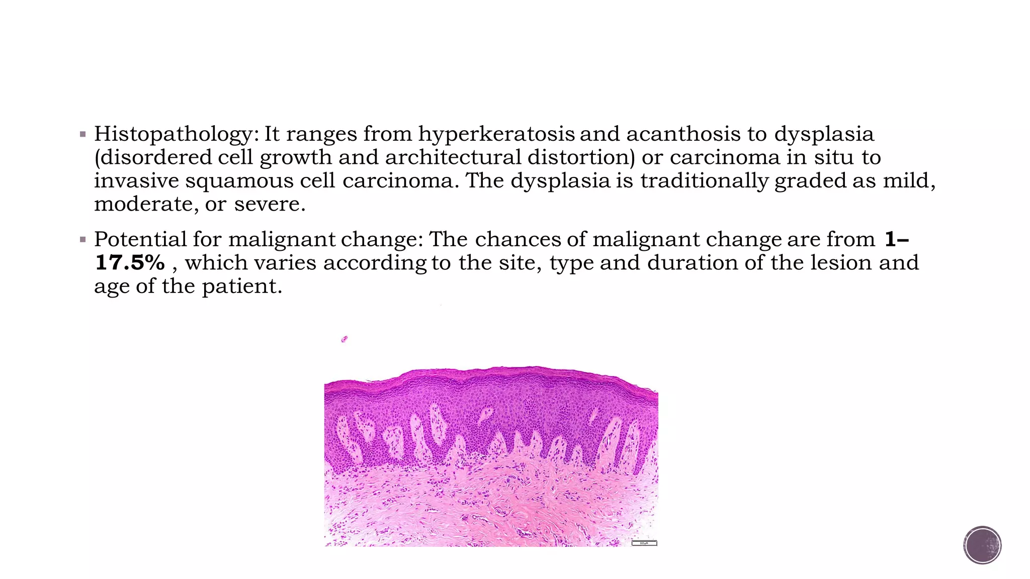
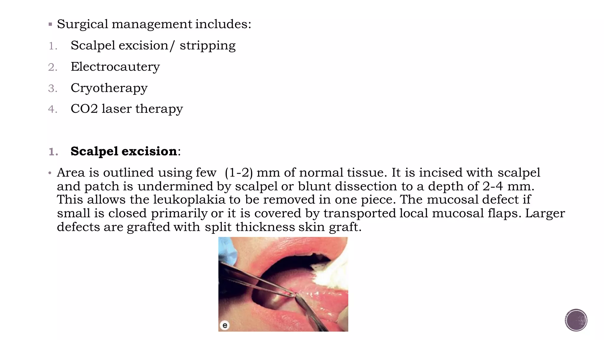
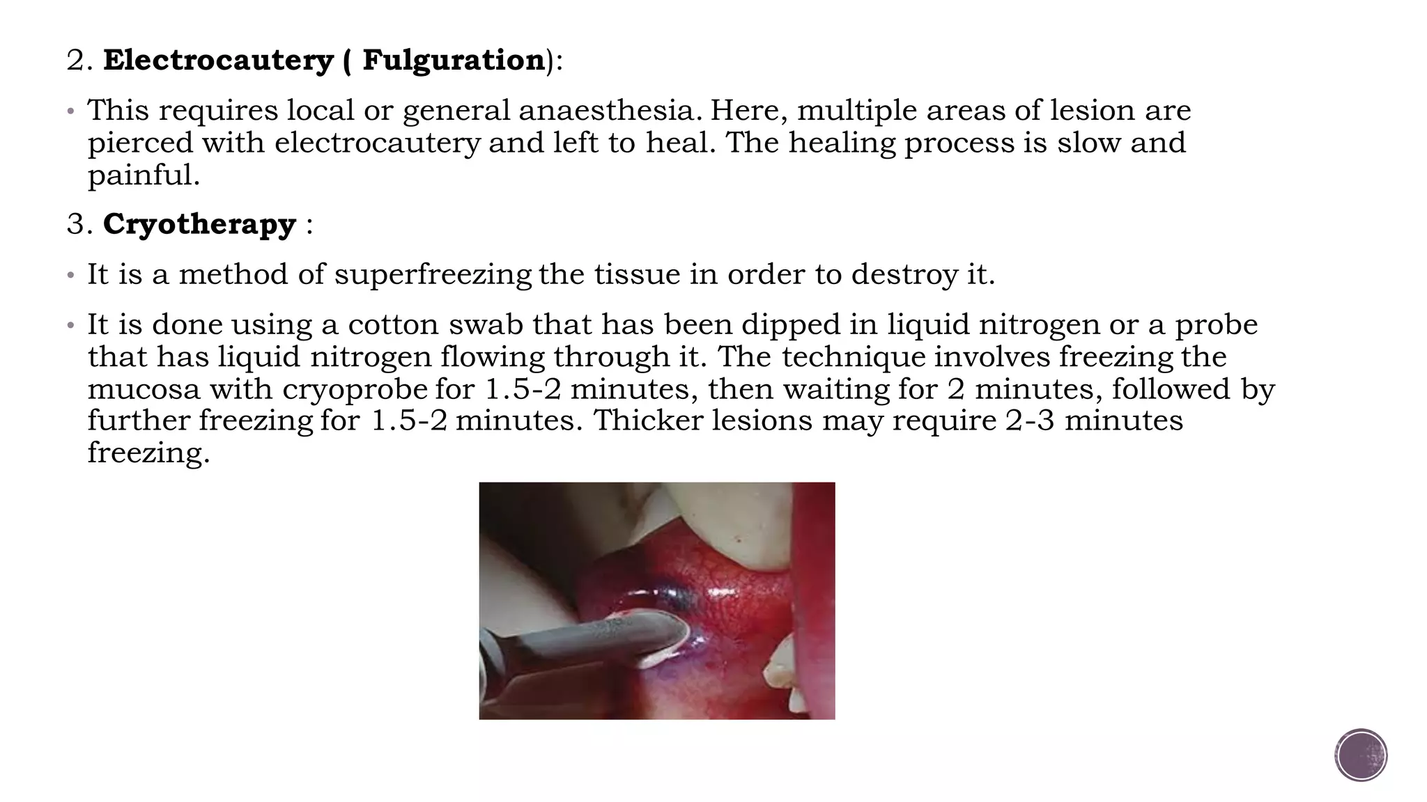
Chairperson: Dr. Ravi D
Moderator: Dr. Balaji NK
Presenter: Dr. Shreya Sinha
▪ Introduction
▪ Premalignant lesions Vs premalignant conditions
▪ Progression of oral cancer
▪ Rate of malignant transformation
▪ Leukoplakia
▪ Erythroplakia
▪ Oral Submucous fibrosis
▪ Oral Lichen planus
▪ Candidiasis
▪ Actinic chelitis
▪ Screening protocol
▪ Others
▪ These are lesions and conditions that are not malignant but have the
potential to become malignant.
▪ Stratified squamous epithelium lines the majority of the oral cavity, and
aberrancy in the epithelium gives rise to premalignant lesions.
▪ Previously, various terms like Pre-cancerous, Pre-malignant, Intra epithelial
dysplasia, Intra epithelial neoplasia were used.
▪ Recently, a more precise phrase, “Oral potentially malignant disorders” has
been adopted by the WHO collaborating center.
Pre-malignant lesions
▪ A morphologically altered tissue in
which malignancy is more likely to
occur than its apparently normal
counterpart.
▪ Leukoplakia
▪ Erythroplakia
▪ Submucous fibrosis
▪ Lichen planus
▪ Actinic chelitis
Pre-malignant conditions
▪ A generalised state associated with a
significantly increased risk of
malignancy.
▪ Plummer Vinson syndrome
▪ Syphilis
▪ The most feared complicationof oral premalignant disorders is progression to carcinoma.
▪ A recent meta-analysis revealed that the rate of malignant transformation of all oral
premalignant disorders over a period of 5 years combined was around 7.9%. In contrast,
individual transformation rates were:
▪ lichen planusis around 1.4%,
▪ leukoplakia 9.5%,
▪ oral submucousfibrosis 5.2%,
▪ erythroplakia 33.1%.
▪ It refers to a white patch in the mucosa that does not rub off and cannot be
clinically identified as another entity.
▪ It is a diagnosis of exclusion.
▪ Risk factors include: chronic smoking, tobacco abuse, areca nut and betel
nut chewing, alcohol abuse, chronic trauma (friction induced hyperkeratosis),
chronic sun exposure, sanguinaria (herbal root extract in some mouthwashes
and toothpaste), male sex, people in fourth decade of life, etc.
▪ Clinical forms are:
1. Homogenous leukoplakia: There is a smooth or wrinkled white patch, well
demarcated, has a consistent pattern throughout and is less often associated
with malignancy.
2. Nodular (Speckled) leukoplakia: There are nodular white patches with
erythematous base.
3. Erosive leukoplakia (Erythroleukoplakia) presents white patches which has
erosions and fissures and is interspersed with red patches (erythroplakia).
4. Proliferative verrucous leukoplakia: This uncommon variant of leukoplakia
is multifocal and persistent and occurs more often in women. A thin flat white
patch progresses to leathery thickened and papillary to verrucous appearance.
homogenous homogenous speckled
erythroleukoplakia proliferative
verrucous
▪ Histopathology: It ranges from hyperkeratosis and acanthosis to dysplasia
(disordered cell growth and architectural distortion) or carcinoma in situ to
invasive squamous cell carcinoma. The dysplasia is traditionally graded as mild,
moderate, or severe.
▪ Potential for malignant change: The chances of malignant change are from 1–
17.5% , which varies according to the site, type and duration of the lesion and
age of the patient.
▪ Risk factors associated with malignant transformation of oral leukoplakia are:
1. Female sex
2. Long standing leukoplakia
3. Leukoplakia in non smokers
4. Leukoplakia over tongue or floor of mouth
5. Size >200 mm2
6. Non homogenous type of leukoplakia
7. Leukoplakia with presence of epithelial dysplasia
A clinical staging system (OL system) for oral leukoplakia on the lines of TNM
staging was recommended by WHO in 2005, taking the size (L) and histopathological
features (P) into consideration.
▪ Management:
1. Biopsy: An incisional biopsy (multiple biopsies in extensive lesions) must be taken
from suspicious areas (such as erythematous, granular, ulcerated and indurated)
to know the grades of dysplasia and rule out malignancy.
2. Medical management
a) L-ascorbic acid (vitamin C)
b) a-tocoferol (vitamin E)
c) Retinoic acid (vitamin A)
d) Vitamin A derivative, isotretinoin and 13-cis retinoic acid: 28,500 IU per day.
e) Beta carotene : 150,000 IU of beta carotene twice per week for 6 months.
f) Bleomycin: topical bleomycin has been used in the treatment of oral leukoplakia in
dosage of 0.5% per day for 12-15 days or 1% per day for 14 days. It prevents the
progression though dysplasia to carcinoma. Bleomycin is a glycopeptide antibiotic
that acts by binding to guanosine-cytosine rich portion of the DNA via association
of the S tripeptide and by partial intercalation of the bithiazole rings.
▪ Surgical management includes:
1. Scalpel excision/ stripping
2. Electrocautery
3. Cryotherapy
4. CO2 laser therapy
1. Scalpel excision:
• Area is outlined using few (1-2) mm of normal tissue. It is incised with scalpel
and patch is undermined by scalpel or blunt dissection to a depth of 2-4 mm.
This allows the leukoplakia to be removed in one piece. The mucosal defect if
small is closed primarily or it is covered by transported local mucosal flaps. Larger
defects are grafted with split thickness skin graft.
2. Electrocautery ( Fulguration):
• This requires local or general anaesthesia. Here, multiple areas of lesion are
pierced with electrocautery and left to heal. The healing process is slow and
painful.
3. Cryotherapy :
• It is a method of superfreezing the tissue in order to destroy it.
• It is done using a cotton swab that has been dipped in liquid nitrogen or a probe
that has liquid nitrogen flowing through it. The technique involves freezing the
mucosa with cryoprobe for 1.5-2 minutes, then waiting for 2 minutes, followed by
further freezing for 1.5-2 minutes. Thicker lesions may require 2-3 minutes
freezing.
4. CO2 Laser therapy:
• CO2 laser beam wavelength – 10.6 micro meter
• Well absorbed by water and hence by soft tissues
• The absorbed energy causes vaporisation of the intra and
extracellular fluid and destruction of cell membrane. The cell debris
are released and burned in the laser beam, depositing a carbonised
layer on the tissue surfaces.
• Two techniques are used to remove the leukoplakia using CO2 laser:
a) Excision- to excise a patch, the laser is used to cut around the
margins, which can be held in tissue forceps while the laser
undermines the leukoplakic patch.
b) Vaporisation- it is done by moving the laser beam back and forth
across the surface of the lesion. It has the risk of leaving small bits
of abnormal tissue which are deep under thickly keratinised
tissue.
Surgical excision of lesions using laser offers several advantages over scalpel
excision which includes
▪ bloodless surgical and postsurgical events;
▪ the ability to precisely coagulate, vaporise, or cut tissue;
▪ minimal swelling and scarring;
▪ reduction of surgical time, postsurgical pain with high patient acceptance.
▪ Differentia diagnosis:
Linea alba buccalis Leukoedema
Fordyce granules Traumatic keratosis
Geographic tongue
▪ It is a fiery red patch that cannot be clinically or pathologically classified as
another entity.
▪ Also known as Erythroplasia of Queyart.
▪ These lesions may have a flat, macular, velvety appearance and may be speckled
with white spots representing foci of keratosis.
▪ It is much less common than leukoplakia, but has a much greater probability
(91%) of showing signs of dysplasia or malignancy at the time of diagnosis.
▪ The rate of malignant transformation over a period of 5 years was found to be
33.1%
▪ Differential diagnosis:
1. Erythematous (atrophic) candidiasis
2. Kaposi’s sarcoma
3. Contact stomatitis
4. Vascular malformation
5. Squamous cell carcinoma
6. Geographic tongue/ erythema migrans
▪ Management:
• It is the same as that for Invasive carcinoma or Carcinoma in-situ and
includes surgery, radiation and cauterisation.
• Surgical excision if proven dysplastic /malignant.
▪ It is an insidious painless oral cavity disease, which is characterized by juxta
epithelial inflammatory reaction, followed by fibro-elastic change of lamina
propria with epithelial atrophy, leading to stiffness of oral mucosa and
causing trismus and inability to eat.
▪ It appears as white fibrotic bands extending from retromolar trigone to soft
palate, buccal mucosa, tongue and pharynx.
▪ It is common in Indian population. Incidence is 0.2-0.5 %.
▪ Etiology:
1. Prolonged local irritation due to paan, betel nut, tobacco, smoking of cigarette
or bidis, excessive amount of spices and chillies in food.
2. Dietary deficiency of iron, vitamin B-complex and vitamin A .
3. Cell mediated immune process: Some consider it a cell mediated immune
reaction to arecoline.
4. Racial: Disease usually affects Indians or people of Indian origin living abroad.
▪ Pathogenesis:
oral mucosa
Betel quid habit
Constant irritation
Chronic inflammation
Activated T cell and macrophages at the site
Increase in cytokines- IL6, TNF, IFN-a
Increse in growth factors- TGF-B
Increased susceptibility due to
deficiency of iron & vitamin B12
cytokines
Increase collagen
production
Decrease collagen
degradation
Increased collagen
(insoluble form – Cross
linking of insoluble collagen)
Fibrosis
Oral submucous fibrosis
▪ The malignant transformation has been observed in 3–7.6% of cases.
▪ Initial stage-
• Burning sensation on eating spicy
food
• Blisters or vesicles on the palate
• Ulceration or recurrent stomatitis
• Excessive salivation
• Dryness of mouth
▪ Later stage-
• Difficulty in opening mouth
• Inability to whistle or blow
• Difficulty swallowing
• Referred pain to the ear
• Change in voice tone due to vocal
cord involvement
• Deafness due to occlusuion of
eustachian tube
▪ Clinical features:
▪ Clinical and functional staging:
❑ Clinical staging:
Stage 1- faucial bands only
Stage 2- faucial and buccal bands
Stage 3- faucial, buccal and labial bands
❑Functional staging:
A- mouth opening >= 20 mm
B- mouth opening 11-19 mm
C- mouth opening <= 10 mm
▪ Management:
1. Local steroids/hyluronidase: Topical injection of steroids, which may be
combined with hyluronidase, into the area of fibrous bands (injection
dexamethasone 4 mg and hylase 1500 IU in one ml, intraoral submucosal
biweekly at different sites for 8–10 weeks). This brings significant improvement
in symptoms and relieves trismus. Steroids suppress inflammatory
reactions, thereby preventing fibrosis by decreasing fibroblastic proliferation
and deposition of collagen.
2. Use of Placentrix 2ml solution at interval of 3 days.
3. Avoidance of irritant factors
4. Systemic immunomodulators:
• Levamisole 150mg for 3 weeks orally
• Dapsone 75mg OD for 90 days orally
5. Vitamins and minerals: Treatment of existent anemia or vitamin
deficiencies. Vitamin A, zinc and antioxidants therapy has shown some
beneficial effect. They are effective in controlling the burning sensation and
ulceration in OSMF
6. Physiotherapy (jaw opening exercises)
▪ Surgical management:
Fibrotomy (scalpel, electrocautery, laser)
Coronoidectomy and Temporalis myotomy
Extraction of 3rd molars
reconstruction
▪ Grafts and flaps tried for reconstruction in the
management of oral submucous fibrosis:
• Bilateral tongue flap
• Nasolabial flaps (most common)
• Island palatal mucoperiosteal flap
• Bilateral radial forearm free flap
• Buccal pad of fat graft
• Temporalis fascia graft
• Split skin graft
▪ It is a mucocutaneous immunologic disorder that occurs due to T-cell
lymphocytic reaction to surface epithelial antigen.
▪ Oral lesions may present in various forms such as reticular, plaque, atrophic,
erosive and bullous. Concomitant presence of various forms is not uncommon.
▪ Premalignant nature of this lesion is still controversial.
▪ In cases of erosive lichen planus or atrophic lichen planus, there is risk of
malignant change.
▪ Risk factors include:
Autoimmunity, genetic background, dentures, infectious agents like
Hepatitis C virus, bowel diseases like Chron’s and Ulcerative colitis, stress,
diabetes and hypertension.
▪ Pathogenesis
unknown antigen
antigen presenting cells
CD 4 lymphocytes
Interferon gamma produced
Basal cell degeneration with sub-epithelial
lymphocytic infiltration
▪ Treatment:
1. Corticosteroids: Topical, intralesional injections and systemic.
2. Other alternatives: immunomodulatory drugs like Hydroxychloroquine,
azathioprine and retinoids for at least 3-6 months.
3. Surgical: Resection of the lesion, lasers and cryotherapy.
▪ Infection caused by Candida albicans has two forms: thrush and chronic
hypertrophic candidiasis.
▪ Certain strains of Candida have been shown to produce Nitrosamines
(chemical carcinogen), hence these lesions carry a risk of malignant
transformation.
▪ Risk factors:
1. Systemic: Diabetes, antibiotics, age and immunosuppression.
2. Local: Corticosteroids, xerostomia, heavy smoking and dentures.
▪ Clinical features: Symptoms range from none to burning, dysgeusia,
sensitivity and generalized discomfort. Odynophagia indicates involvement of
pharynx.
Acute Pseudomembranous
Candidiasis (Thrush)
Chronic Hyperplastic
Candidiasis
▪ Diagnosis: Microscopic evaluation of lesion smears-
1. Potassium hydroxide preparation to demonstrate hyphae
2. Periodic acid Schiff (PAS) stain
3. Culture on Sabouraud’s corn meal or Potato agar medium
4. Biopsy with PAS, Gomori’s methenamine silver(GMS), or other fungal stains of
microscopic sections.
▪ Treatment:
1. Topical therapy-
• Nystatin oral suspension (100,000 units/mL); rinse 5 ml and swallow 4 times
per day
• Clotrimazole solution 1% ; rinse 5 mL and swallow 4 times per day
2. Systemic therapy-
• Fluconazole 100mg, 2 tablets on day 1, followed by 1 tablet on days 2-7, and
1 tablet every other day for days 8-21
• Ketoconazole 200mg, 1 tablet per day for 21 days
▪ It is a premalignant lesion that affects the vermilion border of the lower lip.
▪ It usually results due to chronic sun exposure or UV radiation exposure.
▪ In early stage, redness and swelling of the lips is found alongwith
desquamation of epithelium.
▪ As the disease progresses, erosions and scrub covered ulcers may develop.
▪ Treatment includes:
1. Topical: 5-fluorouracil, Imiquimod, trichloroacetic acid, etc.
2. Laser treatment
3. Vermilionectomy or surgical excision
4. Phototherapy
▪ Some other lesions are:
1. Palatal lesions in reverse smokers
2. Hereditary conditions like Dyskeratosis congenita and Epidermolysis
bullosa.
3. In the year 2007, the WHO and the Cancer meetings also decided to
include the Oral manifestations of Graft Versus Host Disease and
Lichenoid lesions like Lichen pemphigoid, Oral lichen planus, Atypical
lichenoid stomatitis, lichenoid dysplasia, etc. under potentially
malignant disorders of oral cavity.
▪ It is recommended that all adult patients should be screened annually even if
medical and dental histories elicit no risk factors. Known at risk patients
should be screened every 6 months.
1. Biopsy protocol
• Apart from incisional and excisional biopsy used for definitive diagnosis of
oral cancers, Brush biopsy is another technique that can be used as a
preliminary diagnostic tool.
• It uses a stiff brush to obtain a full thickness sampling of epithelial cells for
examination in patients with mucosal lesions.
• It is a simple, relatively inexpensive, high sensitive and less invasive to
perform, but the existence of false positives have been pointed out in this
method.
• Studies have suggested that reduced nuclear size and increased cytoplasm
size are useful early indicators of malignant transformation, and thus
exfoliative cytology is of value for monitoring clinically suspected lesions.
2. Toluidine blue staining
• Vital staining with toluidine blue (as a 1% rinse or application) has been
suggested as a means of surveillance in patients at risk of developing oral
cancer, and for those who have had a confirmed neoplasm on other parts of
aerodigestive tract.
• It is most useful in secondary care for delineating the extent of lesions and for
surveillance of patients at risk of recurrent disease.
3. Autofluorescence
• This phenomenon is based on the interaction of various fluorescent tissue
compounds (fluorophores) that occur naturally in the body. When excited by an
appropriate light stimulus, these compounds emit visible fluorescent light in the
violet to green region of the spectrum.
• VELscope is a portable device comprising of light source and a viewing hand piece.
The technology is based on direct visualisation of tissue fluorescence.
• Both keratinised and nonkeratinized squamous epithelium with a normal
submucosa show a typical homogenous, pale green fluorescence. Sharply
circumscribed areas of decreased auto fluorescence might indicate areas of
musosal abnormalities that should be monitored or investigated by tissue biopsy.
• Microlux is a handheld device that used light emitting diodes( LEDs) as the
elimination source. Prior to exposing the mucosa to light, the patient rinses for 30-
60 seconds with 1% acetic acid. Upon illumination, abnormal tissue will appear
white( aceto-white).
4. Salivary biomarkers
• Abnormal DNA, RNA and protein molecules released by the malignant cells can be
easily obtained from the saliva.
• It includes :
a) Non organic compounds- Na, Ca, F, Mg
b) Peptides- Defensin 1
c) Proteins- IL-1, IL-6, IL-8, TNF-a, Endothelin-1
d) DNAs- p53 gene codon 63
e) mRNAs- IL-1B, H3F3A, etc
f) Oxidative stress related molecules- glutathione, peroxidase
g) Metabolites- lactic acid, valine, etc
h)Glycosylation related molecules- sialic acid, a-L-fucosidase, etc
Linea alba buccalis leukoedema Fordyce granules
Traumatic keratosis
Geographic tongue
Oral Potentially Malignant Disorders

Oral Potentially Malignant Disorders

  • 1.
    Chairperson: Dr. RaviD Moderator: Dr. Balaji NK Presenter: Dr. Shreya Sinha
  • 2.
    ▪ Introduction ▪ Premalignantlesions Vs premalignant conditions ▪ Progression of oral cancer ▪ Rate of malignant transformation ▪ Leukoplakia ▪ Erythroplakia ▪ Oral Submucous fibrosis ▪ Oral Lichen planus ▪ Candidiasis ▪ Actinic chelitis ▪ Screening protocol ▪ Others
  • 3.
    ▪ These arelesions and conditions that are not malignant but have the potential to become malignant. ▪ Stratified squamous epithelium lines the majority of the oral cavity, and aberrancy in the epithelium gives rise to premalignant lesions. ▪ Previously, various terms like Pre-cancerous, Pre-malignant, Intra epithelial dysplasia, Intra epithelial neoplasia were used. ▪ Recently, a more precise phrase, “Oral potentially malignant disorders” has been adopted by the WHO collaborating center.
  • 4.
    Pre-malignant lesions ▪ Amorphologically altered tissue in which malignancy is more likely to occur than its apparently normal counterpart. ▪ Leukoplakia ▪ Erythroplakia ▪ Submucous fibrosis ▪ Lichen planus ▪ Actinic chelitis Pre-malignant conditions ▪ A generalised state associated with a significantly increased risk of malignancy. ▪ Plummer Vinson syndrome ▪ Syphilis
  • 7.
    ▪ The mostfeared complicationof oral premalignant disorders is progression to carcinoma. ▪ A recent meta-analysis revealed that the rate of malignant transformation of all oral premalignant disorders over a period of 5 years combined was around 7.9%. In contrast, individual transformation rates were: ▪ lichen planusis around 1.4%, ▪ leukoplakia 9.5%, ▪ oral submucousfibrosis 5.2%, ▪ erythroplakia 33.1%.
  • 8.
    ▪ It refersto a white patch in the mucosa that does not rub off and cannot be clinically identified as another entity. ▪ It is a diagnosis of exclusion. ▪ Risk factors include: chronic smoking, tobacco abuse, areca nut and betel nut chewing, alcohol abuse, chronic trauma (friction induced hyperkeratosis), chronic sun exposure, sanguinaria (herbal root extract in some mouthwashes and toothpaste), male sex, people in fourth decade of life, etc.
  • 9.
    ▪ Clinical formsare: 1. Homogenous leukoplakia: There is a smooth or wrinkled white patch, well demarcated, has a consistent pattern throughout and is less often associated with malignancy. 2. Nodular (Speckled) leukoplakia: There are nodular white patches with erythematous base. 3. Erosive leukoplakia (Erythroleukoplakia) presents white patches which has erosions and fissures and is interspersed with red patches (erythroplakia). 4. Proliferative verrucous leukoplakia: This uncommon variant of leukoplakia is multifocal and persistent and occurs more often in women. A thin flat white patch progresses to leathery thickened and papillary to verrucous appearance.
  • 10.
  • 11.
    ▪ Histopathology: Itranges from hyperkeratosis and acanthosis to dysplasia (disordered cell growth and architectural distortion) or carcinoma in situ to invasive squamous cell carcinoma. The dysplasia is traditionally graded as mild, moderate, or severe. ▪ Potential for malignant change: The chances of malignant change are from 1– 17.5% , which varies according to the site, type and duration of the lesion and age of the patient.
  • 12.
    ▪ Risk factorsassociated with malignant transformation of oral leukoplakia are: 1. Female sex 2. Long standing leukoplakia 3. Leukoplakia in non smokers 4. Leukoplakia over tongue or floor of mouth 5. Size >200 mm2 6. Non homogenous type of leukoplakia 7. Leukoplakia with presence of epithelial dysplasia
  • 13.
    A clinical stagingsystem (OL system) for oral leukoplakia on the lines of TNM staging was recommended by WHO in 2005, taking the size (L) and histopathological features (P) into consideration.
  • 14.
    ▪ Management: 1. Biopsy:An incisional biopsy (multiple biopsies in extensive lesions) must be taken from suspicious areas (such as erythematous, granular, ulcerated and indurated) to know the grades of dysplasia and rule out malignancy. 2. Medical management a) L-ascorbic acid (vitamin C) b) a-tocoferol (vitamin E) c) Retinoic acid (vitamin A) d) Vitamin A derivative, isotretinoin and 13-cis retinoic acid: 28,500 IU per day. e) Beta carotene : 150,000 IU of beta carotene twice per week for 6 months. f) Bleomycin: topical bleomycin has been used in the treatment of oral leukoplakia in dosage of 0.5% per day for 12-15 days or 1% per day for 14 days. It prevents the progression though dysplasia to carcinoma. Bleomycin is a glycopeptide antibiotic that acts by binding to guanosine-cytosine rich portion of the DNA via association of the S tripeptide and by partial intercalation of the bithiazole rings.
  • 15.
    ▪ Surgical managementincludes: 1. Scalpel excision/ stripping 2. Electrocautery 3. Cryotherapy 4. CO2 laser therapy 1. Scalpel excision: • Area is outlined using few (1-2) mm of normal tissue. It is incised with scalpel and patch is undermined by scalpel or blunt dissection to a depth of 2-4 mm. This allows the leukoplakia to be removed in one piece. The mucosal defect if small is closed primarily or it is covered by transported local mucosal flaps. Larger defects are grafted with split thickness skin graft.
  • 16.
    2. Electrocautery (Fulguration): • This requires local or general anaesthesia. Here, multiple areas of lesion are pierced with electrocautery and left to heal. The healing process is slow and painful. 3. Cryotherapy : • It is a method of superfreezing the tissue in order to destroy it. • It is done using a cotton swab that has been dipped in liquid nitrogen or a probe that has liquid nitrogen flowing through it. The technique involves freezing the mucosa with cryoprobe for 1.5-2 minutes, then waiting for 2 minutes, followed by further freezing for 1.5-2 minutes. Thicker lesions may require 2-3 minutes freezing.
  • 17.
    4. CO2 Lasertherapy: • CO2 laser beam wavelength – 10.6 micro meter • Well absorbed by water and hence by soft tissues • The absorbed energy causes vaporisation of the intra and extracellular fluid and destruction of cell membrane. The cell debris are released and burned in the laser beam, depositing a carbonised layer on the tissue surfaces. • Two techniques are used to remove the leukoplakia using CO2 laser: a) Excision- to excise a patch, the laser is used to cut around the margins, which can be held in tissue forceps while the laser undermines the leukoplakic patch. b) Vaporisation- it is done by moving the laser beam back and forth across the surface of the lesion. It has the risk of leaving small bits of abnormal tissue which are deep under thickly keratinised tissue.
  • 18.
    Surgical excision oflesions using laser offers several advantages over scalpel excision which includes ▪ bloodless surgical and postsurgical events; ▪ the ability to precisely coagulate, vaporise, or cut tissue; ▪ minimal swelling and scarring; ▪ reduction of surgical time, postsurgical pain with high patient acceptance.
  • 19.
    ▪ Differentia diagnosis: Lineaalba buccalis Leukoedema Fordyce granules Traumatic keratosis
  • 20.
  • 21.
    ▪ It isa fiery red patch that cannot be clinically or pathologically classified as another entity. ▪ Also known as Erythroplasia of Queyart. ▪ These lesions may have a flat, macular, velvety appearance and may be speckled with white spots representing foci of keratosis. ▪ It is much less common than leukoplakia, but has a much greater probability (91%) of showing signs of dysplasia or malignancy at the time of diagnosis. ▪ The rate of malignant transformation over a period of 5 years was found to be 33.1%
  • 23.
    ▪ Differential diagnosis: 1.Erythematous (atrophic) candidiasis 2. Kaposi’s sarcoma 3. Contact stomatitis 4. Vascular malformation 5. Squamous cell carcinoma 6. Geographic tongue/ erythema migrans ▪ Management: • It is the same as that for Invasive carcinoma or Carcinoma in-situ and includes surgery, radiation and cauterisation. • Surgical excision if proven dysplastic /malignant.
  • 24.
    ▪ It isan insidious painless oral cavity disease, which is characterized by juxta epithelial inflammatory reaction, followed by fibro-elastic change of lamina propria with epithelial atrophy, leading to stiffness of oral mucosa and causing trismus and inability to eat. ▪ It appears as white fibrotic bands extending from retromolar trigone to soft palate, buccal mucosa, tongue and pharynx. ▪ It is common in Indian population. Incidence is 0.2-0.5 %.
  • 25.
    ▪ Etiology: 1. Prolongedlocal irritation due to paan, betel nut, tobacco, smoking of cigarette or bidis, excessive amount of spices and chillies in food. 2. Dietary deficiency of iron, vitamin B-complex and vitamin A . 3. Cell mediated immune process: Some consider it a cell mediated immune reaction to arecoline. 4. Racial: Disease usually affects Indians or people of Indian origin living abroad.
  • 26.
    ▪ Pathogenesis: oral mucosa Betelquid habit Constant irritation Chronic inflammation Activated T cell and macrophages at the site Increase in cytokines- IL6, TNF, IFN-a Increse in growth factors- TGF-B Increased susceptibility due to deficiency of iron & vitamin B12
  • 27.
    cytokines Increase collagen production Decrease collagen degradation Increasedcollagen (insoluble form – Cross linking of insoluble collagen) Fibrosis Oral submucous fibrosis
  • 29.
    ▪ The malignanttransformation has been observed in 3–7.6% of cases.
  • 30.
    ▪ Initial stage- •Burning sensation on eating spicy food • Blisters or vesicles on the palate • Ulceration or recurrent stomatitis • Excessive salivation • Dryness of mouth ▪ Later stage- • Difficulty in opening mouth • Inability to whistle or blow • Difficulty swallowing • Referred pain to the ear • Change in voice tone due to vocal cord involvement • Deafness due to occlusuion of eustachian tube ▪ Clinical features:
  • 31.
    ▪ Clinical andfunctional staging: ❑ Clinical staging: Stage 1- faucial bands only Stage 2- faucial and buccal bands Stage 3- faucial, buccal and labial bands ❑Functional staging: A- mouth opening >= 20 mm B- mouth opening 11-19 mm C- mouth opening <= 10 mm
  • 32.
    ▪ Management: 1. Localsteroids/hyluronidase: Topical injection of steroids, which may be combined with hyluronidase, into the area of fibrous bands (injection dexamethasone 4 mg and hylase 1500 IU in one ml, intraoral submucosal biweekly at different sites for 8–10 weeks). This brings significant improvement in symptoms and relieves trismus. Steroids suppress inflammatory reactions, thereby preventing fibrosis by decreasing fibroblastic proliferation and deposition of collagen. 2. Use of Placentrix 2ml solution at interval of 3 days.
  • 33.
    3. Avoidance ofirritant factors 4. Systemic immunomodulators: • Levamisole 150mg for 3 weeks orally • Dapsone 75mg OD for 90 days orally 5. Vitamins and minerals: Treatment of existent anemia or vitamin deficiencies. Vitamin A, zinc and antioxidants therapy has shown some beneficial effect. They are effective in controlling the burning sensation and ulceration in OSMF 6. Physiotherapy (jaw opening exercises)
  • 34.
    ▪ Surgical management: Fibrotomy(scalpel, electrocautery, laser) Coronoidectomy and Temporalis myotomy Extraction of 3rd molars reconstruction
  • 35.
    ▪ Grafts andflaps tried for reconstruction in the management of oral submucous fibrosis: • Bilateral tongue flap • Nasolabial flaps (most common) • Island palatal mucoperiosteal flap • Bilateral radial forearm free flap • Buccal pad of fat graft • Temporalis fascia graft • Split skin graft
  • 36.
    ▪ It isa mucocutaneous immunologic disorder that occurs due to T-cell lymphocytic reaction to surface epithelial antigen. ▪ Oral lesions may present in various forms such as reticular, plaque, atrophic, erosive and bullous. Concomitant presence of various forms is not uncommon. ▪ Premalignant nature of this lesion is still controversial. ▪ In cases of erosive lichen planus or atrophic lichen planus, there is risk of malignant change.
  • 37.
    ▪ Risk factorsinclude: Autoimmunity, genetic background, dentures, infectious agents like Hepatitis C virus, bowel diseases like Chron’s and Ulcerative colitis, stress, diabetes and hypertension. ▪ Pathogenesis unknown antigen antigen presenting cells CD 4 lymphocytes Interferon gamma produced Basal cell degeneration with sub-epithelial lymphocytic infiltration
  • 38.
    ▪ Treatment: 1. Corticosteroids:Topical, intralesional injections and systemic. 2. Other alternatives: immunomodulatory drugs like Hydroxychloroquine, azathioprine and retinoids for at least 3-6 months. 3. Surgical: Resection of the lesion, lasers and cryotherapy.
  • 39.
    ▪ Infection causedby Candida albicans has two forms: thrush and chronic hypertrophic candidiasis. ▪ Certain strains of Candida have been shown to produce Nitrosamines (chemical carcinogen), hence these lesions carry a risk of malignant transformation. ▪ Risk factors: 1. Systemic: Diabetes, antibiotics, age and immunosuppression. 2. Local: Corticosteroids, xerostomia, heavy smoking and dentures. ▪ Clinical features: Symptoms range from none to burning, dysgeusia, sensitivity and generalized discomfort. Odynophagia indicates involvement of pharynx.
  • 40.
  • 41.
    ▪ Diagnosis: Microscopicevaluation of lesion smears- 1. Potassium hydroxide preparation to demonstrate hyphae 2. Periodic acid Schiff (PAS) stain 3. Culture on Sabouraud’s corn meal or Potato agar medium 4. Biopsy with PAS, Gomori’s methenamine silver(GMS), or other fungal stains of microscopic sections. ▪ Treatment: 1. Topical therapy- • Nystatin oral suspension (100,000 units/mL); rinse 5 ml and swallow 4 times per day • Clotrimazole solution 1% ; rinse 5 mL and swallow 4 times per day
  • 42.
    2. Systemic therapy- •Fluconazole 100mg, 2 tablets on day 1, followed by 1 tablet on days 2-7, and 1 tablet every other day for days 8-21 • Ketoconazole 200mg, 1 tablet per day for 21 days
  • 43.
    ▪ It isa premalignant lesion that affects the vermilion border of the lower lip. ▪ It usually results due to chronic sun exposure or UV radiation exposure. ▪ In early stage, redness and swelling of the lips is found alongwith desquamation of epithelium. ▪ As the disease progresses, erosions and scrub covered ulcers may develop.
  • 45.
    ▪ Treatment includes: 1.Topical: 5-fluorouracil, Imiquimod, trichloroacetic acid, etc. 2. Laser treatment 3. Vermilionectomy or surgical excision 4. Phototherapy
  • 46.
    ▪ Some otherlesions are: 1. Palatal lesions in reverse smokers 2. Hereditary conditions like Dyskeratosis congenita and Epidermolysis bullosa. 3. In the year 2007, the WHO and the Cancer meetings also decided to include the Oral manifestations of Graft Versus Host Disease and Lichenoid lesions like Lichen pemphigoid, Oral lichen planus, Atypical lichenoid stomatitis, lichenoid dysplasia, etc. under potentially malignant disorders of oral cavity.
  • 47.
    ▪ It isrecommended that all adult patients should be screened annually even if medical and dental histories elicit no risk factors. Known at risk patients should be screened every 6 months. 1. Biopsy protocol • Apart from incisional and excisional biopsy used for definitive diagnosis of oral cancers, Brush biopsy is another technique that can be used as a preliminary diagnostic tool. • It uses a stiff brush to obtain a full thickness sampling of epithelial cells for examination in patients with mucosal lesions. • It is a simple, relatively inexpensive, high sensitive and less invasive to perform, but the existence of false positives have been pointed out in this method.
  • 48.
    • Studies havesuggested that reduced nuclear size and increased cytoplasm size are useful early indicators of malignant transformation, and thus exfoliative cytology is of value for monitoring clinically suspected lesions. 2. Toluidine blue staining • Vital staining with toluidine blue (as a 1% rinse or application) has been suggested as a means of surveillance in patients at risk of developing oral cancer, and for those who have had a confirmed neoplasm on other parts of aerodigestive tract. • It is most useful in secondary care for delineating the extent of lesions and for surveillance of patients at risk of recurrent disease.
  • 49.
    3. Autofluorescence • Thisphenomenon is based on the interaction of various fluorescent tissue compounds (fluorophores) that occur naturally in the body. When excited by an appropriate light stimulus, these compounds emit visible fluorescent light in the violet to green region of the spectrum. • VELscope is a portable device comprising of light source and a viewing hand piece. The technology is based on direct visualisation of tissue fluorescence. • Both keratinised and nonkeratinized squamous epithelium with a normal submucosa show a typical homogenous, pale green fluorescence. Sharply circumscribed areas of decreased auto fluorescence might indicate areas of musosal abnormalities that should be monitored or investigated by tissue biopsy.
  • 50.
    • Microlux isa handheld device that used light emitting diodes( LEDs) as the elimination source. Prior to exposing the mucosa to light, the patient rinses for 30- 60 seconds with 1% acetic acid. Upon illumination, abnormal tissue will appear white( aceto-white). 4. Salivary biomarkers • Abnormal DNA, RNA and protein molecules released by the malignant cells can be easily obtained from the saliva. • It includes : a) Non organic compounds- Na, Ca, F, Mg b) Peptides- Defensin 1 c) Proteins- IL-1, IL-6, IL-8, TNF-a, Endothelin-1
  • 51.
    d) DNAs- p53gene codon 63 e) mRNAs- IL-1B, H3F3A, etc f) Oxidative stress related molecules- glutathione, peroxidase g) Metabolites- lactic acid, valine, etc h)Glycosylation related molecules- sialic acid, a-L-fucosidase, etc
  • 52.
    Linea alba buccalisleukoedema Fordyce granules
  • 53.