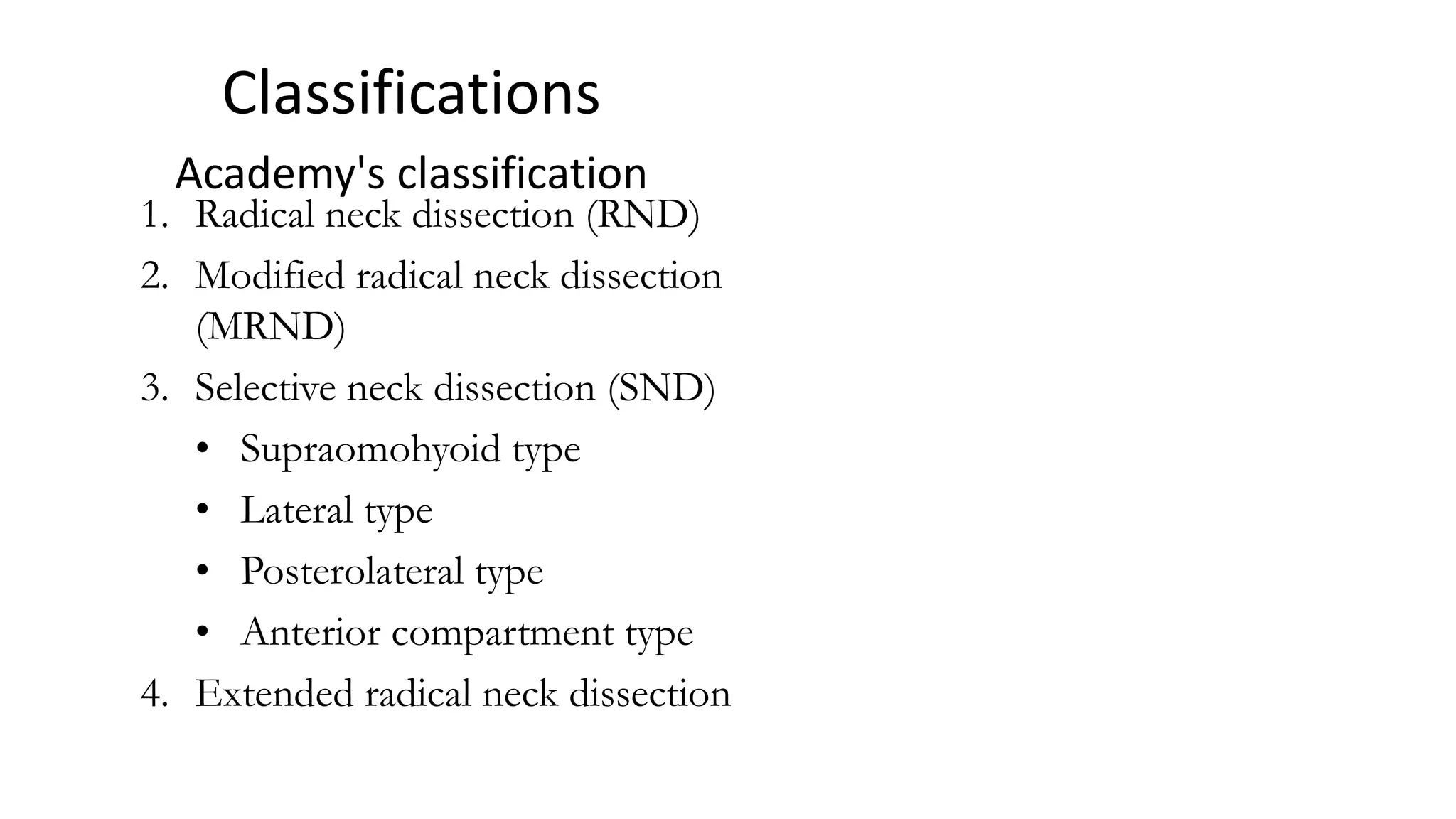
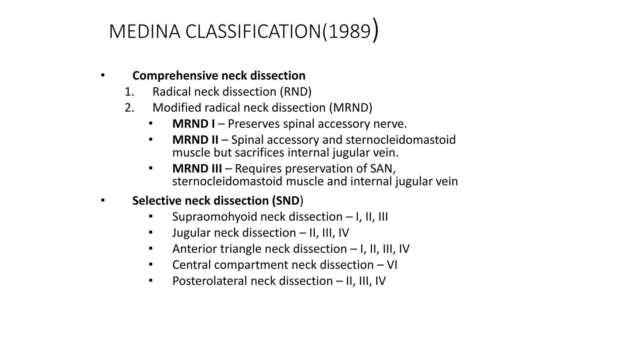
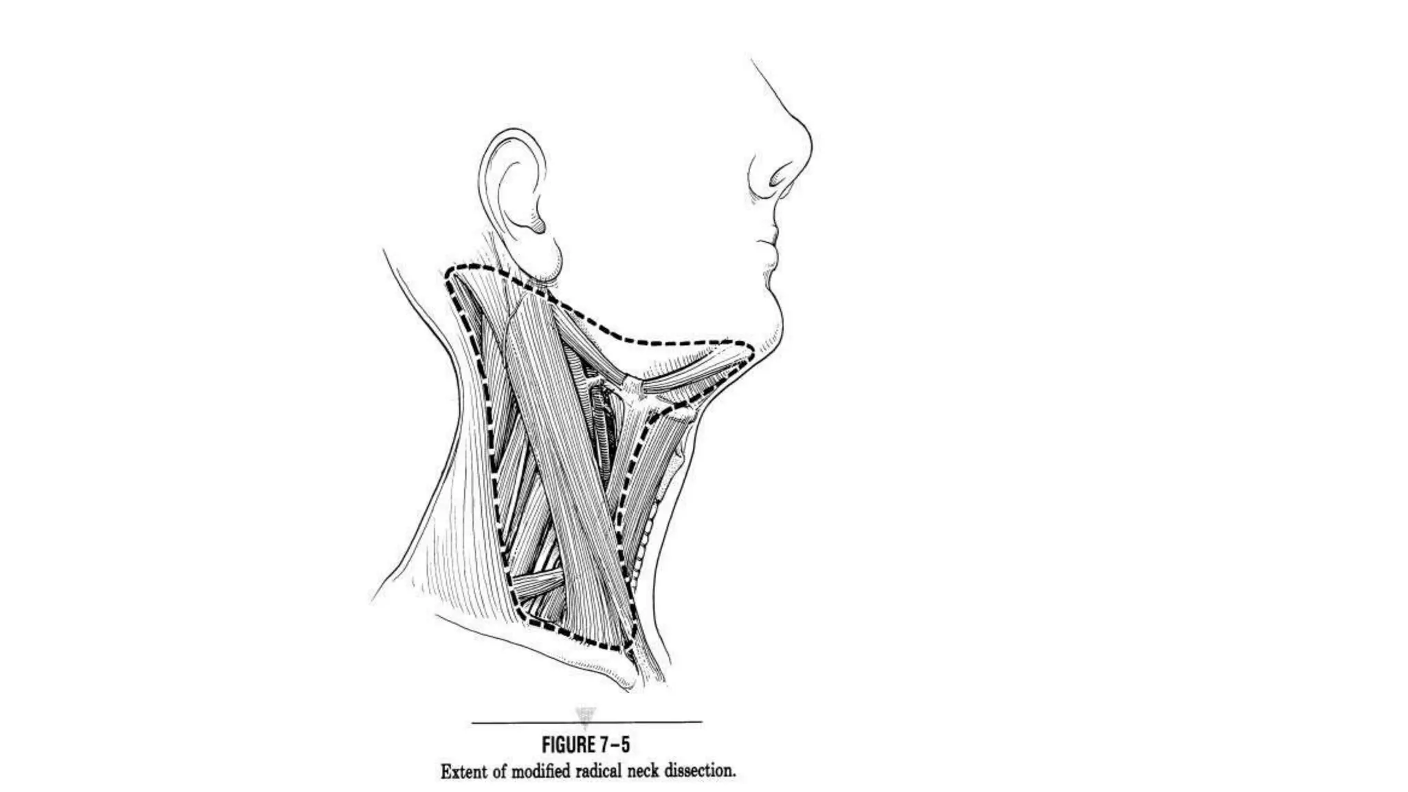
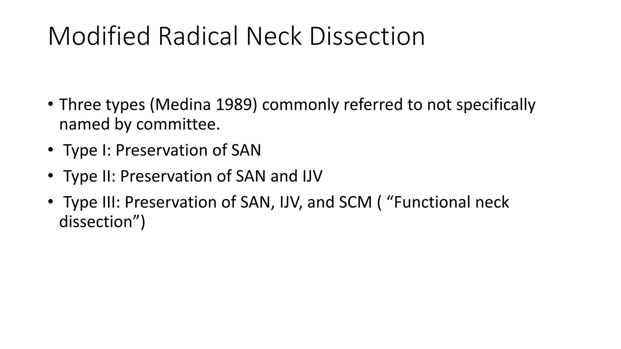
NECK DISSECTION
Dr Stanley John Cheriyan
Oral & Maxillofacial Surgery
Introduction
• The term "neck dissection" refers to the removal of lymph nodes and
lymph node bearing tissues of neck from the inferior border of the
mandible to the clavicle ,as a treatment of head and neck malignancy.
How does tumor spread ?
• Spread of disease of oral cavity to neck -- palpable lymphadenopathy.
• Systemic homogenous spread rarely occurs in the lymphatics of the
neck.
• early eradication of local and regional disease can prevent future
systemic metastasis.
The regional lymph node groups draining a specific primary site
is first echelon lymph nodes
The first echelon lymph nodes at highest risk from primary
tumors in the oral cavity
Risk for nodal metastasis
• Various factors
• Site
• Size
• T stage
• Location of primary tumour
• Histomorphologic characteristics of primary tumor
Risk of nodal metastases increases in relation to location of the
primary tumor
What is the rationale of treatment of
squamous cell carcinoma ?
• SCC has a distinct predilection for lymphatic spread
before distant systemic metastasis.
• early detection and eradication of local and regional
lymphatics prevents future metastasis
Memorial sloan kettering Cancer centre leveling
system
Division of neck levels by sublevels
• IA – submental nodes
• IB – submandibular nodes
• IIA & IIB – together
comprising the upper jugular
nodes
• III – middle jugular nodes
• IV – lower jugular nodes
• VA – Spinal accessory nodes
• VB – Transverse cervical and
supraclavicular nodes
• VI – anterior compartment
nodes
Clinical assessment and staging
• Important parameter for surgical management of the
neck is the staging of the neck itself.
• Results of the staging procedure have lower level of
certainty than biopsies
Assessment
• Clinical examination: 72% to 76% sensitivity
• CT and MRI : 84% to 92%
TNM Staging AJCC 8th Edition
TX Primary tumour cannot be assessed
T0 No evidence of primary tumour
Tis Carcinoma in situ
T1 Tumour 2 cm or less in greatest dimension
T2 Tumour more than 2 cm but not more than 4 cm in greatest
dimension
T3 Tumour more than 4 cm in greatest dimension
T4a (lip) Tumour invades through cortical bone, inferior alveolar nerve,
floor of mouth, or skin (chin or nose)
T4a (oral
cavity)
Tumour invades through cortical bone, into deep/extrinsic muscle
of tongue (genioglossus, hyoglossus, palatoglossus, and
styloglossus), maxillary sinus, or skin of face
T4b (lip
and oral
cavity)
Tumour invades masticator space, pterygoid plates, or skull base;
or encases internal carotid artery
T — Primary tumour 1,2
NX Regional lymph nodes cannot be assessed
N0 No regional lymph node metastasis
N1 Metastasis in a single ipsilateral lymph node, 3 cm or less in
greatest dimension
N2 Metastasis as specified in N2a, 2b, 2c below
N2a Metastasis in a single ipsilateral lymph node, more than 3 cm
but not more than 6 cm in greatest dimension
N2b Metastasis in multiple ipsilateral lymph nodes, none more than
6 cm in greatest dimension
N2c Metastasis in bilateral or contralateral lymph nodes, none more
than 6 cm in greatest dimension
N3 Metastasis in a lymph node more than 6 cm in greatest
dimension
N - Regional Lymph Nodes##
M - Distant metastasis
M0 No distant metastasis
M1 Distant metastasis
Stage 0 Tis N0 M0
Stage I T1 N0 M0
Stage II T2 N0 M0
Stage III T1, T2 N1 M0
T3 N0, N1 M0
Stage IVA T1, T2, T3 N2 M0
T4a N0, N1, N2 M0
Stage IVB Any T N3 M0
T4b Any N M0
Stage IVC Any T Any N M1
Stage grouping
TNM STAGING
• First reported by Pierre Denoix in the 1940s.
• The International Union against cancer (UICC) and AJCC eventually
adapted the system
• It is important to realize that the TNM staging system is simply an
anatomic staging system
• TNM Staging describes tumor burden in only two dimensions
A study of correlation of tumor thickness with risk
of occult nodal metastasis –Spiro et al*
*Spiro RH,Huvos AG, Wong GY ,Spiro JD, Strong EW .Predictive value of tumor
thickness in SCC confined to the tongue and floor of the mouth Am J Surg 1986;
152: 345-350
Patterns of cervical lymphatic metastasis
• lymphatic flow in the neck - consistent pattern -
upper neck and then to the lower neck.
• This orderly lymphatic flow has been demonstrated
by the work of Fisch and Sigel*
*Cervical lymphatic system as visualized by lymphography  Annals of
Otology, Rhinology and Laryngology 73: 869-872.
Studies on Patterns of cervical lymph node
metastasis – Jatin P Shah
* The percentages represents percent of patients with
N0 neck
Distribution of nodal metastasis in patients with N+ neck
Studies on Patterns of cervical lymph node
metastasis – Jatin P Shah
Further studies to support
• low level of metastasis to level V was confirmed -study by Davidson
et al.
• an incidence of level V metastasis of 1% was found among 666 RND
performed
• Inference: Metastasis to level V never occurred in the absence of
clinically palpable nodes in other levels of neck.
History of neck dissections
Dr George Crile (1864-1943)
In 1906 paper
“Exicision of cancer of the
head and neck ”
Gold standard procedure :
“Radical Neck dissection”
Dr. Hayes Martin (1892-1977)
In 1951 paper
“Neck Dissection”
“Routine prophylactic RND was
impractical”
Historical perspective on neck dissection
• RND should not be used for N0 neck, a philosophy
that was largely observed in 2006.
• Nahum et al described a syndrome of pain following
RND – “Shoulder Syndrome”*.
*Nahum AM, Mullally W, Marmor L : A Syndrome resulting from radical neck
dissection. Arch otolaryngol 74 : 82,1961
Historical perspective on neck dissection
• 1880 – Kocher –proposed removal of nodal metastasis
• 1906 – George crile –RND
• 1933 & 1941 – Blair and Martin popularised RND
• 1953 – Pietrantoni - recommended sparing SAN
• 1967-- Bocca and Pignataro described FND
• 1975- Bocca established oncologic safety compared to
RND
• 1980- Ballantyne –concept of selective neck dissection
1. Radical neck dissection (RND)
2. Modified radical neck dissection
(MRND)
3. Selective neck dissection (SND)
• Supraomohyoid type
• Lateral type
• Posterolateral type
• Anterior compartment type
4. Extended radical neck dissection
Classifications
Academy's classification
MEDINA CLASSIFICATION(1989)
• Comprehensive neck dissection
1. Radical neck dissection (RND)
2. Modified radical neck dissection (MRND)
• MRND I – Preserves spinal accessory nerve.
• MRND II – Spinal accessory and sternocleidomastoid
muscle but sacrifices internal jugular vein.
• MRND III – Requires preservation of SAN,
sternocleidomastoid muscle and internal jugular vein
• Selective neck dissection (SND)
• Supraomohyoid neck dissection – I, II, III
• Jugular neck dissection – II, III, IV
• Anterior triangle neck dissection – I, II, III, IV
• Central compartment neck dissection – VI
• Posterolateral neck dissection – II, III, IV
Spiro’s classification
• Radical (4 or 5 node levels resected)
• Conventional RND
• MRND
• Extended RND
• Selective (3 node levels resected)
• SOHND
• Jugular dissection (level II-IV)
• Any other 3 levels
• Limited (no more than 2 node levels resected)
• Para tracheal node dissection
• Mediastinal node dissection
• Any other 1 or 2 node levels resected
Incisions
• Incisions classified into
• Vertical
• Horizontal
• The incisions used for neck dissections are
• Tri-radiate incision and its modification
• Hayes martin double ‘Y’ incision
• McFee incision
Basic needs of an incision are
• Good exposure of the neck and primary disease
• Ensure viability of the skin flaps. Avoid acute angles
• Protect carotid artery even in the cases of wound infection
• Facilitate reconstruction
• Adapt to the condition of patient especially after radiotherapy
• It should be cosmetically acceptable
Differences between incisions
Transverse incision Vertical incision
Have cosmetic advantage as
they follow natural folds of
the skin
They intersect the natural
folds of the skin
Recovery of scar tissue in
these folds are rapid and
successful
They tend to contract along
their long axis – leads to
deformity and restricted
action.
Easy to modify
Tri-radiate incision and its modifications
• Advantages
• Incision provides good exposure
to surgical site.
• Disadvantages
• Flap necrosis is high due to
disruption of vasculature of skin
flaps
• Occurrence of flap separation at
the trifurcation site.
Modification of Tri-radiate incision
• Schobinger (1957)
• Cramer & Culf (1969)
• Conley (1970)
Schobinger (1957)
‘Vertical limb instead of
being straight should be
curved posteriorly ’
Conley (1970)
Suggested a posteriorly
curving vertical incision
rather than a horizontal
incision
Hayes Martin Incision
• It is a paired ‘Y’ incision.
• Here the submandibular
component is met by a
vertical limb which below
becomes continuous with
an inverted ‘Y’ in the
suprascapular region.
• This flap most often gets
cyanosed.
• Flap necrosis and carotid
exposure is more in this
type of incision.
McFee Incision
• It avoids a vertical limb.
• Two horizontal incisions
are used one in
submandibular region
and other in the
suprascapular region.
Apron flaps
• Described by Latyschevsky and
Freund 1960.
• Only a horizontal incision from
mastoid to mentum gently curving
inferiorly upto upper border of the
thyroid cartilage is used.
• Advantages
• Carotid artery is well protected
• Disadvantages
• It will damage the ascending arterial
and venous recovery
• Venous congestion and oedema might
develop at the bottom corner
Hockey stick incision
• Lahey et al (1940) described.
• Modified for RND by Eckert
& Byars 1952.
• It has a longitudinal and
transverse incision
• B/L hockey stick incision
allows the deglovement of
the whole neck.
Radical Neck Dissection
Radical neck dissection
• Current indications for classical radical neck dissection.
• N3 disease
• Multiple gross metastases involving multiple levels.
• Recurrent metastatic disease in a previously irradiated neck.
• Grossly apparent extranodal spread with invasion of the spinal accessory
nerve and /or internal jugular vein at the base of the skull
• Involvement of accessory chain lymph nodes by metastatic disease.
Modified Radical Neck Dissection (MRND)
• Excision of same lymph node bearing regions as RND with
preservation of one or more nonlymphatic structures (SAN, SCM, IJV)
• Spared structure specifically named
• MRND is analogous to the “functional neck dissection” described by
Bocca
Modified Radical Neck Dissection
• Three types (Medina 1989) commonly referred to not specifically
named by committee.
• Type I: Preservation of SAN
• Type II: Preservation of SAN and IJV
• Type III: Preservation of SAN, IJV, and SCM ( “Functional neck
dissection”)
Incision
Dissection of the posterior triangle begins at the anterior border of
trapezius
Dissection of the posterior triangle medially leads to exposure of brachial plexus,
phrenic nerve and cutaneous roots of the cervical plexus
Specimen reflected posteriorly and anterior flap elevated to expose the sternal head
of SCMM
Sternocleidomastoid muscle is detached from the sternum and clavicle and retracted
cephalad to expose the carotid sheath
Internal jugular vein is ligated and divided after common carotid and vagus nerve is
exposed and retracted medially
Dissection proceeds cephalad along the carotid sheath up the skull base
The upper skin flap is now elevated preseving the mandibular branch of the facial
nerve
Surgical field following RND
Two suction drains inserted
Contraindications for RND
• Uncontrollable cancer of the primary site
• Evidence of distant metastasis
• Fixed nodes unchanged by radiotherapy or
chemotherapy
• Life expectancy of less than 3 months
Selective Neck Dissection
Definition
• Cervical lymphadenectomy with preservation of one or more lymph
node groups.
Four common subtypes
• Supraomohyoid neck dissection
• Posterolateral neck dissection
• Lateral neck dissection
• Anterior neck dissection
SELECTIVE NECK
DISSECTION
• Also known as an elective neck dissection
• Rate of occult metastasis in clinically negative neck 20-30%
• Indication: primary lesion with 20% or greater risk of occult
metastasis
• Studies by Fisch and Sigel (1964) demonstrated predictable routes of
lymphatic spread from mucosal surfaces of the H&N
Supraomohyoid neck dissection
• Indications
• Oral cavity carcinoma with N0
neck
• Boundaries – Vermillion
border of lips to junction of
hard and soft palate,
circumvallate papillae
• Subsites - Lips, buccal mucosa,
upper and lower alveolar
ridges, retromolar trigone,
hard palate, and anterior 2/3s
of the tongue and FOM
• Medina recommends SOHND
with T2-T4NO ,TXN1 (palpable
node is <3cm, mobile, and in
levels I or II)
SND: Lateral Type
Indications
• N0 neck in carcinomas of the oropharynx, hypopharynx,
supraglottis, and larynx
• Rationale – Hypopharynx- Occult metastases in 30-35%
Johnson (1994)
• Medial pyriform (MP) vs. lateral pyriform carcinomas (LP)
• MP – 15% failed in the contralateral neck
• LP – 5% failed in the contralateral neck
• Johnson advocates bilateral SNDs for N0 MP carcinomas and
ipsilateral SND for N0 LP carcinomas
Bilateral SND is often indicated in the majority of
hypopharyngeal tumors because of extensive submucosal
spread and involvement of multiple subsites.
Posterolateral neck dissection
SND: Posterolateral Type
Definition
 En bloc excision of lymph bearing tissues in Levels II-IV and
additional node groups – suboccipital and postauricular.
 Indications :
 Cutaneous malignancies
 Melanoma
 Squamous cell carcinoma
 Merkel cell carcinoma
 Soft tissue sarcomas of the scalp and neck
Jugular neck dissection
Extended radical neck dissection
• Definition
• Any previous dissection which
includes removal of one or more
additional lymph node groups
and/or non-lymphatic structures.
• Usually performed with N+ necks in
MRND or RND when metastases
invade structures usually preserved
Complications of RND
• Intra operative complications
• Post operative complications
• Late complications
Intra-operative complications
• Injury to phrenic nerve
• Injury to vagus nerve
• Brachial plexus injury
• Carotid injury
• Hypoglossal nerve injury
• Lingual nerve injury
Post operative complications
• Haemorrhage
• Lymph leak
• Dysphagia
• Carotid blow out
• Facial edema
Complications of ligating bilateral internal jugular
vein simultaneously
Late complications
• Shoulder droop
• Shoulder pain
• Brachial neuralgia
• Neuroma
• Strictures
References
• Stell and Marans Head and Neck Surgery and Oncology - 5th Edition
• Charles W. Cummings, John M. Fredrickson, Lee A. Harker, Charles J. Krause,
David E. Schurller. Neck Dissection. Otolaryngology- Head and neck surgery.
Vol. II, 2nd edition. 1993: 1649-1672.
• Ian A. McGregor, Frances M. McGregor. Neck dissection. Cancer of the face
and mouth – Pathology and management for surgeons. Churchill
Livingstone.1986: 282- 320.
• Ian T. Jackson. Inrtra oral tumour and cervical lymphadenectomy. Grabb &
Smith’s Plastic Surgery. Sherrel J. Aston, Robert W. Beasley, Charles H. M.
Thorne. 5th edition. Lippincott- Raven . 1997 : 439 –452.
• L. H. Sobin & Ch Wittekind. TNM Classification of malignant tumours. 5th
edition. UICC, A John Wiley & Sons Inc. Publication. 1997.. Surg. 1999: 28 : 197
– 202.
• P. Hermanek, R. V. P. Hutter, L. H. Sobin & Ch Wittekind. TNM atlas.
Illustrated guide to the TNM / pTNM classification of malignant
tumours. 4th edition. Springer. 1997.
• Aydin Acar, Gürsel Dursun, Ömer Aydin,Yücel Akbaş. J incision in neck
dissections. The journal of Laryngology and otology. 1998: 112: 55 -
60.
• Susumu Omura, Hiroki Bukawa, Ryoichi Kawabe, Shinjiro Aoki,
Kiyohide Fujita. Comparision between hockey stick and reverse hockey
stick incision: gently curved single linear neck incisions for oral cancer.
Int. J. Oral Maxillofac
THANK YOU

Neck Dissection.pptx

  • 1.
    NECK DISSECTION Dr StanleyJohn Cheriyan Oral & Maxillofacial Surgery
  • 2.
    Introduction • The term"neck dissection" refers to the removal of lymph nodes and lymph node bearing tissues of neck from the inferior border of the mandible to the clavicle ,as a treatment of head and neck malignancy.
  • 3.
    How does tumorspread ? • Spread of disease of oral cavity to neck -- palpable lymphadenopathy. • Systemic homogenous spread rarely occurs in the lymphatics of the neck. • early eradication of local and regional disease can prevent future systemic metastasis.
  • 4.
    The regional lymphnode groups draining a specific primary site is first echelon lymph nodes
  • 5.
    The first echelonlymph nodes at highest risk from primary tumors in the oral cavity
  • 6.
    Risk for nodalmetastasis • Various factors • Site • Size • T stage • Location of primary tumour • Histomorphologic characteristics of primary tumor
  • 7.
    Risk of nodalmetastases increases in relation to location of the primary tumor
  • 8.
    What is therationale of treatment of squamous cell carcinoma ? • SCC has a distinct predilection for lymphatic spread before distant systemic metastasis. • early detection and eradication of local and regional lymphatics prevents future metastasis
  • 9.
    Memorial sloan ketteringCancer centre leveling system
  • 10.
    Division of necklevels by sublevels • IA – submental nodes • IB – submandibular nodes • IIA & IIB – together comprising the upper jugular nodes • III – middle jugular nodes • IV – lower jugular nodes • VA – Spinal accessory nodes • VB – Transverse cervical and supraclavicular nodes • VI – anterior compartment nodes
  • 11.
    Clinical assessment andstaging • Important parameter for surgical management of the neck is the staging of the neck itself. • Results of the staging procedure have lower level of certainty than biopsies
  • 12.
    Assessment • Clinical examination:72% to 76% sensitivity • CT and MRI : 84% to 92%
  • 13.
    TNM Staging AJCC8th Edition TX Primary tumour cannot be assessed T0 No evidence of primary tumour Tis Carcinoma in situ T1 Tumour 2 cm or less in greatest dimension T2 Tumour more than 2 cm but not more than 4 cm in greatest dimension T3 Tumour more than 4 cm in greatest dimension T4a (lip) Tumour invades through cortical bone, inferior alveolar nerve, floor of mouth, or skin (chin or nose) T4a (oral cavity) Tumour invades through cortical bone, into deep/extrinsic muscle of tongue (genioglossus, hyoglossus, palatoglossus, and styloglossus), maxillary sinus, or skin of face T4b (lip and oral cavity) Tumour invades masticator space, pterygoid plates, or skull base; or encases internal carotid artery T — Primary tumour 1,2
  • 14.
    NX Regional lymphnodes cannot be assessed N0 No regional lymph node metastasis N1 Metastasis in a single ipsilateral lymph node, 3 cm or less in greatest dimension N2 Metastasis as specified in N2a, 2b, 2c below N2a Metastasis in a single ipsilateral lymph node, more than 3 cm but not more than 6 cm in greatest dimension N2b Metastasis in multiple ipsilateral lymph nodes, none more than 6 cm in greatest dimension N2c Metastasis in bilateral or contralateral lymph nodes, none more than 6 cm in greatest dimension N3 Metastasis in a lymph node more than 6 cm in greatest dimension N - Regional Lymph Nodes## M - Distant metastasis M0 No distant metastasis M1 Distant metastasis
  • 15.
    Stage 0 TisN0 M0 Stage I T1 N0 M0 Stage II T2 N0 M0 Stage III T1, T2 N1 M0 T3 N0, N1 M0 Stage IVA T1, T2, T3 N2 M0 T4a N0, N1, N2 M0 Stage IVB Any T N3 M0 T4b Any N M0 Stage IVC Any T Any N M1 Stage grouping
  • 16.
    TNM STAGING • Firstreported by Pierre Denoix in the 1940s. • The International Union against cancer (UICC) and AJCC eventually adapted the system • It is important to realize that the TNM staging system is simply an anatomic staging system • TNM Staging describes tumor burden in only two dimensions
  • 17.
    A study ofcorrelation of tumor thickness with risk of occult nodal metastasis –Spiro et al* *Spiro RH,Huvos AG, Wong GY ,Spiro JD, Strong EW .Predictive value of tumor thickness in SCC confined to the tongue and floor of the mouth Am J Surg 1986; 152: 345-350
  • 18.
    Patterns of cervicallymphatic metastasis • lymphatic flow in the neck - consistent pattern - upper neck and then to the lower neck. • This orderly lymphatic flow has been demonstrated by the work of Fisch and Sigel* *Cervical lymphatic system as visualized by lymphography  Annals of Otology, Rhinology and Laryngology 73: 869-872.
  • 19.
    Studies on Patternsof cervical lymph node metastasis – Jatin P Shah * The percentages represents percent of patients with N0 neck
  • 20.
    Distribution of nodalmetastasis in patients with N+ neck Studies on Patterns of cervical lymph node metastasis – Jatin P Shah
  • 21.
    Further studies tosupport • low level of metastasis to level V was confirmed -study by Davidson et al. • an incidence of level V metastasis of 1% was found among 666 RND performed • Inference: Metastasis to level V never occurred in the absence of clinically palpable nodes in other levels of neck.
  • 22.
    History of neckdissections
  • 23.
    Dr George Crile(1864-1943) In 1906 paper “Exicision of cancer of the head and neck ” Gold standard procedure : “Radical Neck dissection”
  • 24.
    Dr. Hayes Martin(1892-1977) In 1951 paper “Neck Dissection” “Routine prophylactic RND was impractical”
  • 25.
    Historical perspective onneck dissection • RND should not be used for N0 neck, a philosophy that was largely observed in 2006. • Nahum et al described a syndrome of pain following RND – “Shoulder Syndrome”*. *Nahum AM, Mullally W, Marmor L : A Syndrome resulting from radical neck dissection. Arch otolaryngol 74 : 82,1961
  • 26.
    Historical perspective onneck dissection • 1880 – Kocher –proposed removal of nodal metastasis • 1906 – George crile –RND • 1933 & 1941 – Blair and Martin popularised RND • 1953 – Pietrantoni - recommended sparing SAN • 1967-- Bocca and Pignataro described FND • 1975- Bocca established oncologic safety compared to RND • 1980- Ballantyne –concept of selective neck dissection
  • 27.
    1. Radical neckdissection (RND) 2. Modified radical neck dissection (MRND) 3. Selective neck dissection (SND) • Supraomohyoid type • Lateral type • Posterolateral type • Anterior compartment type 4. Extended radical neck dissection Classifications Academy's classification
  • 28.
    MEDINA CLASSIFICATION(1989) • Comprehensiveneck dissection 1. Radical neck dissection (RND) 2. Modified radical neck dissection (MRND) • MRND I – Preserves spinal accessory nerve. • MRND II – Spinal accessory and sternocleidomastoid muscle but sacrifices internal jugular vein. • MRND III – Requires preservation of SAN, sternocleidomastoid muscle and internal jugular vein • Selective neck dissection (SND) • Supraomohyoid neck dissection – I, II, III • Jugular neck dissection – II, III, IV • Anterior triangle neck dissection – I, II, III, IV • Central compartment neck dissection – VI • Posterolateral neck dissection – II, III, IV
  • 29.
    Spiro’s classification • Radical(4 or 5 node levels resected) • Conventional RND • MRND • Extended RND • Selective (3 node levels resected) • SOHND • Jugular dissection (level II-IV) • Any other 3 levels • Limited (no more than 2 node levels resected) • Para tracheal node dissection • Mediastinal node dissection • Any other 1 or 2 node levels resected
  • 30.
    Incisions • Incisions classifiedinto • Vertical • Horizontal • The incisions used for neck dissections are • Tri-radiate incision and its modification • Hayes martin double ‘Y’ incision • McFee incision
  • 31.
    Basic needs ofan incision are • Good exposure of the neck and primary disease • Ensure viability of the skin flaps. Avoid acute angles • Protect carotid artery even in the cases of wound infection • Facilitate reconstruction • Adapt to the condition of patient especially after radiotherapy • It should be cosmetically acceptable
  • 32.
    Differences between incisions Transverseincision Vertical incision Have cosmetic advantage as they follow natural folds of the skin They intersect the natural folds of the skin Recovery of scar tissue in these folds are rapid and successful They tend to contract along their long axis – leads to deformity and restricted action. Easy to modify
  • 33.
    Tri-radiate incision andits modifications • Advantages • Incision provides good exposure to surgical site. • Disadvantages • Flap necrosis is high due to disruption of vasculature of skin flaps • Occurrence of flap separation at the trifurcation site.
  • 34.
    Modification of Tri-radiateincision • Schobinger (1957) • Cramer & Culf (1969) • Conley (1970)
  • 35.
    Schobinger (1957) ‘Vertical limbinstead of being straight should be curved posteriorly ’
  • 36.
    Conley (1970) Suggested aposteriorly curving vertical incision rather than a horizontal incision
  • 37.
    Hayes Martin Incision •It is a paired ‘Y’ incision. • Here the submandibular component is met by a vertical limb which below becomes continuous with an inverted ‘Y’ in the suprascapular region. • This flap most often gets cyanosed. • Flap necrosis and carotid exposure is more in this type of incision.
  • 38.
    McFee Incision • Itavoids a vertical limb. • Two horizontal incisions are used one in submandibular region and other in the suprascapular region.
  • 39.
    Apron flaps • Describedby Latyschevsky and Freund 1960. • Only a horizontal incision from mastoid to mentum gently curving inferiorly upto upper border of the thyroid cartilage is used. • Advantages • Carotid artery is well protected • Disadvantages • It will damage the ascending arterial and venous recovery • Venous congestion and oedema might develop at the bottom corner
  • 40.
    Hockey stick incision •Lahey et al (1940) described. • Modified for RND by Eckert & Byars 1952. • It has a longitudinal and transverse incision • B/L hockey stick incision allows the deglovement of the whole neck.
  • 41.
  • 42.
    Radical neck dissection •Current indications for classical radical neck dissection. • N3 disease • Multiple gross metastases involving multiple levels. • Recurrent metastatic disease in a previously irradiated neck. • Grossly apparent extranodal spread with invasion of the spinal accessory nerve and /or internal jugular vein at the base of the skull • Involvement of accessory chain lymph nodes by metastatic disease.
  • 43.
    Modified Radical NeckDissection (MRND) • Excision of same lymph node bearing regions as RND with preservation of one or more nonlymphatic structures (SAN, SCM, IJV) • Spared structure specifically named • MRND is analogous to the “functional neck dissection” described by Bocca
  • 45.
    Modified Radical NeckDissection • Three types (Medina 1989) commonly referred to not specifically named by committee. • Type I: Preservation of SAN • Type II: Preservation of SAN and IJV • Type III: Preservation of SAN, IJV, and SCM ( “Functional neck dissection”)
  • 46.
  • 47.
    Dissection of theposterior triangle begins at the anterior border of trapezius
  • 48.
    Dissection of theposterior triangle medially leads to exposure of brachial plexus, phrenic nerve and cutaneous roots of the cervical plexus
  • 49.
    Specimen reflected posteriorlyand anterior flap elevated to expose the sternal head of SCMM
  • 50.
    Sternocleidomastoid muscle isdetached from the sternum and clavicle and retracted cephalad to expose the carotid sheath
  • 51.
    Internal jugular veinis ligated and divided after common carotid and vagus nerve is exposed and retracted medially
  • 52.
    Dissection proceeds cephaladalong the carotid sheath up the skull base
  • 53.
    The upper skinflap is now elevated preseving the mandibular branch of the facial nerve
  • 54.
  • 55.
  • 56.
    Contraindications for RND •Uncontrollable cancer of the primary site • Evidence of distant metastasis • Fixed nodes unchanged by radiotherapy or chemotherapy • Life expectancy of less than 3 months
  • 57.
    Selective Neck Dissection Definition •Cervical lymphadenectomy with preservation of one or more lymph node groups. Four common subtypes • Supraomohyoid neck dissection • Posterolateral neck dissection • Lateral neck dissection • Anterior neck dissection
  • 58.
    SELECTIVE NECK DISSECTION • Alsoknown as an elective neck dissection • Rate of occult metastasis in clinically negative neck 20-30% • Indication: primary lesion with 20% or greater risk of occult metastasis • Studies by Fisch and Sigel (1964) demonstrated predictable routes of lymphatic spread from mucosal surfaces of the H&N
  • 59.
    Supraomohyoid neck dissection •Indications • Oral cavity carcinoma with N0 neck • Boundaries – Vermillion border of lips to junction of hard and soft palate, circumvallate papillae • Subsites - Lips, buccal mucosa, upper and lower alveolar ridges, retromolar trigone, hard palate, and anterior 2/3s of the tongue and FOM • Medina recommends SOHND with T2-T4NO ,TXN1 (palpable node is <3cm, mobile, and in levels I or II)
  • 60.
    SND: Lateral Type Indications •N0 neck in carcinomas of the oropharynx, hypopharynx, supraglottis, and larynx • Rationale – Hypopharynx- Occult metastases in 30-35% Johnson (1994) • Medial pyriform (MP) vs. lateral pyriform carcinomas (LP) • MP – 15% failed in the contralateral neck • LP – 5% failed in the contralateral neck • Johnson advocates bilateral SNDs for N0 MP carcinomas and ipsilateral SND for N0 LP carcinomas Bilateral SND is often indicated in the majority of hypopharyngeal tumors because of extensive submucosal spread and involvement of multiple subsites.
  • 61.
  • 62.
    SND: Posterolateral Type Definition En bloc excision of lymph bearing tissues in Levels II-IV and additional node groups – suboccipital and postauricular.  Indications :  Cutaneous malignancies  Melanoma  Squamous cell carcinoma  Merkel cell carcinoma  Soft tissue sarcomas of the scalp and neck
  • 63.
  • 64.
    Extended radical neckdissection • Definition • Any previous dissection which includes removal of one or more additional lymph node groups and/or non-lymphatic structures. • Usually performed with N+ necks in MRND or RND when metastases invade structures usually preserved
  • 65.
    Complications of RND •Intra operative complications • Post operative complications • Late complications
  • 66.
    Intra-operative complications • Injuryto phrenic nerve • Injury to vagus nerve • Brachial plexus injury • Carotid injury • Hypoglossal nerve injury • Lingual nerve injury
  • 67.
    Post operative complications •Haemorrhage • Lymph leak • Dysphagia • Carotid blow out • Facial edema
  • 68.
    Complications of ligatingbilateral internal jugular vein simultaneously
  • 69.
    Late complications • Shoulderdroop • Shoulder pain • Brachial neuralgia • Neuroma • Strictures
  • 70.
    References • Stell andMarans Head and Neck Surgery and Oncology - 5th Edition • Charles W. Cummings, John M. Fredrickson, Lee A. Harker, Charles J. Krause, David E. Schurller. Neck Dissection. Otolaryngology- Head and neck surgery. Vol. II, 2nd edition. 1993: 1649-1672. • Ian A. McGregor, Frances M. McGregor. Neck dissection. Cancer of the face and mouth – Pathology and management for surgeons. Churchill Livingstone.1986: 282- 320. • Ian T. Jackson. Inrtra oral tumour and cervical lymphadenectomy. Grabb & Smith’s Plastic Surgery. Sherrel J. Aston, Robert W. Beasley, Charles H. M. Thorne. 5th edition. Lippincott- Raven . 1997 : 439 –452. • L. H. Sobin & Ch Wittekind. TNM Classification of malignant tumours. 5th edition. UICC, A John Wiley & Sons Inc. Publication. 1997.. Surg. 1999: 28 : 197 – 202.
  • 71.
    • P. Hermanek,R. V. P. Hutter, L. H. Sobin & Ch Wittekind. TNM atlas. Illustrated guide to the TNM / pTNM classification of malignant tumours. 4th edition. Springer. 1997. • Aydin Acar, Gürsel Dursun, Ömer Aydin,Yücel Akbaş. J incision in neck dissections. The journal of Laryngology and otology. 1998: 112: 55 - 60. • Susumu Omura, Hiroki Bukawa, Ryoichi Kawabe, Shinjiro Aoki, Kiyohide Fujita. Comparision between hockey stick and reverse hockey stick incision: gently curved single linear neck incisions for oral cancer. Int. J. Oral Maxillofac
  • 72.

Editor's Notes

  • #43 Radical Neck Dissection • Indications – Extensive cervical involvement or matted lymph nodes with gross extracapsular spread and invasion into the SAN, IJV, or SCM
  • #50 
  • #64 Most commonly performed SND • Definition – En bloc removal of cervical lymph node groups I-III – Posterior limit is the cervical plexus and posterior border of the SCM – Inferior limit is the omohyoid muscle overlying the IJV