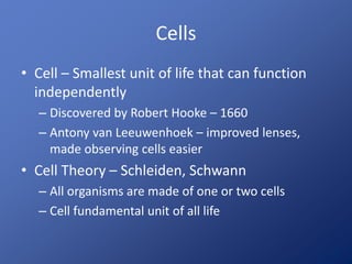
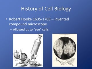
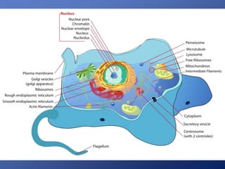
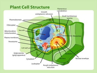
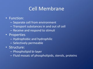
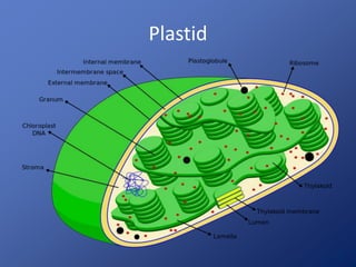
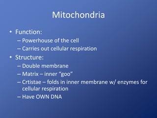
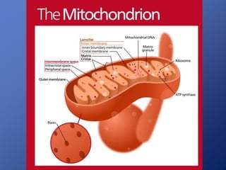
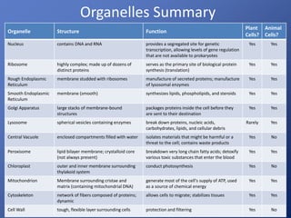
Cell Biology
Cells
• Cell – Smallest unit of life that can function
independently
– Discovered by Robert Hooke – 1660
– Antony van Leeuwenhoek – improved lenses,
made observing cells easier
• Cell Theory – Schleiden, Schwann
– All organisms are made of one or two cells
– Cell fundamental unit of all life
History of Cell Biology
• Robert Hooke 1635-1703 – invented
compound microscope
– Allowed us to “see” cells
History of Cell Biology
• Anton von
Leeuwenhoek 1673-
1723 – used higher
magnification lenses
to see microbes
Microscopy
Why do we need
microscopes?
Microscopes
• Microscope uses an energy source (light, electrons,
ect.) to view objects under magnification
– Can view things that you cannon view with the naked eye
– Light Microscopes – uses light to view things in real color,
item must be thin enough to get light through
• Compound – uses 2 or more lenses to focus visible light through a
specimen, magnify 1600 times
• Confocal – focus white or laser light through a lens to the object.
– Electron Microscopes – uses electrons, MUCH higher
magnification
• Transmission – Sends beam of electrons through a sample
• Scanning – Sends beam of electrons over the surface of object
Dark Field
Cross PolarizedBright Field
Phase Contrast
Electron Microscopy
Scanning Electron Microscopy (SEM)
Advantage: shows details of cell surfaces
Electron Microscopy
Transmission Electron Microscopy (TEM)
Advantage: shows detail of internal cell structures
Cell Structures
• All cells have these structures:
– DNA
– RNA
– Ribosomes – make proteins
– Proteins
– Cytoplasm – fluid filling of the cell
– Cell Membrane – makes a boundary between
inside and outside of cell
Types of Cells – Prokaryotic
• Lack a true nucleus and membrane bound
organelles
• Domains Bacteria and Archaea
• Structures:
– Nucleoid – contains cells DNA, NOT bound by a
membrane
– Cell Wall – rigid, outside of cell membrane, gives
cell its shape (rod-shaped, round, spiral)
– Flagella – whip-like tail for movement
Types of Cells - Eukaryotic
• All other cells except for bacteria, archaea
– Plants, animals, protists, fungi
• Domain – Eukarya
• Have MEMBRANE BOUND organelles, larger
than prokaryotic
• 2 basic types of eukaryotic cells
– Animal
– Plant
Cell Membrane
• Function:
– Separate cell from environment
– Transport substances in and out of cell
– Receive and respond to stimuli
• Properties
– Hydrophobic and hydrophilic
– Selectively permeable
• Structure:
– Phospholipid bi-layer
– Fluid mosaic of phospholipids, sterols, proteins
• Transport Proteins
• Enzymes
• Recognition Proteins
• Adhesion Proteins
• Receptor Proteins
Membrane Proteins
Animal cell membrane
• Maintain structure
• Maintain internal pressure
Cell Wall
Nucleus
• Function:
– “Brain” of the cell
– Contains DNA, mRNA
• Structure
– Nucleolus – assemble ribosomes
– Nuclear envelope – separates nucleus from
cytoplasm
– Nuclear pores – holes in envelop with channels for
mRNA to leave nucleus
Nucleus
Nuclear envelope
Nucleolus
Nuclear pore
1) Nuclear envelope
2) Outer ring
3) Spokes
4) Plug
Endoplasmic Reticulum and
Ribosomes
• Rough Endoplasmic Reticulum (ER)
– Function: Help make cell membrane and secretory
proteins for various bodily functions
• In pancreas, insulin; leukocytes, antibodies
– Structure: Connected to nuclear envelop and cell
membrane with ribosomes on outside
• Smooth ER
– Function: Make and store proteins, carbohydrates, lipids
• In liver, enzymes for detox; muscle, proteins for contraction
– Structure: Same as smooth ER – ribosomes
• Ribosomes
– Function: Assemble proteins for the cell
– Structure: Large and small subunits
Endoplasmic Reticulum and Ribosomes
Golgi Apparatus
• Function:
– Process and complete protein production
– Sorts and packages proteins to send to cell
membrane and out or as membrane proteins
• Structure:
– Stack of flat membrane enclosed sacs
Golgi Apparatus
Lysosomes
• Function:
– Digestion
– Enzymes to break down and recycle food, bacteria, old
organelles
• Structure:
– Made by Golgi Apparatus
– Fuse with vesicles that have things that need to be
digested
• Number of lysosomes depends on type of cell
– White blood cells, liver cells - lots
Peroxisomes
• Function:
– Dispose of toxic
substances
– Protect cells from
toxic byproducts
• Structure:
– DIFFERENT from
lysosomes that
originate from ER NOT
Golgi body
Peroxisomes (green)
Vacuoles
• Function:
– Same as lysosomes
– Replace lysosomes in plant cells
– Growth
– Maintain pressure
– Holds pigments
– Contractile vacuole – protists (pump water, digest)
• Structure:
– Contains water solution of enzymes, sugars, salts,
weak acids
Vacuoles
• Various Functions http://www.youtube.com/watch?v=iG6Dd3
COug4
Contractile vacuole
Central vacuole
Cytoskeleton
• Function:
– Transportation within the cell
– Support
– Cell division
– Connectivity
– Movement – Cilia and flagella
• Structure:
– Network of protein tubules and tracks
• Microfilaments
• Intermediate filaments
• Microtubules – organized by centrosomes
The Cytoskeleton
• Microfilaments
• Functions
– Structure
– Cell motility (muscles)
The Cytoskeleton
• Intermediate Filaments
– Functions:
• Cell shape
• Anchor organelles
Keratin intermediate filaments (red)
The Cytoskeleton
• Microtubules
– Functions:
• Support
• Tracks
• Cell division
Cilia and Flagella
http://www.youtube.com/watch?v=QGAm6hMysTA
http://www.youtube.com/watch?v=7kM_kRPrcrk
http://www.youtube.com/watch?v=09kLIsNfaO8&NR=1
Cilia Flagella
Cytoplasm
• Cytoplasm - intracellular fluid that surrounds
organelles
– ~55% cell volume
– 70-90% water + ions, glucose, amino acids, fatty
acids, proteins, lipids, ATP, waste products, some
cells have large macromolecules for storage
Endosymbiotic Theory
Chloroplast
• Function:
– Site of photosynthesis
– Only 1 type of plastid – all have different pigments
• Structure
– Double membrane
– Stroma – inner fluid
– Thylakoid – stacked disks with grana
• Have photosynthetic pigments (chlorophyll)
– Have OWN DNA and ribosomes
Plastid
Mitochondria
• Function:
– Powerhouse of the cell
– Carries out cellular respiration
• Structure:
– Double membrane
– Matrix – inner “goo”
– Crtistae – folds in inner membrane w/ enzymes for
cellular respiration
– Have OWN DNA
Cell Junctions
Type Function Example Location
Plasmodesmata enable direct, regulated, symplastic intercellular
transport of substances between cells
plant cell walls
Tight Junctions hold cells together; help to maintain the polarity of
cells; prevent the passage of molecules and ions
through the space between plasma membranes of
adjacent cells
the kidney and liver
Anchoring
(adhering)
Junctions
serve as a bridge connecting the actin cytoskeleton
of neighboring cells through direct interaction
epithelial and
endothelial tissues
Gap Junctions connects the cytoplasm of two cells, which allows
various molecules, ions, and electrical impluses to
directly pass through a regulated gate between cells
nerves
Tight Junctions
Anchoring (Adhering) Junction
Gap Junctions
Organelles Summary
Organelle Structure Function
Plant
Cells?
Animal
Cells?
Nucleus contains DNA and RNA provides a segregated site for genetic
transcription, allowing levels of gene regulation
that are not available to prokaryotes
Yes Yes
Ribosome highly complex; made up of dozens of
distinct proteins
serves as the primary site of biological protein
synthesis (translation)
Yes Yes
Rough Endoplasmic
Reticulum
membrane studded with ribosomes manufacture of secreted proteins; manufacture
of lysosomal enzymes
Yes Yes
Smooth Endoplasmic
Reticulum
membrane (smooth) synthesizes lipids, phospholipids, and steroids Yes Yes
Golgi Apparatus large stacks of membrane-bound
structures
packages proteins inside the cell before they
are sent to their destination
Yes Yes
Lysosome spherical vesicles containing enzymes break down proteins, nucleic acids,
carbohydrates, lipids, and cellular debris
Rarely Yes
Central Vacuole enclosed compartments filled with water isolates materials that might be harmful or a
threat to the cell; contains waste products
Yes No
Peroxisome lipid bilayer membrane; crystalloid core
(not always present)
breakdown very long chain fatty acids; detoxify
various toxic substances that enter the blood
Yes Yes
Chloroplast outer and inner membrane surrounding
thylakoid system
conduct photosynthesis Yes No
Mitochondrion Membrane surrounding cristae and
matrix (containing mitochondrial DNA)
generate most of the cell's supply of ATP, used
as a source of chemical energy
Yes Yes
Cytoskeleton network of fibers composed of proteins;
dynamic
allows cells to migrate; stabilizes tissues Yes Yes
Cell Wall tough, flexible layer surrounding cells protection and filtering Yes No

4. cell structure and function

  • 1.
  • 2.
    Cells • Cell –Smallest unit of life that can function independently – Discovered by Robert Hooke – 1660 – Antony van Leeuwenhoek – improved lenses, made observing cells easier • Cell Theory – Schleiden, Schwann – All organisms are made of one or two cells – Cell fundamental unit of all life
  • 3.
    History of CellBiology • Robert Hooke 1635-1703 – invented compound microscope – Allowed us to “see” cells
  • 4.
    History of CellBiology • Anton von Leeuwenhoek 1673- 1723 – used higher magnification lenses to see microbes
  • 5.
    Microscopy Why do weneed microscopes?
  • 6.
    Microscopes • Microscope usesan energy source (light, electrons, ect.) to view objects under magnification – Can view things that you cannon view with the naked eye – Light Microscopes – uses light to view things in real color, item must be thin enough to get light through • Compound – uses 2 or more lenses to focus visible light through a specimen, magnify 1600 times • Confocal – focus white or laser light through a lens to the object. – Electron Microscopes – uses electrons, MUCH higher magnification • Transmission – Sends beam of electrons through a sample • Scanning – Sends beam of electrons over the surface of object
  • 7.
  • 8.
    Electron Microscopy Scanning ElectronMicroscopy (SEM) Advantage: shows details of cell surfaces
  • 9.
    Electron Microscopy Transmission ElectronMicroscopy (TEM) Advantage: shows detail of internal cell structures
  • 10.
    Cell Structures • Allcells have these structures: – DNA – RNA – Ribosomes – make proteins – Proteins – Cytoplasm – fluid filling of the cell – Cell Membrane – makes a boundary between inside and outside of cell
  • 11.
    Types of Cells– Prokaryotic • Lack a true nucleus and membrane bound organelles • Domains Bacteria and Archaea • Structures: – Nucleoid – contains cells DNA, NOT bound by a membrane – Cell Wall – rigid, outside of cell membrane, gives cell its shape (rod-shaped, round, spiral) – Flagella – whip-like tail for movement
  • 12.
    Types of Cells- Eukaryotic • All other cells except for bacteria, archaea – Plants, animals, protists, fungi • Domain – Eukarya • Have MEMBRANE BOUND organelles, larger than prokaryotic • 2 basic types of eukaryotic cells – Animal – Plant
  • 15.
    Cell Membrane • Function: –Separate cell from environment – Transport substances in and out of cell – Receive and respond to stimuli • Properties – Hydrophobic and hydrophilic – Selectively permeable • Structure: – Phospholipid bi-layer – Fluid mosaic of phospholipids, sterols, proteins
  • 16.
    • Transport Proteins •Enzymes • Recognition Proteins • Adhesion Proteins • Receptor Proteins Membrane Proteins Animal cell membrane
  • 17.
    • Maintain structure •Maintain internal pressure Cell Wall
  • 18.
    Nucleus • Function: – “Brain”of the cell – Contains DNA, mRNA • Structure – Nucleolus – assemble ribosomes – Nuclear envelope – separates nucleus from cytoplasm – Nuclear pores – holes in envelop with channels for mRNA to leave nucleus
  • 19.
    Nucleus Nuclear envelope Nucleolus Nuclear pore 1)Nuclear envelope 2) Outer ring 3) Spokes 4) Plug
  • 20.
    Endoplasmic Reticulum and Ribosomes •Rough Endoplasmic Reticulum (ER) – Function: Help make cell membrane and secretory proteins for various bodily functions • In pancreas, insulin; leukocytes, antibodies – Structure: Connected to nuclear envelop and cell membrane with ribosomes on outside • Smooth ER – Function: Make and store proteins, carbohydrates, lipids • In liver, enzymes for detox; muscle, proteins for contraction – Structure: Same as smooth ER – ribosomes • Ribosomes – Function: Assemble proteins for the cell – Structure: Large and small subunits
  • 21.
  • 22.
    Golgi Apparatus • Function: –Process and complete protein production – Sorts and packages proteins to send to cell membrane and out or as membrane proteins • Structure: – Stack of flat membrane enclosed sacs
  • 23.
  • 24.
    Lysosomes • Function: – Digestion –Enzymes to break down and recycle food, bacteria, old organelles • Structure: – Made by Golgi Apparatus – Fuse with vesicles that have things that need to be digested • Number of lysosomes depends on type of cell – White blood cells, liver cells - lots
  • 25.
    Peroxisomes • Function: – Disposeof toxic substances – Protect cells from toxic byproducts • Structure: – DIFFERENT from lysosomes that originate from ER NOT Golgi body Peroxisomes (green)
  • 26.
    Vacuoles • Function: – Sameas lysosomes – Replace lysosomes in plant cells – Growth – Maintain pressure – Holds pigments – Contractile vacuole – protists (pump water, digest) • Structure: – Contains water solution of enzymes, sugars, salts, weak acids
  • 27.
    Vacuoles • Various Functionshttp://www.youtube.com/watch?v=iG6Dd3 COug4 Contractile vacuole Central vacuole
  • 28.
    Cytoskeleton • Function: – Transportationwithin the cell – Support – Cell division – Connectivity – Movement – Cilia and flagella • Structure: – Network of protein tubules and tracks • Microfilaments • Intermediate filaments • Microtubules – organized by centrosomes
  • 29.
    The Cytoskeleton • Microfilaments •Functions – Structure – Cell motility (muscles)
  • 30.
    The Cytoskeleton • IntermediateFilaments – Functions: • Cell shape • Anchor organelles Keratin intermediate filaments (red)
  • 31.
    The Cytoskeleton • Microtubules –Functions: • Support • Tracks • Cell division
  • 32.
  • 33.
    Cytoplasm • Cytoplasm -intracellular fluid that surrounds organelles – ~55% cell volume – 70-90% water + ions, glucose, amino acids, fatty acids, proteins, lipids, ATP, waste products, some cells have large macromolecules for storage
  • 34.
  • 35.
    Chloroplast • Function: – Siteof photosynthesis – Only 1 type of plastid – all have different pigments • Structure – Double membrane – Stroma – inner fluid – Thylakoid – stacked disks with grana • Have photosynthetic pigments (chlorophyll) – Have OWN DNA and ribosomes
  • 36.
  • 37.
    Mitochondria • Function: – Powerhouseof the cell – Carries out cellular respiration • Structure: – Double membrane – Matrix – inner “goo” – Crtistae – folds in inner membrane w/ enzymes for cellular respiration – Have OWN DNA
  • 39.
    Cell Junctions Type FunctionExample Location Plasmodesmata enable direct, regulated, symplastic intercellular transport of substances between cells plant cell walls Tight Junctions hold cells together; help to maintain the polarity of cells; prevent the passage of molecules and ions through the space between plasma membranes of adjacent cells the kidney and liver Anchoring (adhering) Junctions serve as a bridge connecting the actin cytoskeleton of neighboring cells through direct interaction epithelial and endothelial tissues Gap Junctions connects the cytoplasm of two cells, which allows various molecules, ions, and electrical impluses to directly pass through a regulated gate between cells nerves
  • 40.
  • 41.
  • 42.
  • 43.
    Organelles Summary Organelle StructureFunction Plant Cells? Animal Cells? Nucleus contains DNA and RNA provides a segregated site for genetic transcription, allowing levels of gene regulation that are not available to prokaryotes Yes Yes Ribosome highly complex; made up of dozens of distinct proteins serves as the primary site of biological protein synthesis (translation) Yes Yes Rough Endoplasmic Reticulum membrane studded with ribosomes manufacture of secreted proteins; manufacture of lysosomal enzymes Yes Yes Smooth Endoplasmic Reticulum membrane (smooth) synthesizes lipids, phospholipids, and steroids Yes Yes Golgi Apparatus large stacks of membrane-bound structures packages proteins inside the cell before they are sent to their destination Yes Yes Lysosome spherical vesicles containing enzymes break down proteins, nucleic acids, carbohydrates, lipids, and cellular debris Rarely Yes Central Vacuole enclosed compartments filled with water isolates materials that might be harmful or a threat to the cell; contains waste products Yes No Peroxisome lipid bilayer membrane; crystalloid core (not always present) breakdown very long chain fatty acids; detoxify various toxic substances that enter the blood Yes Yes Chloroplast outer and inner membrane surrounding thylakoid system conduct photosynthesis Yes No Mitochondrion Membrane surrounding cristae and matrix (containing mitochondrial DNA) generate most of the cell's supply of ATP, used as a source of chemical energy Yes Yes Cytoskeleton network of fibers composed of proteins; dynamic allows cells to migrate; stabilizes tissues Yes Yes Cell Wall tough, flexible layer surrounding cells protection and filtering Yes No

Editor's Notes

  • #4 Left: “Hooke Microscope” by Robert Hooke. (Public Domain). http://commons.wikimedia.org/wiki/File:Hooke-microscope.png Right: “Suber cells and mimosa leaves” by Robert Hooke. (Public Domain). http://commons.wikimedia.org/wiki/File:RobertHookeMicrographia1665.jpg
  • #5 “Leeuwenhoek simple microscope copy” by Wellcome Images. Licensed under a CC-BY 4.0 International license. http://commons.wikimedia.org/wiki/File:Leeuwenhoek_simple_microscope_(copy),_Leyden,_1901-1930_Wellcome_L0057739.jpg
  • #6 Image content by Lumen Learning.
  • #8 Bright field - "Paper Micrograph Bright" by Zephyris - Own work. Licensed under CC BY-SA 3.0 via Commons - https://commons.wikimedia.org/wiki/File:Paper_Micrograph_Bright.png#/media/File:Paper_Micrograph_Bright.png Cross Polarized - "Paper Micrograph Cross-Polarised" by Zephyris - Own work. Licensed under CC BY-SA 3.0 via Commons - https://commons.wikimedia.org/wiki/File:Paper_Micrograph_Cross-Polarised.png#/media/File:Paper_Micrograph_Cross-Polarised.png Dark Field - "Paper Micrograph Dark" by Zephyris - Own work. Licensed under CC BY-SA 3.0 via Commons - https://commons.wikimedia.org/wiki/File:Paper_Micrograph_Dark.png#/media/File:Paper_Micrograph_Dark.png Phase Contrast - "Paper Micrograph Phase" by Zephyris - Own work. Licensed under CC BY-SA 3.0 via Commons - https://commons.wikimedia.org/wiki/File:Paper_Micrograph_Phase.png#/media/File:Paper_Micrograph_Phase.png
  • #9 “Misc Pollen Colorized” by Dartmouth Electron Microscope Facility. Released into public domain by copyright holder. https://commons.wikimedia.org/wiki/File:Misc_pollen_colorized.jpg
  • #10 “Staphylococcus aureus” by Eric Erbe and Christopher Pooley. (Public Domain). https://commons.wikimedia.org/wiki/File:Staphylococcus_aureus,_50,000x,_USDA,_ARS,_EMU.jpg
  • #14 “Animal Cell Structure” by LadyofHats. Released into public domain by copyright holder. https://commons.wikimedia.org/wiki/File:Animal_cell_structure_en.svg
  • #15 “Plant Cell Structure” by LadyofHats. Released into public domain by copyright holder. https://commons.wikimedia.org/wiki/File:Plant_cell_structure.png
  • #17 “Cell Membrane Detailed Diagram” by LadyofHats. Released into public domain by copyright holder. https://commons.wikimedia.org/wiki/File:Cell_membrane_detailed_diagram_en.svg
  • #18 “Plant Cell Wall Diagram” by LadyofHats. Released into public domain by copyright holder. https://commons.wikimedia.org/wiki/File:Plant_cell_wall_diagram.svg
  • #20 Top left: from OpenStax Top right: “Nuclear Pores” by Magnus Manske. Licensed under a CC-BY-SA 3.0 Unported license. http://commons.wikimedia.org/wiki/File:Nuclear_pores.png Bottom: “Micrograph of a Cell Nucleus” by US NIGMS/NIH. (Public Domain). http://commons.wikimedia.org/wiki/File:Micrograph_of_a_cell_nucleus.png
  • #22 https://commons.wikimedia.org/wiki/File%3ABlausen_0350_EndoplasmicReticulum.png By BruceBlaus. When using this image in external sources it can be cited as: Blausen.com staff. "Blausen gallery 2014". Wikiversity Journal of Medicine. DOI:10.15347/wjm/2014.010. ISSN 20018762. (Own work) [CC BY 3.0 (http://creativecommons.org/licenses/by/3.0)], via Wikimedia Commons
  • #24 Image from Open Stax
  • #26 “Lipid bodies and peroxisomes” by The Journal of Cell Biology. Licensed under a CC-BY-NC-SA 3.0 Unported license. https://www.flickr.com/photos/thejcb/4077865657/in/photostream/
  • #27 “Lipid bodies and peroxisomes” by The Journal of Cell Biology. Licensed under a CC-BY-NC-SA 3.0 Unported license. https://www.flickr.com/photos/thejcb/4077865657/in/photostream/
  • #28 Left: “Epidermis Peel” by BlueRidgeKitties. Licensed under a CC-BY-NC-SA 2.0 Generic license. https://www.flickr.com/photos/blueridgekitties/8259412733 Right: “enhanced phase paramecium” by Jasper Nance. Licensed under a CC-BY-NC-ND 2.0 Generic license. https://www.flickr.com/photos/nebarnix/309954509/in/photostream/
  • #30 Model: “Adherens Junctions structural proteins” by LadyofHats. Released into public domain by copyright holder. https://commons.wikimedia.org/wiki/File:Adherens_Junctions_structural_proteins.svg Microfilaments: “MEF microfillaments” by Y tambe. Licensed under a CC-BY-SA 3.0 Unported license. https://commons.wikimedia.org/wiki/File:MEF_microfilaments.jpg
  • #31 Keratin intermediate filaments: “Epithelial cells” by John Schmidt. Licensed under a CC-BY-SA 3.0 Unported license. https://en.wikipedia.org/wiki/File:Epithelial-cells.jpg Filament: “IF id” by Zlir’a. Licensed under a CC-BY-SA 3.0 Unported license. https://commons.wikimedia.org/wiki/File:IF_id.svg
  • #32 “Microtubules” by Boumphreyfr. Licensed under a CC-BY-SA 3.0 Unported license. https://commons.wikimedia.org/wiki/File:Microtubules.png
  • #33 Cilia: “Bronchiolar Epithelium” by Charles Daghlian. Released into the public domain by copyright holder. https://en.wikipedia.org/wiki/File:Bronchiolar_epithelium_3_-_SEM.jpg Flagellum: “Flagellum” by Pearson Scott. Released into the public domain by copyright holder. https://commons.wikimedia.org/wiki/File:Flagellum_(PSF).png
  • #35 “Serial Endosymbiosis” by Kelvinsong. Licensed under a CC-BY-SA 3.0 Unported license. https://commons.wikimedia.org/wiki/File:Serial_endosymbiosis.svg. Adapted by Lumen Learning.
  • #37 "Scheme Chloroplast-en" by User:Miguelsierra, adapted by User:Vossman - Own work. Licensed under GFDL via Wikimedia Commons - https://commons.wikimedia.org/wiki/File:Scheme_Chloroplast-en.svg#/media/File:Scheme_Chloroplast-en.svg
  • #39 “Mitochondrion” by Kelvinsong. Available under a CC-0 1.0 Universal Public Domain Dedication. https://commons.wikimedia.org/wiki/File:Mitochondrion_(standalone_version)-en.svg
  • #40 “Plasmodesma” https://en.wikipedia.org/wiki/Plasmodesma “Tight Junction” https://en.wikipedia.org/wiki/Tight_junction “Adherens Junction” https://en.wikipedia.org/wiki/Adherens_junction “Gap Junction” https://en.wikipedia.org/wiki/Gap_junction
  • #41 “Cellular Tight Junction” by LadyofHats. Released into public domain by copyright holder. https://commons.wikimedia.org/wiki/File:Cellular_tight_junction-en.svg
  • #42 “Desmosome cell Junction” by LadyofHats. Released into public domain by copyright holder. https://commons.wikimedia.org/wiki/File:Desmosome_cell_junction_en.svg
  • #43 “Gap Cell Junction” by LadyofHats. Released into public domain by copyright holder. https://commons.wikimedia.org/wiki/File:Gap_cell_junction-en.svg
  • #44 “Cell Nucleus” https://en.wikipedia.org/wiki/Cell_nucleus “Ribosome” https://en.wikipedia.org/wiki/Ribosome “Endoplasmic Reticulum” https://en.wikipedia.org/wiki/Endoplasmic_reticulum “Golgi Apparatus” https://en.wikipedia.org/wiki/Golgi_apparatus “Lysosome” https://en.wikipedia.org/wiki/Lysosome “Vacuole” https://en.wikipedia.org/wiki/Vacuole “Peroxisome” https://en.wikipedia.org/wiki/Peroxisome “Chloroplast” https://en.wikipedia.org/wiki/Chloroplast “Mitochondrion” https://en.wikipedia.org/wiki/Mitochondrion “Cytoskeleton” https://en.wikipedia.org/wiki/Cytoskeleton “Cell Wall” https://en.wikipedia.org/wiki/Cell_wall