Downloaded 26 times

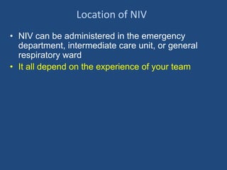
![Hypoxemic Respiratory Failure
• Recognized by an acute fall in P02 or
oxyhemoglobin saturation (for example, a P02
to less than or equal to 60 mm Hg or PO2
/fraction of inspired oxygen [FiO2] less than or
equal to 200
• Conditions causing acute hypoxemic
respiratory failure include alveolar collapse
and flooding with fluid, pus, or blood.](https://image.slidesharecdn.com/3noninvasiveventilation-140519135323-phpapp02/85/3-noninvasive-ventilation-3-320.jpg)

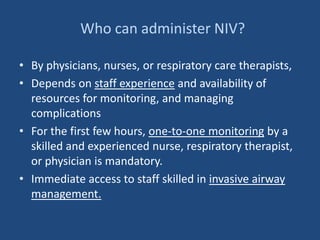
![Acute Lung Injury & Acute Respiratory
Distress Syndrome
• Definitions
– ALI: PO2 /fraction of inspired oxygen [FiO2]in liter
is less than 300
– ARDS: PO2 /fraction of inspired oxygen [FiO2]in
liter is less than 300 ( more severe ALI)
ALI /ARDS is a noncardiogenic form of pulmonary
edema characterized by acute and persistent lung
inflammation and increased vascular permeability
( Damaged and leaky pipes)](https://image.slidesharecdn.com/3noninvasiveventilation-140519135323-phpapp02/85/3-noninvasive-ventilation-5-320.jpg)


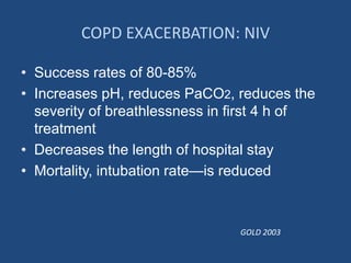


Respiratory failure can occur due to hypoxemic failure, hypercapnic (ventilatory) failure, or impaired airways. Noninvasive ventilation (NIV) delivers breaths through a tight-fitting mask and can treat various causes of respiratory failure without needing intubation. The first 30 minutes of NIV require close monitoring as clinicians work to improve gas exchange and comfort while watching for signs that intubation is needed instead. NIV is effective for acute exacerbations of COPD and cardiogenic pulmonary edema when criteria are met.