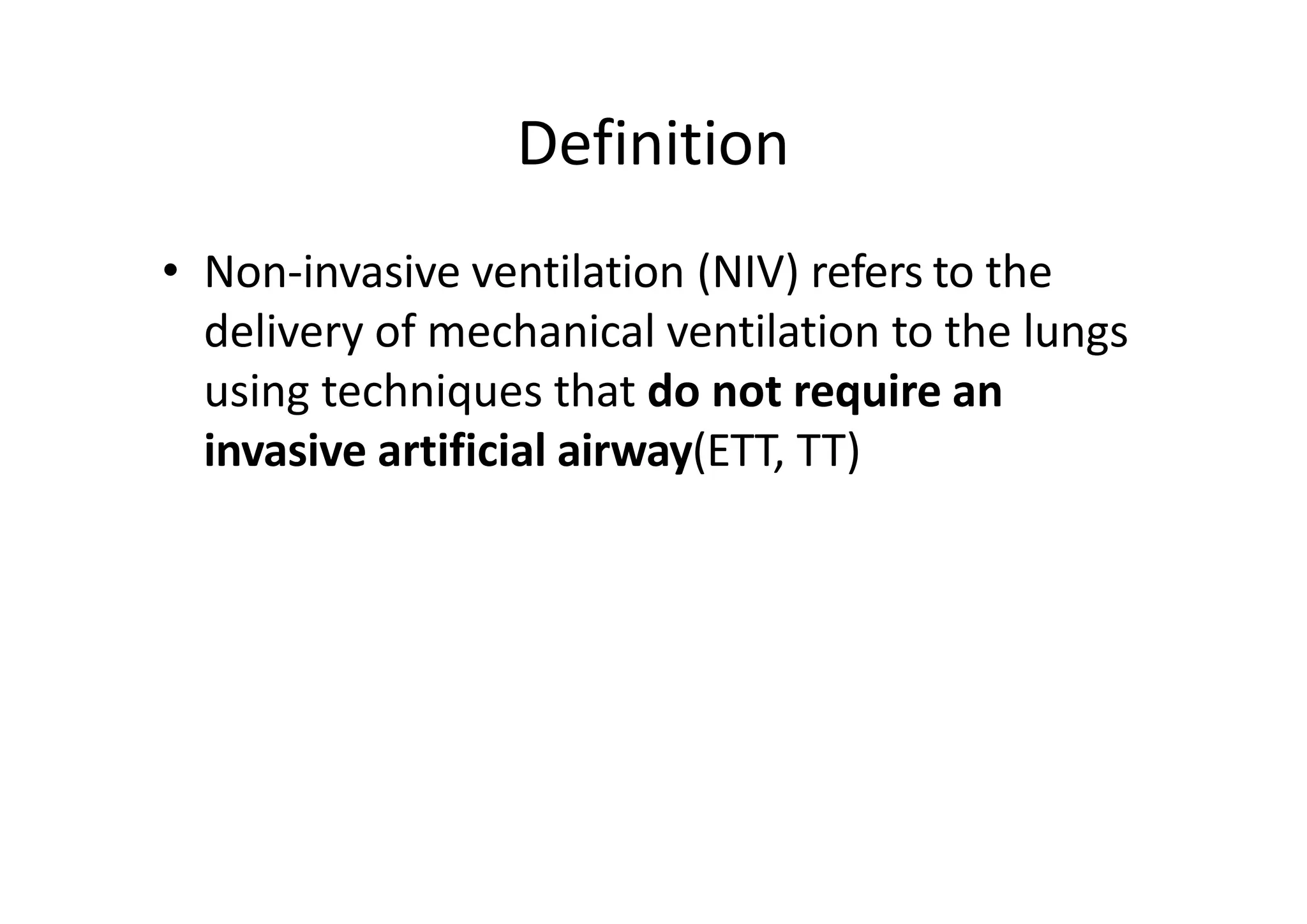
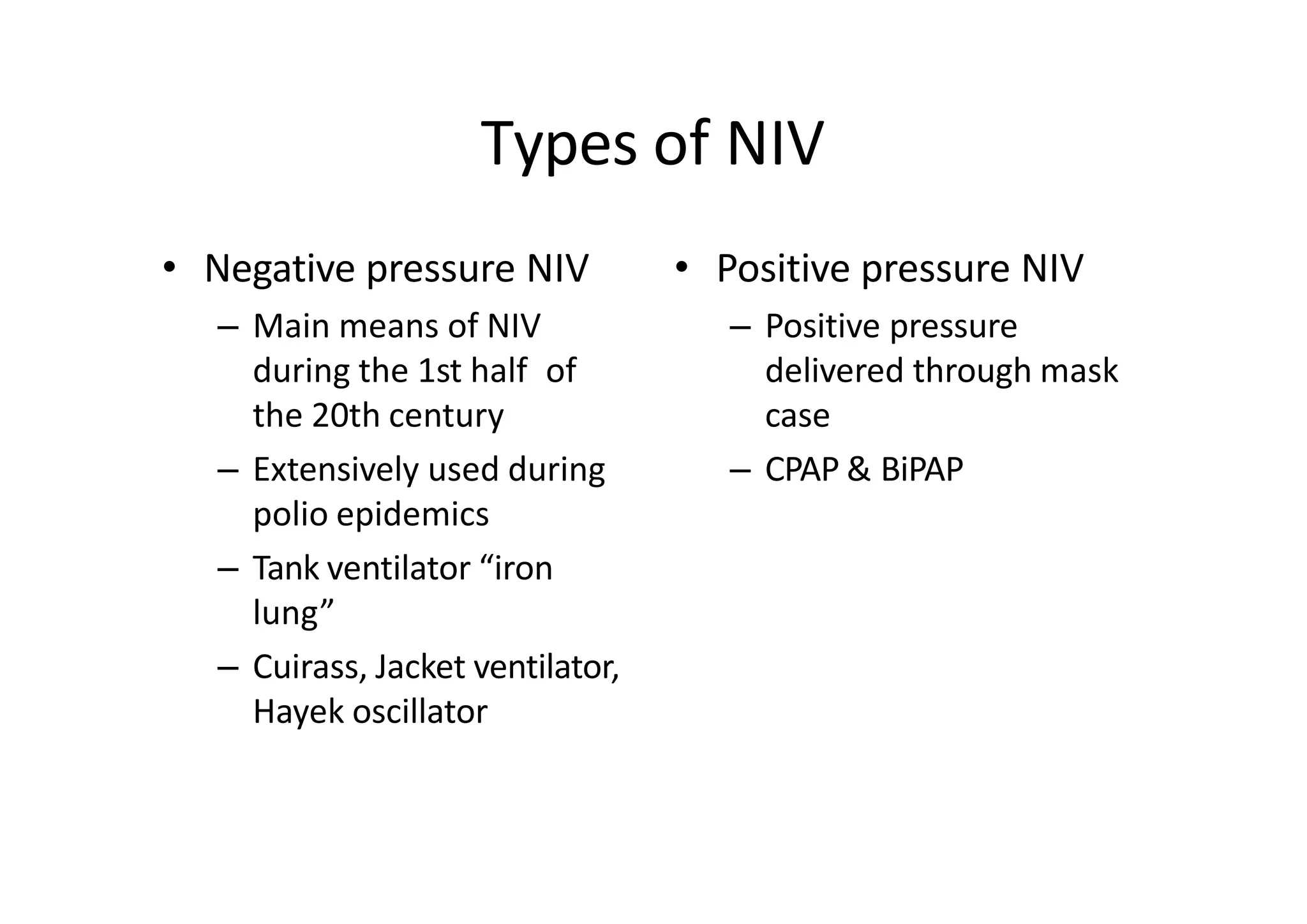
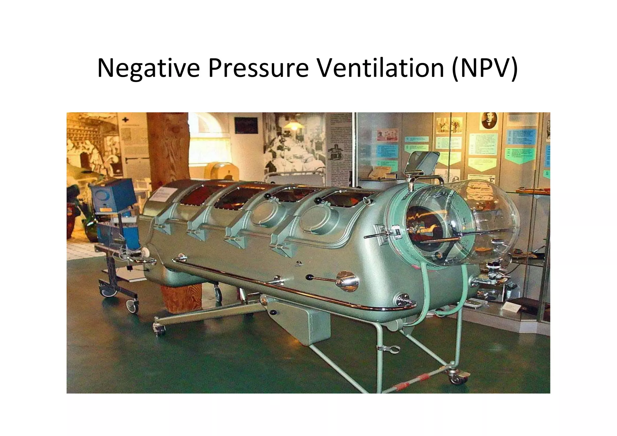
Non invasive ventilation
Overview
• Definition
• Types
• Advantages & disadvantages
• Indications & contraindications
• Interface
• Modes of NIV
• Guidelines for initiation & termination
• Complications
• Conclusion
Definition
• Non‐invasive ventilation (NIV) refers to the
delivery of mechanical ventilation to the lungs
using techniques that do not require an
invasive artificial airway(ETT, TT)
Types of NIV
• Negative pressure NIV
– Main means of NIV
during the 1st half of
the 20th century
– Extensively used during
polio epidemics
– Tank ventilator “iron
lung”
– Cuirass, Jacket ventilator,
Hayek oscillator
• Positive pressure NIV
– Positive pressure
delivered through mask
case
– CPAP & BiPAP
Negative Pressure Ventilation (NPV)
• Negative pressure ventilators apply a negative pressure
intermittently around the patient’s body or chest wall
• The patient’s head (upper airway) is exposed to room
air
• Negative pressure is applied intermittently to the
thoracic area resulting in a pressure drop around the
thorax
• This negative pressure is transmitted to the pleural
space and alveoli creating a pressure gradient between
the inside of the lungs and the mouth
Negative Pressure Ventilation (NPV)
NIV ‐ Advantages
• Non invasiveness
– Flexibility in initiating and removing mechanical
ventilation
– Allows intermittent application
– Improves patient comfort
– Reduces the need for sedation
– Oral patency
• Preserves speech, swallowing and expectoration,
reduces the need for nagastric tubes
Advantages
• Avoid the resistive work imposed by the
endotracheal tube
• Avoids the complications of endotracheal
intubation
– Early (local trauma, aspiration)
– Late (injury to hypopharynx, larynx, and trachea,
nosocomial infections)
• Reduces infectious complications‐ pneumonia,
sinusitis, sepsis
• Less cost
Mechanism of action
• Reduction in inspiratory muscle work and
avoidance of respiratory muscle fatigue
• Tidal volume is increased
• CPAP counterbalances the inspiratory threshold
work related to intrinsic PEEP.
• NIV improves respiratory system compliance by
reversing microatelectasis of the lung
• Enhanced cardiovascular function
– Afterload reduction d/t increased intrathoracic
pressure
Pathophysiology of acute
hypercapnia
Pathophysiology of acute hypoxemic
respiratory failure
Disadvantages
• System
– Slower correction of gas exchange abnormalities
– Increased initial time commitment
– Gastric distension (occurs in <2% patients)
• Mask
– Air leakage
– Transient hypoxemia from accidental removal
– Eye irritation
– Facial skin necrosis –most common complication
• Lack of airway access and protection
– Suctioning of secretions
– Aspiration
Levels of evidence
• A
– Multiple RCTs
– Recommended
• B
– Atleast one RCT
– Weaker evidence
• C
– Case series/reports
– Can be tried but with close monitoring
Indications
• Airway obstruction
– COPD (Evidence A)
– Facilitation of weaning in COPD (Evidence A)
– Asthma (B)
– Extubation failure in COPD (B)
– Cystic Fibrosis (C)
– OSA/obesity hypoventilation (C)
– Upper airway obstruction (C)
NIV & stable COPD
• NIV increasingly used in stable very severe
COPD
• NIV+O2 therapy – in selected patients with
pronounced daytime hypercapnia
• Clear benefits in both survival & risk of
hospital admission in patients with both COPD
& OSA
Global initiative for Chronic Obstructive Lung Disease(GOLD)
update 2013
Cochrane Database Syst Rev.
2004;(3):CD004104.
Concludes
• Data from good quality RCTs show benefit of
NIV as 1st line intervention in addition to
usual medical care to ARF 2° to an acute
exacerbation of COPD in all suitable patients
• Use early in the course of respiratory failure
and before severe ensues, as a means of
reducing the likelihood of endotracheal
intubation, treatment failure and mortality
NIV in COPD exacerbation
• Multiple RCTs support a success rate of 80‐
85%
• Has been shown to improve respiratory
acidosis ( pH & pCO2 )
• Respiratory rate, WOB, severity of
breathlessness, complications like VAP,length
of hospital stay (Evidence A)
• Mortality & intubation rates are
reduced(Evidence A)
GOLD update 2013
NIV in COPD exacerbation
• Indications for NIV – atleast one of the
following
– Respiratory acidosis (pH<7.35 &/or PaCO2)
– Severe dyspnea with clinical signs s/o
respiratory muscle fatigue, increased WOB or
both
• Use of respiratory accessory muscles
• Paradoxical motion of abdomen
• Intercostal retraction
GOLD update 2013
• 12 trials of moderate to good quality
• Compared to the IPPV strategy, NPPV significantly
decreased
– Mortality(RR 0.55)
– VAP(RR 0.29)
– Length of stay in an ICU(WMD* ‐6.27) &
hospital(WMD ‐7.19 day)
– Total duration of ventilation(WMD ‐5.64 day)
– Duration of endotracheal mechanical
ventilation(WMD ‐ 7.81 days)
Cochrane Database Syst Rev. 2010;(8):CD004127.
*Weighted mean difference
• Compared to IPPV Noninvasive weaning had
no effect on weaning failures or the duration
of ventilation related to weaning
• Concluded
– a consistent, positive effect on mortality and
ventilator associated pneumonia
Respir Care. 2012 Oct;57(10):1619–25.
Indications
• Hypoxemic respiratory failure
– Acute pulmonary edema‐ CPAP (Evidence A)
– Immunocompromised patients (Evidence A)
– Postoperative patients (B)
– ARDS (C)
– Pneumonia (C)
– Trauma or burns (C)
– Restrictive thoracic disorders (C)
– Do not intubate patients (C)
– During bronchoscopy (C)
Cochrane Database Syst Rev. 2013;5:CD005351.
Concludes
• Included 32 studies
• NIV is an effective and safe intervention for
the treatment of adult patients with acute
cardiogenic pulmonary oedema
• Evidence to date on the potential benefit of
NIV in reducing mortality is entirely derived
from small‐trials and further large‐scale trials
are needed
Indications
• Immunocompromised patients
– Particularly exposed to infectious risk related to
invasive ventilation
– Multiple RCTs support whenever possible, NIV
should be tried first in immunocompromised
patients with hypoxemic RF
Respir Care. 2011 Oct 1;56(10):1583–8.
Indications
• Postsurgical period
– Shown benefits after lung resectional surgery
,CABG , gastroplasty, high risk procedures like
thoracoabdominal aortic procedures
– Should be considered in selected postoperative
patients at high risk of pulmonary complications
or with frank respiratory failure especially in the
setting of underlying COPD or pulmonary edema
Indications
• Restrictive Lung Disease
– Patients with restriction related to an underlying
neuromuscular disease and superimposed ARF
may benefit from a trial of NIV
– Small case series have reported that using NIPPV
in patients with myasthenic crises may avoid
intubation
PATIENT SELECTION
• Step 1
– An etiology of respiratory failure likely to respond
favourably to NIV
• Step 2
– Identify patients in need of ventilatory assistance by using
clinical and blood gas criteria
– Moderate to severe dyspnea, tachypnea, and impending
respiratory muscle fatigue
– COPD with RR >24 bpm
– Hypoxemic respiratory failure with RR >30‐35 bpm
• Step 3
– Exclude patients for whom NIV would be unsafe
PREDICTORS OF NIV
SUCCESS IN ACUTE
RESPIRATORY FAILURE
• Lower acuity of illness (APACHE score)
• Ability to cooperate; better neurologic score
• Ability to coordinate breathing with ventilator
• Less air leakage; intact dentition
• Hypercarbia, but not too severe (PaCO2 between
45 and 92 mm Hg)
• Acidemia, but not too severe (pH between 7.1
and 7.35)
• Improvements in gas exchange and heart and
respiratory rates within first 2 hours
Exclusion Criteria for NIV
• Respiratory arrest or need for immediate intubation
• Hemodynamic instability
• Inability to protect the airway (impaired cough or
swallowing)
• Excessive secretions
• Agitated and confused patient
• Facial deformities or conditions that prevent mask
from fitting
• Uncooperative or unmotivated patient
• Brain injury with unstable respiratory drive
• Untreated pneumothorax
Contraindications to NIV
•
•
•
•
•
Cardiac or respiratory arrest
Non respiratory organ failure
Severe encephalopathy (eg, GCS <10)
Severe upper gastrointestinal bleeding
Hemodynamic instability or unstable cardiac
arrhythmia
•
•
•
•
•
Facial or neurological surgery, trauma, or deformity
Upper airway obstruction
Inability to cooperate/protect airway
Inability to clear secretions
High risk for aspiration
Equipment for NIV‐ Ventilators
• Portable NIV machines
– Advantages
• Portability
• Ease of use
• Better compensation for leaks
• Better exhalation
– Disadvantages
• Cannot develop pressures >30cm H2O
• Lack O2 blenders
• Lack of sophisticated alarm systems, battery backup
• Critical care ventilators
Ventilators for NIV
• Volume controlled home ventilators
– First machines to be used
– Limited ability to compensate for leaks
• Bilevel ventilators
– Most commonly used
– EPAP & IPAP
– Single limb circuit
– Good efficiency & leak compensation
• ICU ventilators
– Limited leak compensation
Portable BiPAP machines
• RESMED
• RESPIRONICS‐philips
Modes of NIV
• Pressure modes
– Better tolerated than
volume‐cycled vents
– Constant positive airway
pressure(CPAP)
– Bilevel or biphasic
positive airway pressure
(BiPAP)
– Pressure support
ventilation(PSV)
• Volume modes
– Initial tidal volumes
range from 10 to 15
mL/kg
– Control
– Assist control
– Proportional assist
control
CPAP
• A mode for invasive and noninvasive
mechanical ventilation
• Provides static positive airway pressure
throughout the respiratory cycle‐ both
inspiration & expiration
• Facilitates inhalation by reducing pressure
thresholds to initiate airflow
CPAP
• Creats a "pneumatic splint" for the upper airway,
preventing the soft tissues of the upper airway
from narrowing and collapsing.
• Increase functional residual capacity
– Improve lung compliance
– Open collapsed alveoli
– Improve oxygenation
– Decrease work of breathing
• Decrease left ventricular transmural pressure, ↓
aGerload and ↑CO
CPAP‐ASB
Adaptive servo‐ventilation
• Specifically to treat central sleep apnea (CSA)
• Designed to vary support according to a
patient’s individual breathing rate
• Automatically calculates a target ventilation
(90% of the patient's recent average
ventilation)
• Adjusts the pressure support to achieve it
BiPAP – Spontaneous
• Airway pressure cycles between an inspiratory
positive airway pressure (IPAP) and an expiratory
positive airway pressure (EPAP)
• Patient's inspiratory effort triggers the switch
from EPAP to IPAP
• The limit during inspiration is the set level of IPAP
• The inspiratory phase cycles off, and the machine
switches back to EPAP when it detects a
cessation of patient effort(decrease in
inspiratory flow rate, or a maximum inspiratory
time reached‐ typically 2‐3 seconds)
BiPAP – Spontaneous
• Vt varies breath to breath and is determined by degree
of IPAP, patient effort, and lung compliance
• IPAP is necessarily set higher than EPAP by a minimum
of 5cm H2O
• Difference between the two settings is equivalent to
the amount of PS provided
• Spontaneous mode depends on patient effort to trigger
inhalation. A patient breathing at a low rate can
develop a respiratory acidosis.
• BiPAP can be described as a continuous positive airway
pressure system with a time‐cycled or flow‐cycled
change of the applied CPAP level
BiPAP‐S
BiPAP
• Spontaneous/timed (ST) mode
– The trigger in the ST mode can be the patient's
effort or an elapsed time interval, predetermined
by a set respiratory backup rate.
• If the patient does not initiate a breath in the
prescribed interval, then IPAP is triggered. For machine‐
generated breaths, the ventilator cycles back to EPAP
based on a set inspiratory time.
– For patient‐initiated breaths, the ventilator cycles
as it would in the spontaneous mode.
BiPAP
• Increases in inspiratory pressure are helpful to
alleviate dyspnea
• Increases in expiratory pressure are better to
improve oxygenation
Interface
• The device that makes physical contact between
the patient and the ventilator is termed the
interface
• Types
– Nasal mask
– Nasal pillow
– Oronasal mask
– Full face mask
– Helmet
• Interfaces should be comfortable, offer a good
seal, minimize leak, and limit dead space
Interface
• Selection of a comfortable mask that fits
properly is key to the success of NIV
• Full facemask should be tried first in the acute
setting
• Patient should be allowed to hold the mask in
place initially
• Mask straps are then tightened with the least
tension necessary to avoid excessive air
leakage
Interfaces for delivery of NIV
Oronasal mask interface
• 1st line for acute care
setting
• COPD – mouth
breathing
Nasal masks
• Better tolerated than full face masks for long‐
term &chronic applications
• Less claustrophobia and discomfort and allow
eating, conversation, and expectoration
Nasal masks
• Pressure over nasal
bridge
– forehead spacers
– ultrathin silicon seals or
heat‐sensitive gels that
minimize skin trauma
• Problem‐ Air leakage
through mouth
Nasal pillows
Nasal vs Oronasal mask
Variables Nasal Oronasal
Comfort +++ ++
Claustrophobia + ++
Rebreathing + ++
Lowers CO2 + ++
Permits expectoration ++ +
Permits speech ++ +
Permits eating + ‐
Function if nose obstructed ‐ +
Total face mask
• Equally comfortable
• Similar
– Application times
– Early NIV discontinuation
rates
– Improvements in vital
signs and gas exchange
– Intubation and mortality
rates
CHEST 2011; 139(5):1034‐1041
Helmet vs Facemask
• Helmet
– equally tolerated
– effective in ameliorating
gas exchange
– decreasing inspiratory
effort
– But less efficient in
decreasing inspiratory
effort
– worsened the patient‐
ventilator interaction
Intensive Care Med. 2007 Jan;33(1):74‐81
Protocol for initiation of NIV
• Appropriately monitored location
• Patient in bed or chair sitting at > 30‐degree angle
• A full‐face mask should be used for the first 24 hours,
followed by switching to a nasal mask if preferred by
the patient (Evidence C)
• Encourage patient to hold mask
• Apply harness; avoid excessive strap tension
• Connect interface to ventilator tubing and turn on
ventilator
• Check for air leaks, readjust straps as needed
Protocol for initiation of NIV
• Check for air leaks, readjust straps as needed
• Consider mild sedation (lorazepam 0.5 mg iv)
in agitated patients
• Encouragement, reassurance, and frequent
checks and adjustments as needed
• Monitor occasional blood gases (within 1 to 2
h and then as needed)
Guidelines for providing NIV
• An initial IPAP of 10 cm H2O & EPAP of 4–5 cm
H2O should be used (Evidence A)
• IPAP should be increased by 2–5 cm increments
at a rate of approximately 5 cm H2O every 10
mins, with a usual IPAP target of 20 cm H2O or
until a therapeutic response is achieved or
patient tolerability has been reached (Evidence A)
• O2 should be entrained into the circuit and the
flow adjusted to SpO2 >88–92% (EvidenceB)
BTS: NIV in COPD: management
of acute type 2 respiratory failure
Guidelines for providing NIV
• Bronchodilators
– preferably administered off NIV
– if necessary be entrained between the expiration
port and face mask
– Delivery of both oxygen and nebulised solutions is
affected by NIV pressure settings (Evidence A)
• If a nasogastric tube is in place, a fine bore tube is
preferred to minimise mask leakage(Evidence C)
BTS: NIV in COPD: management
of acute type 2 respiratory failure
Guidelines for providing NIV
• Patient comfort and enhanced compliance are
key factors in determining outcome (Evidence
A)
– Synchrony of ventilation should be checked
frequently.
– A clinical assessment of mask fit to include skin
condition and degree of leak (particularly on to
the corneas) should be performed at the same
time
BTS: NIV in COPD: management
of acute type 2 respiratory failure
OXYGENATION AND HUMIDIFICATION
• Oxygen is titrated to achieve a desired oxygen
saturation > 90% to 92%
– Use of oxygen blenders
– adjusting litre flow delivered via oxygen tubing
connected directly to the mask or ventilator circuit
• Heated blow over vaporiser should be used if
longer application intended
Monitoring
• Subjective responses
– Bed side observation
– Ask about discomfort related to the mask or airflow
• Physiologic response
– ↓ RR, ↓ HR, BP,continuous EKG
– Patient breath in synchrony with the ventilator
– ↓ accessory muscle activity and abdominal paradox
– Monitor air leaks and Vt
Monitoring
• Gas exchange(Evidence C)
– Continuous SpO2 and ECG during the first 12 hours
– ABG
• After 1 hour of NIV therapy and 1 hour after every
subsequent change in settings
• After 4 hours, or earlier in patients who are not
improving clinically
BTS: NIV in COPD: management
of acute type 2 respiratory failure
Guidelines for providing NIV
• Duration of treatment
– Patients who benefit from NIV during the first 4
hours of treatment should receive NIV for as long as
possible (a minimum of 6 hours) during the first 24
hours (Evidence A)
• Treatment should last until the acute cause has
resolved, commonly after about 3 days
• When NIV is successful (pH>7.35,resolution of
cause, normalisation of RR) after 24 hrs/more –
plan weaning
BTS: NIV in COPD: management
of acute type 2 respiratory failure
Assessment of NIV
Weaning strategy(A)
• Continue NIV for 16 hours on day 2
• Continue NIV for 12 hours on day 3 including
6–8 hours overnight use
• Discontinue NIV on day 4, unless continuation
is clinically indicate
BTS: NIV in COPD: management
of acute type 2 respiratory failure
Criteria for Terminating NIV and Switching
to Invasive Mechanical Ventilation
• Worsening pH and PaCO2
• Tachypnea (over 30 bpm)
• Hemodynamic instability
• SpO2 < 90%
• Decreased level of consciousness
• Inability to clear secretions
• Inability to tolerate interface
Complications
• Interface related • Air pressure/flow
related
• Patient related
Conclusion
• NIV is an important tool to tide over an acute
insult in the hands of a experienced operator
• Key factors in success
– Evidence based application for selected etiologies
– Careful patient selection/rejection
– Skilled initiation & application
– Algorithmic approach in initiation, use,
discontinuation
– Patient comfort & avoiding dyssynchrony
– Avoiding complications
Conclusion
• Most importantly
– Decision making on when to switch to invasive
mechanical ventilation in a setting of failure of NIV
THANKS

seminar non invasive ventilation final.pptx

  • 1.
  • 2.
    Overview • Definition • Types •Advantages & disadvantages • Indications & contraindications • Interface • Modes of NIV • Guidelines for initiation & termination • Complications • Conclusion
  • 3.
    Definition • Non‐invasive ventilation(NIV) refers to the delivery of mechanical ventilation to the lungs using techniques that do not require an invasive artificial airway(ETT, TT)
  • 4.
    Types of NIV •Negative pressure NIV – Main means of NIV during the 1st half of the 20th century – Extensively used during polio epidemics – Tank ventilator “iron lung” – Cuirass, Jacket ventilator, Hayek oscillator • Positive pressure NIV – Positive pressure delivered through mask case – CPAP & BiPAP
  • 5.
    Negative Pressure Ventilation(NPV) • Negative pressure ventilators apply a negative pressure intermittently around the patient’s body or chest wall • The patient’s head (upper airway) is exposed to room air • Negative pressure is applied intermittently to the thoracic area resulting in a pressure drop around the thorax • This negative pressure is transmitted to the pleural space and alveoli creating a pressure gradient between the inside of the lungs and the mouth
  • 6.
  • 7.
    NIV ‐ Advantages •Non invasiveness – Flexibility in initiating and removing mechanical ventilation – Allows intermittent application – Improves patient comfort – Reduces the need for sedation – Oral patency • Preserves speech, swallowing and expectoration, reduces the need for nagastric tubes
  • 8.
    Advantages • Avoid theresistive work imposed by the endotracheal tube • Avoids the complications of endotracheal intubation – Early (local trauma, aspiration) – Late (injury to hypopharynx, larynx, and trachea, nosocomial infections) • Reduces infectious complications‐ pneumonia, sinusitis, sepsis • Less cost
  • 9.
    Mechanism of action •Reduction in inspiratory muscle work and avoidance of respiratory muscle fatigue • Tidal volume is increased • CPAP counterbalances the inspiratory threshold work related to intrinsic PEEP. • NIV improves respiratory system compliance by reversing microatelectasis of the lung • Enhanced cardiovascular function – Afterload reduction d/t increased intrathoracic pressure
  • 10.
  • 11.
    Pathophysiology of acutehypoxemic respiratory failure
  • 12.
    Disadvantages • System – Slowercorrection of gas exchange abnormalities – Increased initial time commitment – Gastric distension (occurs in <2% patients) • Mask – Air leakage – Transient hypoxemia from accidental removal – Eye irritation – Facial skin necrosis –most common complication • Lack of airway access and protection – Suctioning of secretions – Aspiration
  • 13.
    Levels of evidence •A – Multiple RCTs – Recommended • B – Atleast one RCT – Weaker evidence • C – Case series/reports – Can be tried but with close monitoring
  • 14.
    Indications • Airway obstruction –COPD (Evidence A) – Facilitation of weaning in COPD (Evidence A) – Asthma (B) – Extubation failure in COPD (B) – Cystic Fibrosis (C) – OSA/obesity hypoventilation (C) – Upper airway obstruction (C)
  • 15.
    NIV & stableCOPD • NIV increasingly used in stable very severe COPD • NIV+O2 therapy – in selected patients with pronounced daytime hypercapnia • Clear benefits in both survival & risk of hospital admission in patients with both COPD & OSA Global initiative for Chronic Obstructive Lung Disease(GOLD) update 2013
  • 16.
    Cochrane Database SystRev. 2004;(3):CD004104.
  • 17.
    Concludes • Data fromgood quality RCTs show benefit of NIV as 1st line intervention in addition to usual medical care to ARF 2° to an acute exacerbation of COPD in all suitable patients • Use early in the course of respiratory failure and before severe ensues, as a means of reducing the likelihood of endotracheal intubation, treatment failure and mortality
  • 18.
    NIV in COPDexacerbation • Multiple RCTs support a success rate of 80‐ 85% • Has been shown to improve respiratory acidosis ( pH & pCO2 ) • Respiratory rate, WOB, severity of breathlessness, complications like VAP,length of hospital stay (Evidence A) • Mortality & intubation rates are reduced(Evidence A) GOLD update 2013
  • 19.
    NIV in COPDexacerbation • Indications for NIV – atleast one of the following – Respiratory acidosis (pH<7.35 &/or PaCO2) – Severe dyspnea with clinical signs s/o respiratory muscle fatigue, increased WOB or both • Use of respiratory accessory muscles • Paradoxical motion of abdomen • Intercostal retraction GOLD update 2013
  • 20.
    • 12 trialsof moderate to good quality • Compared to the IPPV strategy, NPPV significantly decreased – Mortality(RR 0.55) – VAP(RR 0.29) – Length of stay in an ICU(WMD* ‐6.27) & hospital(WMD ‐7.19 day) – Total duration of ventilation(WMD ‐5.64 day) – Duration of endotracheal mechanical ventilation(WMD ‐ 7.81 days) Cochrane Database Syst Rev. 2010;(8):CD004127. *Weighted mean difference
  • 21.
    • Compared toIPPV Noninvasive weaning had no effect on weaning failures or the duration of ventilation related to weaning • Concluded – a consistent, positive effect on mortality and ventilator associated pneumonia
  • 22.
    Respir Care. 2012Oct;57(10):1619–25.
  • 23.
    Indications • Hypoxemic respiratoryfailure – Acute pulmonary edema‐ CPAP (Evidence A) – Immunocompromised patients (Evidence A) – Postoperative patients (B) – ARDS (C) – Pneumonia (C) – Trauma or burns (C) – Restrictive thoracic disorders (C) – Do not intubate patients (C) – During bronchoscopy (C)
  • 24.
    Cochrane Database SystRev. 2013;5:CD005351.
  • 25.
    Concludes • Included 32studies • NIV is an effective and safe intervention for the treatment of adult patients with acute cardiogenic pulmonary oedema • Evidence to date on the potential benefit of NIV in reducing mortality is entirely derived from small‐trials and further large‐scale trials are needed
  • 26.
    Indications • Immunocompromised patients –Particularly exposed to infectious risk related to invasive ventilation – Multiple RCTs support whenever possible, NIV should be tried first in immunocompromised patients with hypoxemic RF
  • 27.
    Respir Care. 2011Oct 1;56(10):1583–8.
  • 28.
    Indications • Postsurgical period –Shown benefits after lung resectional surgery ,CABG , gastroplasty, high risk procedures like thoracoabdominal aortic procedures – Should be considered in selected postoperative patients at high risk of pulmonary complications or with frank respiratory failure especially in the setting of underlying COPD or pulmonary edema
  • 29.
    Indications • Restrictive LungDisease – Patients with restriction related to an underlying neuromuscular disease and superimposed ARF may benefit from a trial of NIV – Small case series have reported that using NIPPV in patients with myasthenic crises may avoid intubation
  • 32.
    PATIENT SELECTION • Step1 – An etiology of respiratory failure likely to respond favourably to NIV • Step 2 – Identify patients in need of ventilatory assistance by using clinical and blood gas criteria – Moderate to severe dyspnea, tachypnea, and impending respiratory muscle fatigue – COPD with RR >24 bpm – Hypoxemic respiratory failure with RR >30‐35 bpm • Step 3 – Exclude patients for whom NIV would be unsafe
  • 33.
    PREDICTORS OF NIV SUCCESSIN ACUTE RESPIRATORY FAILURE • Lower acuity of illness (APACHE score) • Ability to cooperate; better neurologic score • Ability to coordinate breathing with ventilator • Less air leakage; intact dentition • Hypercarbia, but not too severe (PaCO2 between 45 and 92 mm Hg) • Acidemia, but not too severe (pH between 7.1 and 7.35) • Improvements in gas exchange and heart and respiratory rates within first 2 hours
  • 34.
    Exclusion Criteria forNIV • Respiratory arrest or need for immediate intubation • Hemodynamic instability • Inability to protect the airway (impaired cough or swallowing) • Excessive secretions • Agitated and confused patient • Facial deformities or conditions that prevent mask from fitting • Uncooperative or unmotivated patient • Brain injury with unstable respiratory drive • Untreated pneumothorax
  • 35.
    Contraindications to NIV • • • • • Cardiacor respiratory arrest Non respiratory organ failure Severe encephalopathy (eg, GCS <10) Severe upper gastrointestinal bleeding Hemodynamic instability or unstable cardiac arrhythmia • • • • • Facial or neurological surgery, trauma, or deformity Upper airway obstruction Inability to cooperate/protect airway Inability to clear secretions High risk for aspiration
  • 36.
    Equipment for NIV‐Ventilators • Portable NIV machines – Advantages • Portability • Ease of use • Better compensation for leaks • Better exhalation – Disadvantages • Cannot develop pressures >30cm H2O • Lack O2 blenders • Lack of sophisticated alarm systems, battery backup • Critical care ventilators
  • 37.
    Ventilators for NIV •Volume controlled home ventilators – First machines to be used – Limited ability to compensate for leaks • Bilevel ventilators – Most commonly used – EPAP & IPAP – Single limb circuit – Good efficiency & leak compensation • ICU ventilators – Limited leak compensation
  • 38.
    Portable BiPAP machines •RESMED • RESPIRONICS‐philips
  • 39.
    Modes of NIV •Pressure modes – Better tolerated than volume‐cycled vents – Constant positive airway pressure(CPAP) – Bilevel or biphasic positive airway pressure (BiPAP) – Pressure support ventilation(PSV) • Volume modes – Initial tidal volumes range from 10 to 15 mL/kg – Control – Assist control – Proportional assist control
  • 40.
    CPAP • A modefor invasive and noninvasive mechanical ventilation • Provides static positive airway pressure throughout the respiratory cycle‐ both inspiration & expiration • Facilitates inhalation by reducing pressure thresholds to initiate airflow
  • 41.
    CPAP • Creats a"pneumatic splint" for the upper airway, preventing the soft tissues of the upper airway from narrowing and collapsing. • Increase functional residual capacity – Improve lung compliance – Open collapsed alveoli – Improve oxygenation – Decrease work of breathing • Decrease left ventricular transmural pressure, ↓ aGerload and ↑CO
  • 42.
  • 43.
    Adaptive servo‐ventilation • Specificallyto treat central sleep apnea (CSA) • Designed to vary support according to a patient’s individual breathing rate • Automatically calculates a target ventilation (90% of the patient's recent average ventilation) • Adjusts the pressure support to achieve it
  • 44.
    BiPAP – Spontaneous •Airway pressure cycles between an inspiratory positive airway pressure (IPAP) and an expiratory positive airway pressure (EPAP) • Patient's inspiratory effort triggers the switch from EPAP to IPAP • The limit during inspiration is the set level of IPAP • The inspiratory phase cycles off, and the machine switches back to EPAP when it detects a cessation of patient effort(decrease in inspiratory flow rate, or a maximum inspiratory time reached‐ typically 2‐3 seconds)
  • 45.
    BiPAP – Spontaneous •Vt varies breath to breath and is determined by degree of IPAP, patient effort, and lung compliance • IPAP is necessarily set higher than EPAP by a minimum of 5cm H2O • Difference between the two settings is equivalent to the amount of PS provided • Spontaneous mode depends on patient effort to trigger inhalation. A patient breathing at a low rate can develop a respiratory acidosis. • BiPAP can be described as a continuous positive airway pressure system with a time‐cycled or flow‐cycled change of the applied CPAP level
  • 46.
  • 47.
    BiPAP • Spontaneous/timed (ST)mode – The trigger in the ST mode can be the patient's effort or an elapsed time interval, predetermined by a set respiratory backup rate. • If the patient does not initiate a breath in the prescribed interval, then IPAP is triggered. For machine‐ generated breaths, the ventilator cycles back to EPAP based on a set inspiratory time. – For patient‐initiated breaths, the ventilator cycles as it would in the spontaneous mode.
  • 48.
    BiPAP • Increases ininspiratory pressure are helpful to alleviate dyspnea • Increases in expiratory pressure are better to improve oxygenation
  • 49.
    Interface • The devicethat makes physical contact between the patient and the ventilator is termed the interface • Types – Nasal mask – Nasal pillow – Oronasal mask – Full face mask – Helmet • Interfaces should be comfortable, offer a good seal, minimize leak, and limit dead space
  • 50.
    Interface • Selection ofa comfortable mask that fits properly is key to the success of NIV • Full facemask should be tried first in the acute setting • Patient should be allowed to hold the mask in place initially • Mask straps are then tightened with the least tension necessary to avoid excessive air leakage
  • 51.
  • 52.
  • 53.
    Oronasal mask interface •1st line for acute care setting • COPD – mouth breathing
  • 54.
    Nasal masks • Bettertolerated than full face masks for long‐ term &chronic applications • Less claustrophobia and discomfort and allow eating, conversation, and expectoration
  • 55.
    Nasal masks • Pressureover nasal bridge – forehead spacers – ultrathin silicon seals or heat‐sensitive gels that minimize skin trauma • Problem‐ Air leakage through mouth
  • 56.
  • 57.
    Nasal vs Oronasalmask Variables Nasal Oronasal Comfort +++ ++ Claustrophobia + ++ Rebreathing + ++ Lowers CO2 + ++ Permits expectoration ++ + Permits speech ++ + Permits eating + ‐ Function if nose obstructed ‐ +
  • 58.
    Total face mask •Equally comfortable • Similar – Application times – Early NIV discontinuation rates – Improvements in vital signs and gas exchange – Intubation and mortality rates CHEST 2011; 139(5):1034‐1041
  • 59.
    Helmet vs Facemask •Helmet – equally tolerated – effective in ameliorating gas exchange – decreasing inspiratory effort – But less efficient in decreasing inspiratory effort – worsened the patient‐ ventilator interaction Intensive Care Med. 2007 Jan;33(1):74‐81
  • 60.
    Protocol for initiationof NIV • Appropriately monitored location • Patient in bed or chair sitting at > 30‐degree angle • A full‐face mask should be used for the first 24 hours, followed by switching to a nasal mask if preferred by the patient (Evidence C) • Encourage patient to hold mask • Apply harness; avoid excessive strap tension • Connect interface to ventilator tubing and turn on ventilator • Check for air leaks, readjust straps as needed
  • 61.
    Protocol for initiationof NIV • Check for air leaks, readjust straps as needed • Consider mild sedation (lorazepam 0.5 mg iv) in agitated patients • Encouragement, reassurance, and frequent checks and adjustments as needed • Monitor occasional blood gases (within 1 to 2 h and then as needed)
  • 62.
    Guidelines for providingNIV • An initial IPAP of 10 cm H2O & EPAP of 4–5 cm H2O should be used (Evidence A) • IPAP should be increased by 2–5 cm increments at a rate of approximately 5 cm H2O every 10 mins, with a usual IPAP target of 20 cm H2O or until a therapeutic response is achieved or patient tolerability has been reached (Evidence A) • O2 should be entrained into the circuit and the flow adjusted to SpO2 >88–92% (EvidenceB) BTS: NIV in COPD: management of acute type 2 respiratory failure
  • 63.
    Guidelines for providingNIV • Bronchodilators – preferably administered off NIV – if necessary be entrained between the expiration port and face mask – Delivery of both oxygen and nebulised solutions is affected by NIV pressure settings (Evidence A) • If a nasogastric tube is in place, a fine bore tube is preferred to minimise mask leakage(Evidence C) BTS: NIV in COPD: management of acute type 2 respiratory failure
  • 64.
    Guidelines for providingNIV • Patient comfort and enhanced compliance are key factors in determining outcome (Evidence A) – Synchrony of ventilation should be checked frequently. – A clinical assessment of mask fit to include skin condition and degree of leak (particularly on to the corneas) should be performed at the same time BTS: NIV in COPD: management of acute type 2 respiratory failure
  • 65.
    OXYGENATION AND HUMIDIFICATION •Oxygen is titrated to achieve a desired oxygen saturation > 90% to 92% – Use of oxygen blenders – adjusting litre flow delivered via oxygen tubing connected directly to the mask or ventilator circuit • Heated blow over vaporiser should be used if longer application intended
  • 66.
    Monitoring • Subjective responses –Bed side observation – Ask about discomfort related to the mask or airflow • Physiologic response – ↓ RR, ↓ HR, BP,continuous EKG – Patient breath in synchrony with the ventilator – ↓ accessory muscle activity and abdominal paradox – Monitor air leaks and Vt
  • 67.
    Monitoring • Gas exchange(EvidenceC) – Continuous SpO2 and ECG during the first 12 hours – ABG • After 1 hour of NIV therapy and 1 hour after every subsequent change in settings • After 4 hours, or earlier in patients who are not improving clinically BTS: NIV in COPD: management of acute type 2 respiratory failure
  • 68.
    Guidelines for providingNIV • Duration of treatment – Patients who benefit from NIV during the first 4 hours of treatment should receive NIV for as long as possible (a minimum of 6 hours) during the first 24 hours (Evidence A) • Treatment should last until the acute cause has resolved, commonly after about 3 days • When NIV is successful (pH>7.35,resolution of cause, normalisation of RR) after 24 hrs/more – plan weaning BTS: NIV in COPD: management of acute type 2 respiratory failure
  • 69.
  • 70.
    Weaning strategy(A) • ContinueNIV for 16 hours on day 2 • Continue NIV for 12 hours on day 3 including 6–8 hours overnight use • Discontinue NIV on day 4, unless continuation is clinically indicate BTS: NIV in COPD: management of acute type 2 respiratory failure
  • 71.
    Criteria for TerminatingNIV and Switching to Invasive Mechanical Ventilation • Worsening pH and PaCO2 • Tachypnea (over 30 bpm) • Hemodynamic instability • SpO2 < 90% • Decreased level of consciousness • Inability to clear secretions • Inability to tolerate interface
  • 72.
    Complications • Interface related• Air pressure/flow related • Patient related
  • 73.
    Conclusion • NIV isan important tool to tide over an acute insult in the hands of a experienced operator • Key factors in success – Evidence based application for selected etiologies – Careful patient selection/rejection – Skilled initiation & application – Algorithmic approach in initiation, use, discontinuation – Patient comfort & avoiding dyssynchrony – Avoiding complications
  • 74.
    Conclusion • Most importantly –Decision making on when to switch to invasive mechanical ventilation in a setting of failure of NIV
  • 75.