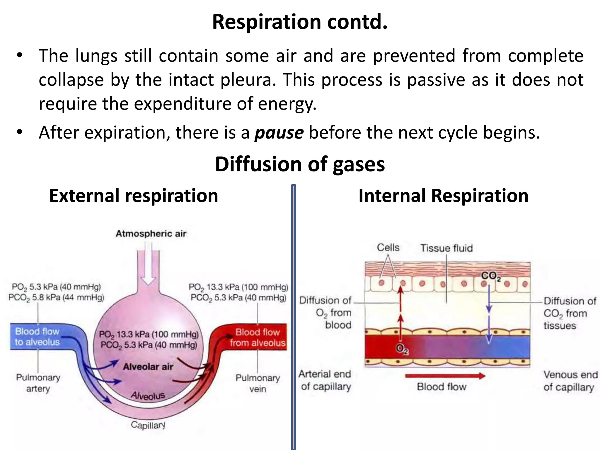
The respiratory system provides oxygen to the body's cells through respiration and removes carbon dioxide. It includes the nose, pharynx, larynx, trachea, bronchi, lungs, and muscles of respiration. Oxygen and carbon dioxide are exchanged between the alveoli and blood in the lungs through diffusion. Respiration is regulated by the respiratory center in the brainstem and chemoreceptors that detect changes in blood gases. Artificial respiration can prevent deaths by reopening the airway and exchanging gases until natural breathing resumes.


![Regulation of Respiration contd.
• Nerve impulses, generated in the peripheral chemoreceptors,
are conveyed by the glossopharyngeal and vagus nerves to the
medulla and stimulate the respiratory center.
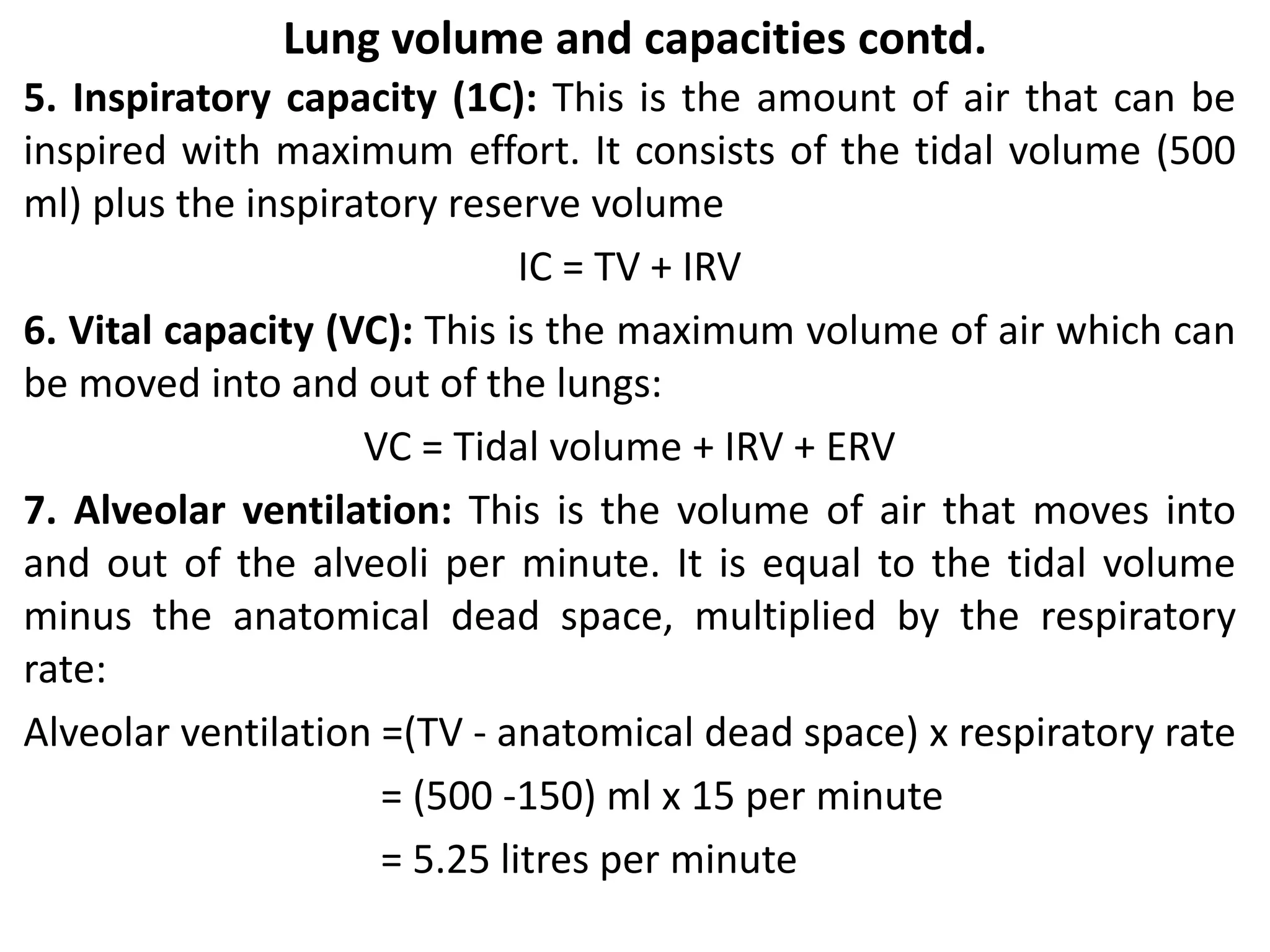
• The rate and depth of breathing are then increased. An increase
in blood acidity (decreased pH or raised [H+]) stimulates the
peripheral chemoreceptors, resulting in increased ventilation,
increased CO2 excretion and increased blood pH.
Other factors that influence respiration: Breathing may be
modified by the higher centres in the brain by:
• Speech, singing
• Emotional displays, e.g. crying, laughing, fear
• Drugs, e.g. sedatives, alcohol
• Sleep](https://image.slidesharecdn.com/4-220919104150-6577ca81/75/5-Respiratory-system-pptx-26-2048.jpg)