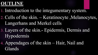
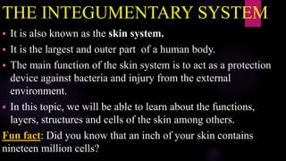
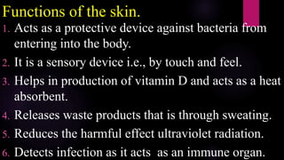
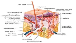
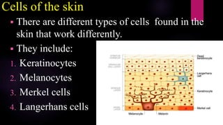
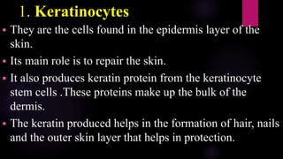
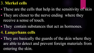
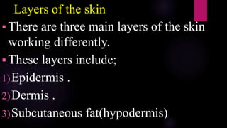
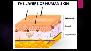
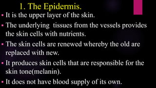
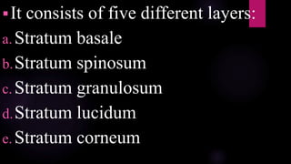
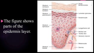
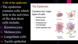
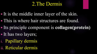
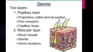
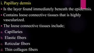
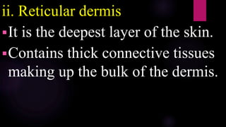
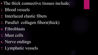
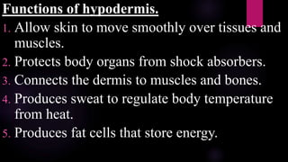
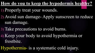
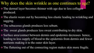
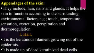
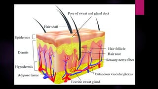
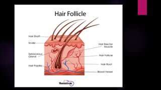
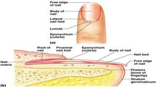
KIPTOO EVA JERUTO
DIPLOMA IN DENTALASSISTING
DDA540/23
THE INTEGUMENTARY SYSTEM
OUTLINE
1. Introduction to the integumentary system.
2. Cells of the skin. – Keratinocyte ,Melanocytes,
Langerhans and Merkel cells
3. Layers of the skin.- Epidermis, Dermis and
Hypodermis
4. Appendages of the skin – Hair, Nail and
Glands
THE INTEGUMENTARY SYSTEM
 It is also known as the skin system.
 It is the largest and outer part of a human body.
 The main function of the skin system is to act as a protection
device against bacteria and injury from the external
environment.
 In this topic, we will be able to learn about the functions,
layers, structures and cells of the skin among others.
Fun fact: Did you know that an inch of your skin contains
nineteen million cells?
Functions of the skin.
1. Acts as a protective device against bacteria from
entering into the body.
2. It is a sensory device i.e., by touch and feel.
3. Helps in production of vitamin D and acts as a heat
absorbent.
4. Releases waste products that is through sweating.
5. Reduces the harmful effect ultraviolet radiation.
6. Detects infection as it acts as an immune organ.
Cells of the skin
 There are different types of cells found in the
skin that work differently.
 They include:
1. Keratinocytes
2. Melanocytes
3. Merkel cells
4. Langerhans cells
1. Keratinocytes
 They are the cells found in the epidermis layer of the
skin.
 Its main role is to repair the skin.
 It also produces keratin protein from the keratinocyte
stem cells .These proteins make up the bulk of the
dermis.
 The keratin produced helps in the formation of hair, nails
and the outer skin layer that helps in protection.
2. Melanocytes.
 They are responsible for the formation of melanin.
 It is dark and dendritic in shape
 There are two types of melanin;
i. Eumelanin- Primarily makes dark colors in your
hair, skin and eyes.
ii. Pheomelanin- Primarily makes pink or red colors
in the body e.g. Lips
3. Merkel cells
 These are the cells that help in the sensitivity of skin
 They are closer to the nerve ending where they
receive a sense of touch.
 They contain substances that act as hormones.
4. Langerhans cells
 They are basically the guards of the skin where they
are able to detect and prevent foreign materials from
entering the skin.
Layers of the skin
 There are three main layers of the skin
working differently.
 These layers include;
1)Epidermis .
2)Dermis .
3)Subcutaneous fat(hypodermis)
1. The Epidermis.
 It is the upper layer of the skin.
 The underlying tissues from the vessels provides
the skin cells with nutrients.
 The skin cells are renewed whereby the old are
replaced with new.
 It produces skin cells that are responsible for the
skin tone(melanin).
 It does not have blood supply of its own.
It consists of five different layers:
a.Stratum basale
b.Stratum spinosum
c.Stratum granulosum
d.Stratum lucidum
e.Stratum corneum
The figure shows
parts of the
epidermis layer.
Layers of the epidermis.
a. Stratum basale
 This is the deepest layer of the epidermis.
 They are responsible for;
I. Creation of new skin cells.
II. Production of keratin from the keratinocyte stem cells.
III. Through production of keratin the following are formed
hair, nails and the outer skin that acts as a protection
device
iv. Melanin production due to the
melanocyte cell.
b. Stratum spinosum
 This layer is found in between the
stratum basale and stratum granulosum
layer.
 Its main function is to help strengthen the
skin.
c. Stratum granulosum
 It is located in between the stratum spinosum and
stratum lucidum.
 The keratin is formed in this layer due to the
keratinocyte granules.
d. Stratum lucidum
 It is found in between stratum granulosum and
stratum corneum.
e. Stratum corneum
It is the outermost layer of epidermis layer.
This is where the keratinocytes are converted
into the corneocytes.
The corneocytes are the strong dead
keratinocytes.
The corneocytes are responsible to protect one
from harm e.g.; ultraviolet radiation, pathogens
and heat
It contains fat that prevents loss of
moisture by keeping water from
entering or leaving the body.
The new keratinocytes cells from the
stratum basale then replaces the
corneocytes that are shed.
FORMULAR FOR EPIDERMIS
LAYER
Come-Stratum corneum
Let’s – Stratum lucidum
Get – Stratum granulosum
Sun – Stratum spinosum
Burned – Stratum basale
Functions of the epidermis
1. It helps in hydration- stratum corneum
2. Production of new skin cells- stratum basale
3. Acts as a protection device- stratum corneum
4. It is responsible for skin color-stratum basale
How does the epidermis help with other organs?
It protects skeletal system, muscles and tissues from
harm
Cells of the epidermis
 The epidermis
contains cells which
help in the activities
of the skin these
cells include;
I. Keratinocytes
II. Melanocytes
III. Langerhans cells
IV.Tactile epithelial
2.The Dermis
 It is the middle inner layer of the skin.
 This is where hair structures are found.
 Its principle component is collagen(protein)
 It has two layers;
i. Papillary dermis
ii. Reticular dermis
i. Papillary dermis
 Is the layer found immediately beneath the epidermis.
 Contains loose connective tissues that is highly
vascularized.
 The loose connective tissues include;
a. Capillaries
b. Elastic fibers
c. Reticular fibers
d. Thin collagen fibers
ii. Reticular dermis
It is the deepest layer of the skin.
Contains thick connective tissues
making up the bulk of the dermis.
 The thick connective tissues include;
1. Blood vessels
2. Interlaced elastic fibers
3. Parallel collagen fibers(thick)
4. Fibroblasts
5. Mast cells
6. Nerve endings
7. Lymphatic vessels
Structures in the dermis.
Connective tissues
Blood capillaries- Supplies nutrients and oxygen and take away
cell waste from the skin
Lymphatic vessels- They destroy bacteria causing infections.
Sweat glands- produces sweat as a waste product out of the skin.
Sebaceous glands- Secrete oil that keep the skin smooth and
protects the skin from bacteria as a waterproof device.
Nerve endings- Acts as a sensing device for feel, touch, harm etc.
Hair follicles- Nourishes the hair to grow.
Tissues of the dermis.
1. Collagen- Strengthens the skin.
2. Elastic tissues- Enables flexibility of the skin by
producing elastin.
3. Reticular fibers- Controls blood pressure of
vascular cells.
Functions of the dermis.
1.Excretion.
Contains sweat gland where sweat is produced and released though the skin.
2.Regulates body temperature.
When the body temperature is high it releases sweat that cools off the body.
3. Produces oil that inhibits bacterial growth on the skin.
4. Growing hair through the hair follicles that contract when body is cold and
scared causing goose bumps.
5. The nerve endings enables one to experience feeling of either touch, hurt
,itches etc.
6.Distributing blood through the blood vessels to the epidermis it also
removes toxins enabling the skin to strengthen.
7.Enables skin protection due to phagocyte cells that consume harmful
substances .
3.The Hypodermis
 It is the deepest layer of the skin.
 It is also known as subcutis or subcutaneous
tissue.
 This is the layer that contains fat cells beneath
the dermis
 The hypodermis thickness varies across the
body i.e.;
o High testosterone- Thick hypodermis in abdomen,
arms, lower back and shoulders.
o High estrogen- Thick hypodermis in butt, hips and
thighs.
Function of the oil in hypodermis.
1.Insulates body from the cold due to fat produced.
2.Acts as a cushion for internal structures e.g.;
muscles and organs when something hits you.
Functions of hypodermis.
1. Allow skin to move smoothly over tissues and
muscles.
2. Protects body organs from shock absorbers.
3. Connects the dermis to muscles and bones.
4. Produces sweat to regulate body temperature
from heat.
5. Produces fat cells that store energy.
How do you to keep the hypodermis healthy?
1) Properly treat your wounds.
2) Avoid sun damage- Apply sunscreen to reduce
sun damage.
3) Take precautions to avoid burns.
4) Keep your body to avoid hypothermia or
frostbite.
Hypothermia- is a systematic cold injury.
Why does the skin wrinkle as one continues to age?
 The dermal layer becomes thinner with age due to less collagen
produced.
 The elastin wears out by becoming less elastic leading to wrinkling and
sagging.
 The sebaceous glands produces less sebum.
 The sweat glands produces less sweat contributing to dry skin.
 Surface area/contact between dermis and epidermis decreases hence,
leading to less supply of blood to the epidermis from dermis and fewer
nutrients making it to the outer skin layer.
 The flattening out of the connecting region makes skin more fragile.
SKIN RECEPTORS.
 Near surface skin receptors.- Detect gentle
pressure, temperature and vibrations and pain.
(naked nerve endings) Dendrites
Deep in skin receptors. – Detect hair motions
and responds to strong pressure vibrations.
Appendages of the skin.
They include; hair, nails and glands. It helps the
skin to function according to the surrounding
environmental factors e.g.; touch, temperature
sensation, excretion, perspiration and
thermoregulation.
1. Hair.
•It is the keratinous filament growing out of the
epidermis.
•It is made up of dead keratinized dead cells.
• It is comprised of the hair shaft and hair follicle
a. Hair shaft
• It is the hair that is visible outside the skin.
• It contains cuticle cells which surround the cortex. The
cortical layer is comprised of keratin providing structure
to the bulk of hair shaft.
b. Hair follicle
• It is the primary structure for hair growth
• This is where the strands of hair originate from.
Terms of the hair
i. Hair root - It is where the rest of the hair is found below the skin.
ii. Hair bulb - It is where the hair root ends .Found in the dermis
iii. Hair matrix - It is the layer in which the basal cells are active
iv. Hair papilla - It is the content surrounded by the hair bulb. It is made up of
connective tissue and contains blood capillaries and nerve endings from the dermis
v. Medulla - It is the central core of the hair surrounded by the cortex .
vi. Cuticle - It is the layer of compressed keratinized cells that is covered by an
outer layer of hard keratinized cells.
vii. Hair loss - It occurs when more hair is shed than is being replaced. This
happens when there is hormonal or dietary changes. It can also be due to aging
process or hormone influence.
Structures of the hair follicle.
• It consists of three layers of cells;
1. Internal root sheath cells
• It extends up to the hair shaft and surrounds the root of
growing hair.
• It is produced in the basal cells of the matrix.
2. External root sheath cells
• It is made up of the basal cells at the base of root hair
and the upper region is keratinous.
• It is an extension of the epidermis that
encloses the hair root.
3.Glassy membrane
• It is a thick connective tissue covering the
hair root and connects to the dermis tissue
The hair follicle has layers of cells that
form from basal cells in hair matrix and
hair root
Hair texture
• This is determined by the shape and structure of the cortex which is
also determined by the shape of the hair follicle.
• There are two types ;
• Straight
• Curly
Hair color
• Melanocyte in the hair papilla creates the melanin pigment that gives
the hair color.
• The different hair color is due to the different types of melanin.
• When a person ages melanin production reduces and their hair tends to
lose its color and becomes grey or white.
How does the hair grow?
When the keratinocytes from the hair bulb is
produced ,new cells are deposited at the hair bulb
where the hair shaft is pushed through the hair
follicle towards the surface. These cells are then
pushed to the skin surface to form the hair shaft that
is visible. The hair that grows out of the skin is
composed of dead keratin. That is why when you
shave or cut your hair it doesn’t have sensation.
Hair growth occurs in three phases;
1. Anagen phase - Cells divide root hair and pushes
the hair shaft up and out.
2. Catagen phase - It marks the transition from the
hair follicle’s active growth.
3. Telogen phase - Hair follicle is at rest and no new
growth occurs.
Then it goes to the first phase.
CATAGEN
PHASE
TELOGEN
PHASE
ANAGEN PHASE
Functions of the hair
1. Communication through nerve signals by making the external hair
shaft stand up when one is frightened(goosebumps). Also when cats
are frightened they raise their fur. Arrector pili responds to nerve
signals(from sympathetic nervous system) by contracting
2. Thermoregulation (traps a layer of air to add insulation).
3. Acts as a sensing device i.e. To the air movement and substances in
the environment, detects presence of insects.
4. Acts as a protective device :
Traps dust from entering the eyes and nose, it also excludes dust particles
that may contain allergens and microbes
Eyelashes and eyebrows protect eyes from dirt and water(blinking).
2. Nail
• They are accessory structures of the integumentary system.
• Nail bed - Is a specialized structure of the epidermis found at the
tip of our fingers and toes. The blood vessel makes the nail bed
appear pink.
• Nail body -
• It is formed on the nail bed.
• It prevents our fingers and toes from mechanical stress.
• It is composed of densely parked keratinocytes.
• Nail fold - Found on the sides of the nail that anchors the nail
body.
• Nail cuticle - It is the meeting point between the nail
fold to the proximal end of the nail body. Also known as
Eponichium.
• Lunula(little moon) - It is the crescent-like shape on the
fingernails. It the part of the nail that doesn’t appear pink
due to the thick layer epithelium at the nail matrix.
• Nail matrix - It is the root of nail that enables it to grow.
• Hyponichium - It is the area beneath the fingernail
extension.it consists of a thickened layer of stratum
corneum.
3. Glands
• They are the organs of the endocrine system.
• They are produced and released to perform a
specific function.
• Their main function is to release ; water, salt or oil
under the skin to the surface of the skin.
• There are four main types of glands: Sebaceous
glands(sebum), Ceruminous glands(wax),
Mammary glands(milk), Sudoriferous
glands(sweat)
1. Sebaceous glands
• They secrete sebum(oil) and gives the face oil and
protects skin from becoming dry which reaches the skin
surface through ducts.
• Sebum is a mixture of lipids that forms a thin film on the
skin.
• It’s part of the pilosebaceous unit that include: hair, hair
follicle and arrector pili muscles.
• They are involved in skin problems such as: acne and
keratosis pilaris
Functions
a.Acts as a protective layer to the skin
b.Prevents fluid loss
c.Plays an antimicrobial role by being
a bimolecular barrier that disrupts
bacterial membranes.
2. Ceruminous glands
• They are ear glands that secrete wax.
• Its main function is to keep the outer surface of
the eardrum flexible and also to prevent the ear
from drying .
• It also assists in cleaning and lubrication.
• Provides protection from bacteria, fungus and
insects.
Wax from the ear that is
secreted by the
ceruminous glands
3. Mammary glands.
• They are glands found on the chest of a human
being.
• They secrete milk when a human being or an
animal gives birth. This occurs when exocrine
glands are enlarged and modifies sweat glands.
Due to the skin receptors when a baby sucks
on the breast of a mother it sends nerve signals
to the brain of a mother and milk is produced.
Components of the mammary gland
Alveoli
• It is lined with milk secreting epithelial cells surrounded
by the myoepithelial cells.
• They join up to form lobules. Each has a lactiferous duct
that drains into the openings in the nipple.
• The myoepithelial cells contract pushing milk from
alveoli through lactiferous duct towards the nipple
where it collects in the widening(sinuses) of the duct
Types of mammary glands
1. Simple mammary glands
• Consists of milk-secreting tissue leading to a single lactiferous duct.
2. Complex mammary gland
• Consists of all simple mammary gland serving one nipple.
• As human beings, we only have complex mammary gland in each
breast.
• The presence of more than two nipples is known as polythelia
• The presence of more than two complex mammary gland is called
polymastia
4. Sudoriferous glands
• It is also known as sweat glands.
• They are located in the dermis.
• They secrete sweat through the skin.
• Sweat produced produces 99% of water.
• There are two types of sweat glands;
• Apocrine sweat glands
• Eccrine sweat glands
i. Apocrine glands which are responsible for odor
as bacteria breaks down the secreted organic
substances. They are found in the armpits, scalp
and genital regions. They become active during
puberty and secrete their substance in to hair
follicles.
ii. Eccrine glands which produces serous fluids that
regulates temperature. They are found on the
palms, feet, armpits and forehead. They excrete
their substances directly to the surface of the skin.
THANK YOU

INTEGUMENTARY SYSTEM BY EVA KIPTOO.pptx

  • 1.
    KIPTOO EVA JERUTO DIPLOMAIN DENTALASSISTING DDA540/23 THE INTEGUMENTARY SYSTEM
  • 2.
    OUTLINE 1. Introduction tothe integumentary system. 2. Cells of the skin. – Keratinocyte ,Melanocytes, Langerhans and Merkel cells 3. Layers of the skin.- Epidermis, Dermis and Hypodermis 4. Appendages of the skin – Hair, Nail and Glands
  • 3.
    THE INTEGUMENTARY SYSTEM It is also known as the skin system.  It is the largest and outer part of a human body.  The main function of the skin system is to act as a protection device against bacteria and injury from the external environment.  In this topic, we will be able to learn about the functions, layers, structures and cells of the skin among others. Fun fact: Did you know that an inch of your skin contains nineteen million cells?
  • 4.
    Functions of theskin. 1. Acts as a protective device against bacteria from entering into the body. 2. It is a sensory device i.e., by touch and feel. 3. Helps in production of vitamin D and acts as a heat absorbent. 4. Releases waste products that is through sweating. 5. Reduces the harmful effect ultraviolet radiation. 6. Detects infection as it acts as an immune organ.
  • 6.
    Cells of theskin  There are different types of cells found in the skin that work differently.  They include: 1. Keratinocytes 2. Melanocytes 3. Merkel cells 4. Langerhans cells
  • 7.
    1. Keratinocytes  Theyare the cells found in the epidermis layer of the skin.  Its main role is to repair the skin.  It also produces keratin protein from the keratinocyte stem cells .These proteins make up the bulk of the dermis.  The keratin produced helps in the formation of hair, nails and the outer skin layer that helps in protection.
  • 8.
    2. Melanocytes.  Theyare responsible for the formation of melanin.  It is dark and dendritic in shape  There are two types of melanin; i. Eumelanin- Primarily makes dark colors in your hair, skin and eyes. ii. Pheomelanin- Primarily makes pink or red colors in the body e.g. Lips
  • 9.
    3. Merkel cells These are the cells that help in the sensitivity of skin  They are closer to the nerve ending where they receive a sense of touch.  They contain substances that act as hormones. 4. Langerhans cells  They are basically the guards of the skin where they are able to detect and prevent foreign materials from entering the skin.
  • 10.
    Layers of theskin  There are three main layers of the skin working differently.  These layers include; 1)Epidermis . 2)Dermis . 3)Subcutaneous fat(hypodermis)
  • 12.
    1. The Epidermis. It is the upper layer of the skin.  The underlying tissues from the vessels provides the skin cells with nutrients.  The skin cells are renewed whereby the old are replaced with new.  It produces skin cells that are responsible for the skin tone(melanin).  It does not have blood supply of its own.
  • 13.
    It consists offive different layers: a.Stratum basale b.Stratum spinosum c.Stratum granulosum d.Stratum lucidum e.Stratum corneum
  • 14.
    The figure shows partsof the epidermis layer.
  • 15.
    Layers of theepidermis. a. Stratum basale  This is the deepest layer of the epidermis.  They are responsible for; I. Creation of new skin cells. II. Production of keratin from the keratinocyte stem cells. III. Through production of keratin the following are formed hair, nails and the outer skin that acts as a protection device
  • 16.
    iv. Melanin productiondue to the melanocyte cell. b. Stratum spinosum  This layer is found in between the stratum basale and stratum granulosum layer.  Its main function is to help strengthen the skin.
  • 17.
    c. Stratum granulosum It is located in between the stratum spinosum and stratum lucidum.  The keratin is formed in this layer due to the keratinocyte granules. d. Stratum lucidum  It is found in between stratum granulosum and stratum corneum.
  • 18.
    e. Stratum corneum Itis the outermost layer of epidermis layer. This is where the keratinocytes are converted into the corneocytes. The corneocytes are the strong dead keratinocytes. The corneocytes are responsible to protect one from harm e.g.; ultraviolet radiation, pathogens and heat
  • 19.
    It contains fatthat prevents loss of moisture by keeping water from entering or leaving the body. The new keratinocytes cells from the stratum basale then replaces the corneocytes that are shed.
  • 20.
    FORMULAR FOR EPIDERMIS LAYER Come-Stratumcorneum Let’s – Stratum lucidum Get – Stratum granulosum Sun – Stratum spinosum Burned – Stratum basale
  • 21.
    Functions of theepidermis 1. It helps in hydration- stratum corneum 2. Production of new skin cells- stratum basale 3. Acts as a protection device- stratum corneum 4. It is responsible for skin color-stratum basale How does the epidermis help with other organs? It protects skeletal system, muscles and tissues from harm
  • 22.
    Cells of theepidermis  The epidermis contains cells which help in the activities of the skin these cells include; I. Keratinocytes II. Melanocytes III. Langerhans cells IV.Tactile epithelial
  • 23.
    2.The Dermis  Itis the middle inner layer of the skin.  This is where hair structures are found.  Its principle component is collagen(protein)  It has two layers; i. Papillary dermis ii. Reticular dermis
  • 25.
    i. Papillary dermis Is the layer found immediately beneath the epidermis.  Contains loose connective tissues that is highly vascularized.  The loose connective tissues include; a. Capillaries b. Elastic fibers c. Reticular fibers d. Thin collagen fibers
  • 26.
    ii. Reticular dermis Itis the deepest layer of the skin. Contains thick connective tissues making up the bulk of the dermis.
  • 27.
     The thickconnective tissues include; 1. Blood vessels 2. Interlaced elastic fibers 3. Parallel collagen fibers(thick) 4. Fibroblasts 5. Mast cells 6. Nerve endings 7. Lymphatic vessels
  • 28.
    Structures in thedermis. Connective tissues Blood capillaries- Supplies nutrients and oxygen and take away cell waste from the skin Lymphatic vessels- They destroy bacteria causing infections. Sweat glands- produces sweat as a waste product out of the skin. Sebaceous glands- Secrete oil that keep the skin smooth and protects the skin from bacteria as a waterproof device. Nerve endings- Acts as a sensing device for feel, touch, harm etc. Hair follicles- Nourishes the hair to grow.
  • 29.
    Tissues of thedermis. 1. Collagen- Strengthens the skin. 2. Elastic tissues- Enables flexibility of the skin by producing elastin. 3. Reticular fibers- Controls blood pressure of vascular cells.
  • 30.
    Functions of thedermis. 1.Excretion. Contains sweat gland where sweat is produced and released though the skin. 2.Regulates body temperature. When the body temperature is high it releases sweat that cools off the body. 3. Produces oil that inhibits bacterial growth on the skin. 4. Growing hair through the hair follicles that contract when body is cold and scared causing goose bumps. 5. The nerve endings enables one to experience feeling of either touch, hurt ,itches etc. 6.Distributing blood through the blood vessels to the epidermis it also removes toxins enabling the skin to strengthen. 7.Enables skin protection due to phagocyte cells that consume harmful substances .
  • 31.
    3.The Hypodermis  Itis the deepest layer of the skin.  It is also known as subcutis or subcutaneous tissue.  This is the layer that contains fat cells beneath the dermis  The hypodermis thickness varies across the body i.e.;
  • 32.
    o High testosterone-Thick hypodermis in abdomen, arms, lower back and shoulders. o High estrogen- Thick hypodermis in butt, hips and thighs. Function of the oil in hypodermis. 1.Insulates body from the cold due to fat produced. 2.Acts as a cushion for internal structures e.g.; muscles and organs when something hits you.
  • 33.
    Functions of hypodermis. 1.Allow skin to move smoothly over tissues and muscles. 2. Protects body organs from shock absorbers. 3. Connects the dermis to muscles and bones. 4. Produces sweat to regulate body temperature from heat. 5. Produces fat cells that store energy.
  • 34.
    How do youto keep the hypodermis healthy? 1) Properly treat your wounds. 2) Avoid sun damage- Apply sunscreen to reduce sun damage. 3) Take precautions to avoid burns. 4) Keep your body to avoid hypothermia or frostbite. Hypothermia- is a systematic cold injury.
  • 35.
    Why does theskin wrinkle as one continues to age?  The dermal layer becomes thinner with age due to less collagen produced.  The elastin wears out by becoming less elastic leading to wrinkling and sagging.  The sebaceous glands produces less sebum.  The sweat glands produces less sweat contributing to dry skin.  Surface area/contact between dermis and epidermis decreases hence, leading to less supply of blood to the epidermis from dermis and fewer nutrients making it to the outer skin layer.  The flattening out of the connecting region makes skin more fragile.
  • 36.
    SKIN RECEPTORS.  Nearsurface skin receptors.- Detect gentle pressure, temperature and vibrations and pain. (naked nerve endings) Dendrites Deep in skin receptors. – Detect hair motions and responds to strong pressure vibrations.
  • 37.
    Appendages of theskin. They include; hair, nails and glands. It helps the skin to function according to the surrounding environmental factors e.g.; touch, temperature sensation, excretion, perspiration and thermoregulation. 1. Hair. •It is the keratinous filament growing out of the epidermis. •It is made up of dead keratinized dead cells.
  • 38.
    • It iscomprised of the hair shaft and hair follicle a. Hair shaft • It is the hair that is visible outside the skin. • It contains cuticle cells which surround the cortex. The cortical layer is comprised of keratin providing structure to the bulk of hair shaft. b. Hair follicle • It is the primary structure for hair growth • This is where the strands of hair originate from.
  • 39.
    Terms of thehair i. Hair root - It is where the rest of the hair is found below the skin. ii. Hair bulb - It is where the hair root ends .Found in the dermis iii. Hair matrix - It is the layer in which the basal cells are active iv. Hair papilla - It is the content surrounded by the hair bulb. It is made up of connective tissue and contains blood capillaries and nerve endings from the dermis v. Medulla - It is the central core of the hair surrounded by the cortex . vi. Cuticle - It is the layer of compressed keratinized cells that is covered by an outer layer of hard keratinized cells. vii. Hair loss - It occurs when more hair is shed than is being replaced. This happens when there is hormonal or dietary changes. It can also be due to aging process or hormone influence.
  • 40.
    Structures of thehair follicle. • It consists of three layers of cells; 1. Internal root sheath cells • It extends up to the hair shaft and surrounds the root of growing hair. • It is produced in the basal cells of the matrix. 2. External root sheath cells • It is made up of the basal cells at the base of root hair and the upper region is keratinous.
  • 41.
    • It isan extension of the epidermis that encloses the hair root. 3.Glassy membrane • It is a thick connective tissue covering the hair root and connects to the dermis tissue The hair follicle has layers of cells that form from basal cells in hair matrix and hair root
  • 42.
    Hair texture • Thisis determined by the shape and structure of the cortex which is also determined by the shape of the hair follicle. • There are two types ; • Straight • Curly Hair color • Melanocyte in the hair papilla creates the melanin pigment that gives the hair color. • The different hair color is due to the different types of melanin. • When a person ages melanin production reduces and their hair tends to lose its color and becomes grey or white.
  • 43.
    How does thehair grow? When the keratinocytes from the hair bulb is produced ,new cells are deposited at the hair bulb where the hair shaft is pushed through the hair follicle towards the surface. These cells are then pushed to the skin surface to form the hair shaft that is visible. The hair that grows out of the skin is composed of dead keratin. That is why when you shave or cut your hair it doesn’t have sensation.
  • 44.
    Hair growth occursin three phases; 1. Anagen phase - Cells divide root hair and pushes the hair shaft up and out. 2. Catagen phase - It marks the transition from the hair follicle’s active growth. 3. Telogen phase - Hair follicle is at rest and no new growth occurs. Then it goes to the first phase.
  • 45.
  • 46.
    Functions of thehair 1. Communication through nerve signals by making the external hair shaft stand up when one is frightened(goosebumps). Also when cats are frightened they raise their fur. Arrector pili responds to nerve signals(from sympathetic nervous system) by contracting 2. Thermoregulation (traps a layer of air to add insulation). 3. Acts as a sensing device i.e. To the air movement and substances in the environment, detects presence of insects. 4. Acts as a protective device : Traps dust from entering the eyes and nose, it also excludes dust particles that may contain allergens and microbes Eyelashes and eyebrows protect eyes from dirt and water(blinking).
  • 49.
    2. Nail • Theyare accessory structures of the integumentary system. • Nail bed - Is a specialized structure of the epidermis found at the tip of our fingers and toes. The blood vessel makes the nail bed appear pink. • Nail body - • It is formed on the nail bed. • It prevents our fingers and toes from mechanical stress. • It is composed of densely parked keratinocytes. • Nail fold - Found on the sides of the nail that anchors the nail body.
  • 50.
    • Nail cuticle- It is the meeting point between the nail fold to the proximal end of the nail body. Also known as Eponichium. • Lunula(little moon) - It is the crescent-like shape on the fingernails. It the part of the nail that doesn’t appear pink due to the thick layer epithelium at the nail matrix. • Nail matrix - It is the root of nail that enables it to grow. • Hyponichium - It is the area beneath the fingernail extension.it consists of a thickened layer of stratum corneum.
  • 52.
    3. Glands • Theyare the organs of the endocrine system. • They are produced and released to perform a specific function. • Their main function is to release ; water, salt or oil under the skin to the surface of the skin. • There are four main types of glands: Sebaceous glands(sebum), Ceruminous glands(wax), Mammary glands(milk), Sudoriferous glands(sweat)
  • 53.
    1. Sebaceous glands •They secrete sebum(oil) and gives the face oil and protects skin from becoming dry which reaches the skin surface through ducts. • Sebum is a mixture of lipids that forms a thin film on the skin. • It’s part of the pilosebaceous unit that include: hair, hair follicle and arrector pili muscles. • They are involved in skin problems such as: acne and keratosis pilaris
  • 54.
    Functions a.Acts as aprotective layer to the skin b.Prevents fluid loss c.Plays an antimicrobial role by being a bimolecular barrier that disrupts bacterial membranes.
  • 55.
    2. Ceruminous glands •They are ear glands that secrete wax. • Its main function is to keep the outer surface of the eardrum flexible and also to prevent the ear from drying . • It also assists in cleaning and lubrication. • Provides protection from bacteria, fungus and insects.
  • 56.
    Wax from theear that is secreted by the ceruminous glands
  • 57.
    3. Mammary glands. •They are glands found on the chest of a human being. • They secrete milk when a human being or an animal gives birth. This occurs when exocrine glands are enlarged and modifies sweat glands. Due to the skin receptors when a baby sucks on the breast of a mother it sends nerve signals to the brain of a mother and milk is produced.
  • 58.
    Components of themammary gland Alveoli • It is lined with milk secreting epithelial cells surrounded by the myoepithelial cells. • They join up to form lobules. Each has a lactiferous duct that drains into the openings in the nipple. • The myoepithelial cells contract pushing milk from alveoli through lactiferous duct towards the nipple where it collects in the widening(sinuses) of the duct
  • 59.
    Types of mammaryglands 1. Simple mammary glands • Consists of milk-secreting tissue leading to a single lactiferous duct. 2. Complex mammary gland • Consists of all simple mammary gland serving one nipple. • As human beings, we only have complex mammary gland in each breast. • The presence of more than two nipples is known as polythelia • The presence of more than two complex mammary gland is called polymastia
  • 60.
    4. Sudoriferous glands •It is also known as sweat glands. • They are located in the dermis. • They secrete sweat through the skin. • Sweat produced produces 99% of water. • There are two types of sweat glands; • Apocrine sweat glands • Eccrine sweat glands
  • 61.
    i. Apocrine glandswhich are responsible for odor as bacteria breaks down the secreted organic substances. They are found in the armpits, scalp and genital regions. They become active during puberty and secrete their substance in to hair follicles. ii. Eccrine glands which produces serous fluids that regulates temperature. They are found on the palms, feet, armpits and forehead. They excrete their substances directly to the surface of the skin.
  • 62.