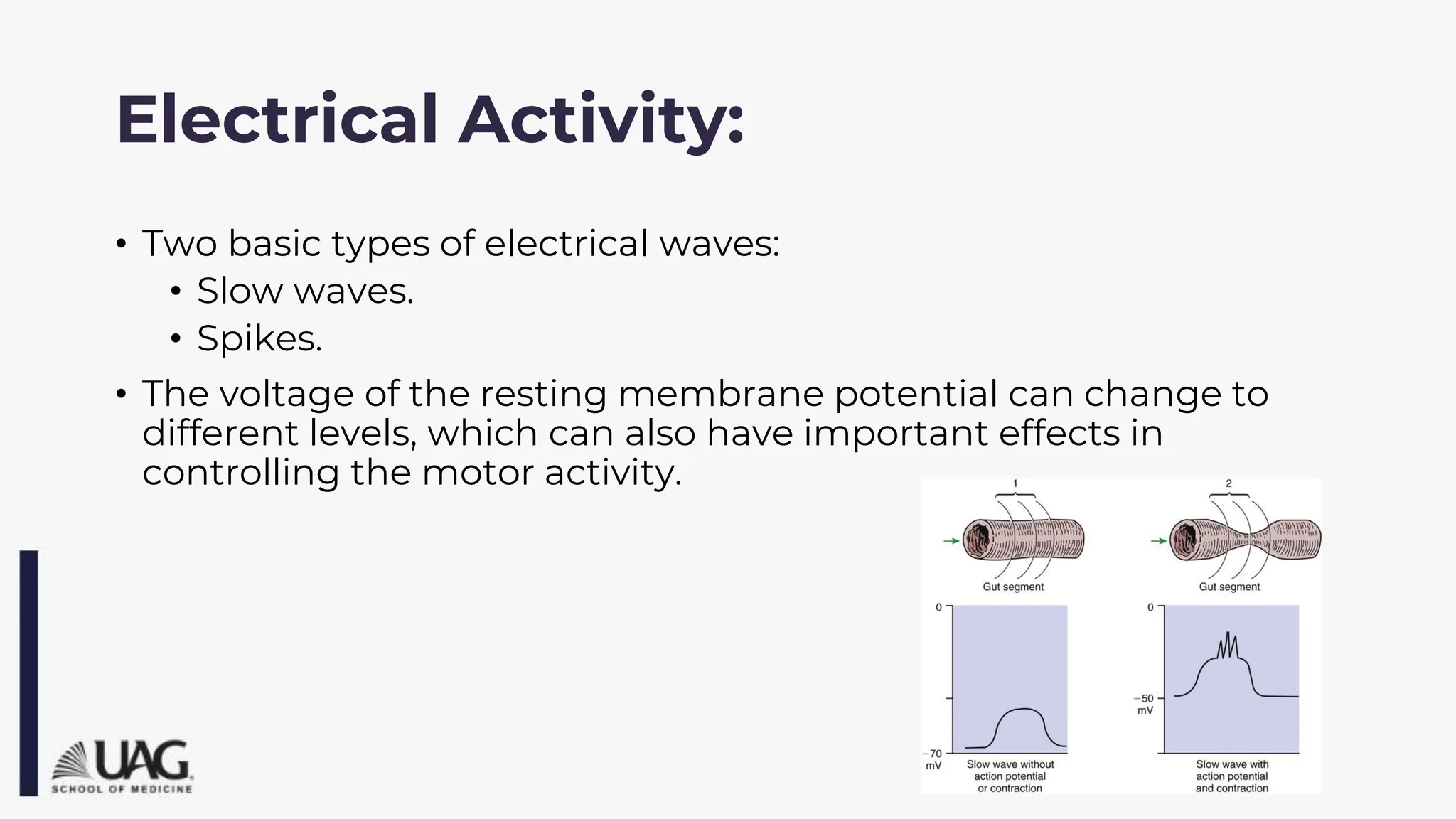
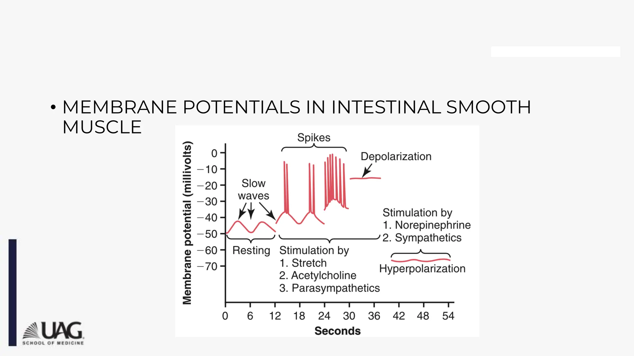
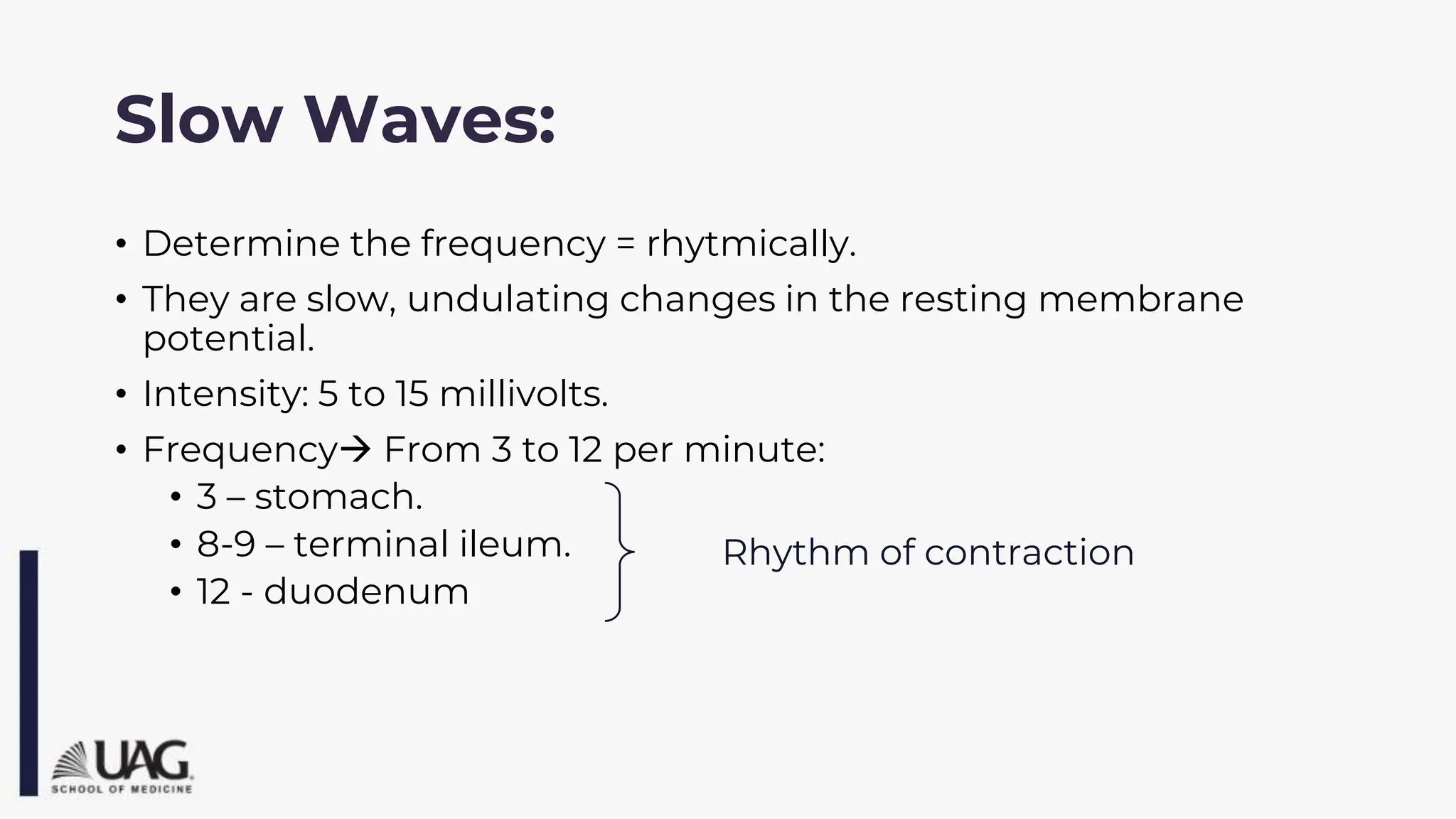
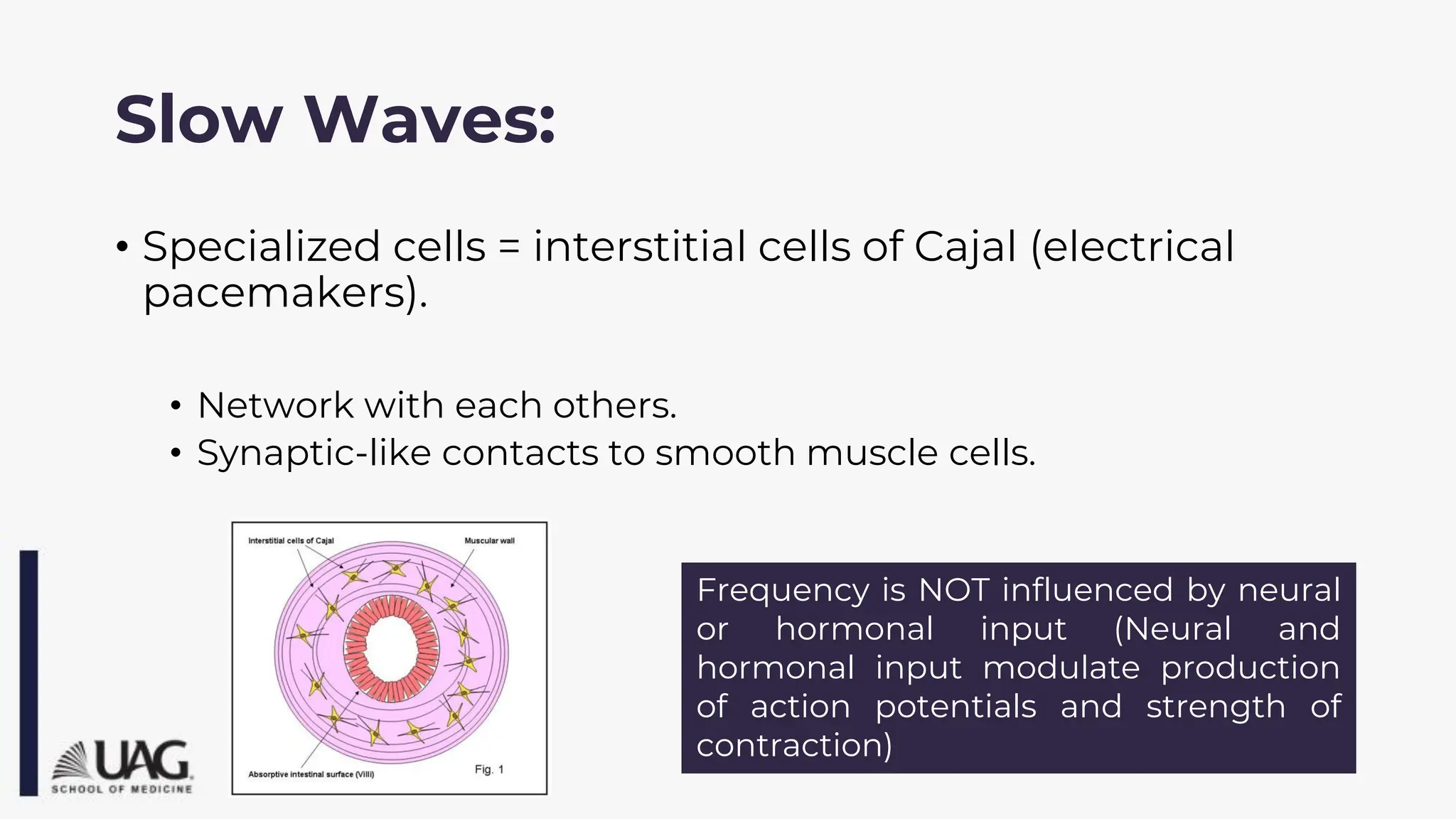
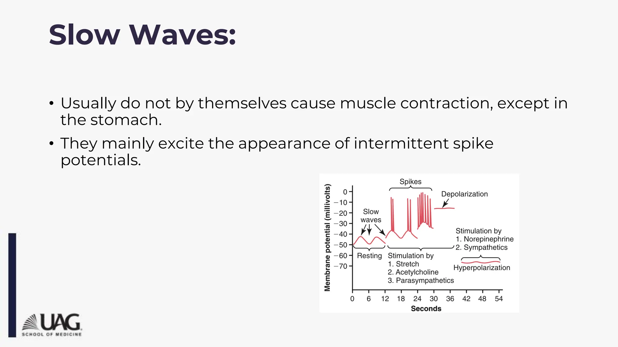
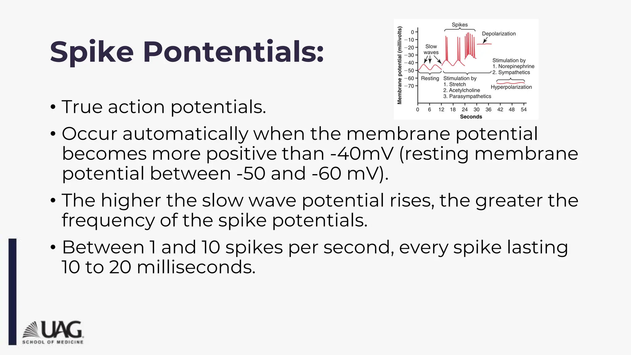
Organization and
regulation of the GI tract
Created : Miguel Alejandro Dávalos Benítez, MD
Modified by: Dr. Ortiz
Objectives:
• Understand the general arrangement of the GI tract including gross
anatomy and histology.
• Know the basic physiology behind the GI tract
• Recognize the characteristics associated with GI tract smooth muscle
• Describe the electrical activity of GI smooth muscle.
• Comprehend the role of the Nervous system in the organization and
functioning of the GI tract
• Know the association of the Autonomic nervous system in the changes in
the physiology of the GI tract.
• Identify the role of the Endocrine system in the physiology od the GI tract.
Generalities:
• The alimentary tract provides the body with a contiual
supply of:
• Water, electrolytes, vitamins and nutrients.
• For this purpose requieres:
• Movement of food through the alimentary tract.
• Secretion of digestive juices and digestion of the food.
• Absorption of water, electrolytes, vitamins and digestive
products.
• Circulation of blood flow through the gastrointestinal organs
to carry away the absorbed substances .
• Control of all these functions.
1
2
3
4
5
Gastrointestinal Smooth Muscle:
• The fibers of the smooth muscle are:
• 200 to 500 micrometers in lenght
• 2 to 10 micrometers in diameter
Arranged in bundles of
1000 parallel fibers.
The muscle fibers
are electrically
connected with
another through
large numbers of
gap junctions →
low resistance
movement of
ions from one
muscle cell to the
next.
Syncytium:
• Each muscle layer functions as a syncytium:
• When an action potential is elicited anywhere within the
muscle mass→ all directions.
• The distance that it travels depens on the excitability of the
muscle.
• Few connections exist between the longitudinal and circular
muscle layers, excitation of one of this layers often excites the
other as well.
Electrical Activity:
• Two basic types of electrical waves:
• Slow waves.
• Spikes.
• The voltage of the resting membrane potential can change to
different levels, which can also have important effects in
controlling the motor activity.
• MEMBRANE POTENTIALS IN INTESTINAL SMOOTH
MUSCLE
Slow Waves:
• Determine the frequency = rhytmically.
• They are slow, undulating changes in the resting membrane
potential.
• Intensity: 5 to 15 millivolts.
• Frequency→ From 3 to 12 per minute:
• 3 – stomach.
• 8-9 – terminal ileum.
• 12 - duodenum
Rhythm of contraction
Slow Waves:
• Specialized cells = interstitial cells of Cajal (electrical
pacemakers).
• Network with each others.
• Synaptic-like contacts to smooth muscle cells.
Frequency is NOT influenced by neural
or hormonal input (Neural and
hormonal input modulate production
of action potentials and strength of
contraction)
• Usually do not by themselves cause muscle contraction, except in
the stomach.
• They mainly excite the appearance of intermittent spike
potentials.
Slow Waves:
Spike Pontentials:
• True action potentials.
• Occur automatically when the membrane potential
becomes more positive than -40mV (resting membrane
potential between -50 and -60 mV).
• The higher the slow wave potential rises, the greater the
frequency of the spike potentials.
• Between 1 and 10 spikes per second, every spike lasting
10 to 20 milliseconds.
Factors that depolarize the
membrane:
• Stretching of the muscle.
• Stimulation by acetiycholine (parasympatic nerves).
• Stimulation by several specific gastrointestinal hormones.
• MORE POSITIVE = EASIER TO DEPOLARIZE = ACTION
Factors that hyperpolarize the
membrane:
• Norepinephrine or epinephrine in the fiber membrane.
• Stimulation of the sympathetic nerves -> secret
norepinephrine.
• MORE NEGATIVE = MORE DIFFICULT TO DEPOLARIZE =
NO ACTION
Calcium:
• Smooth muscle contraction occurs in response to entry
of calcium ions into the muscle fiber.
• Calcium activate the myosin filaments causing
attractive forces between the myosin and actin
filaments → Contraction.
• Slow waves only cause the entry of sodium but no
calcium = no contraction.
• Spike waves cause significant quantities of calcium ion
enter the fibers = contraction.
Tonic Contraction:
• Is continuous.
• Not associated with the basic electrical rhythm of the slow waves.
• Last several minutes or even hours.
• Increase or decrease in intensity, but continues.
• Caused by:
• Continuous repetitive spike potentials.
• Hormones – continuous partial depolarization.
• Continuos entry of calcium ions – unclear.
Neural Control:
Enteric Nervous System:
• It lies entirely in the wall of the gut beginnning in the esophagus
and extending to the anus.
• 100 million of neurons -> control of movements and secretion.
• 2 plexus:
• Myenteric plexus / Auerbach’s plexus→ gastrointestinal
movements.
• Submucosal plexus / Meissner’s plexus→ gastrointestinal
secretion and local blood flow.
Myenteric Plexus:
• Consist of a linear chain of many interconnecting neurons that
extends the entire lenght of the gastrointestinal tract.
• Lies between the longitudinal and circular layers of intestinal
smooth muscle.
• Concerned mainly with controlling muscle activity.
• When is stimulated, its principal effects are:
• Increased tonic contraction, or “tone”.
• Increased intensity.
• Increased rate.
• Increased velocity of conduction → more rapid movement.
• The myenteric plexus should not be considered entirely
excitatory because some of its neurons are inhibitory.
Submucosal Plexus:
• Controlling function in the inner wall.
• Many sensory signals originate from the gastrointestinal
epithelium.
• Help control:
• Local intestinal secretion.
• Local absorption.
• Local contraction.
Parasympathetic Stimulation:
• Divide in:
• Cranial: principally in the vagus nerve (X) -> esophagus,
stomach and pancreas.
• Sacral: 2, 3, 4th sacral segments -> large intestine and all the
way to the anus (defecation reflexes).
• Postganglionic neurons – located mainly in the
myenteric and submucosal plexus.
• Stimulation cause general increase in activity of the entire
enteric nervous system.
INCREASE ACTIVITY
Sympathetic Stimulation:
• Originate in the spinal cord between segments T5 and
L2.
• Innervate essentially all of the gastrointestinal tract.
• The nerve endings secret principally norepinephrine.
• The two principal effects:
• Inhibit intestinal tract smooth muscle.
• Inhibitory effect on the neurons.
INHIBITS
ACTIVITY
Afferent Sensory Nerve Fibers:
• These sensory nerves can be stimulated by:
• Irritation of the gut mucosa.
• Excessive distention of the gut.
• Presence of specific chemical substances in the gut.
• Signals transmitted through the fibers can then cause
excitation or, under other conditions, inhibition of
intestinal movements or intestinal secretion.
Gastrointestinal Reflexes:
• Three types of gastrointestinal reflexes:
• Integrated entirely within the gut wall:
• - Gastrointestinal secretion, peristalsis, mixing, contractions, local
inhibitiry effects.
• From the gut to the prevertebral sympathetic ganglia and back to
the GI tract:
• - Long distance reflexes (gastrocolic, enterogastric, colonoileal
reflexes.)
• From the gut to the spinal cord or brain stem and then back to the GI
tract:
• - Control gastric motor and secretory activity, pain and defecation
reflexes.
Hormonal Control:
Generalities:
• Physiological actions on target cells with specific
receptors for the hormone.
• The effects persist even after all nervous connections
between the site of release and the site of action have
been severed.
Gastrin:
• Stimuli for secretion: Protein, distension, nerve, (acid inhibit release).
• Site of secretion: G cells of the antrum, duodenum, and jejunum.
• Actions: Stimulates gastric acid secretion and mucosa growth.
Cholecystokinin:
• Stimuli for secretion: Protein, fat, acid.
• Site of secretion: I cells of the duodenum, jejunum and ileum.
• Actions:
• Stimulates: Pancreatic enzyme secretion, pancreatic
bicarbonate secretion, gallbladder contraction, growth of
exocrine pancreas.
• Inhibits: Gastric emtying.
Secretin:
• Stimuli for secretion: Fat, acid.
• Site of secretion: S cells of the duodenum, jejunum and ileum.
• Actions:
• Stimulates: Pepsin secretion, pancreatic bicarbonate
secretion, biliary bicarbonate secretion, growth of exocrine
pancreas.
• Inhibits: Gastric acid secretion.
Gastric Inhibitory Peptide:
• Stimuli for secretion: Protein, fat, carbohydrate.
• Site of secretion: K cells of the duodenum, jejunum.
• Actions:
• Stimulates: Insuline release.
• Inhibits: Gastric acid secretion.
Motilin:
• Stimuli for secretion: Fat, acid, nerve.
• Site of secretion: M cells of the duodenum, jejunum.
• Actions:
• Stimulates: Gastric & intestinal motility.
Which statement is false in regards of the electrical
activity of the GI tract smooth muscle?
a. Calcium entry is needed for smooth muscle contraction
b. Spikes last longer than slow waves
c. Interstitial cells of Cajal are needed for electrical generation of
slow waves.
d. Muscle layers in the GI tract wall share electrical connections
Question:
• A physiology experiment is conducted in an isolated rat small intestine. The
intestine is bathed with all essential nutrients, ions, and gases in a glass dish
maintained at a temperature of 37°C. The proximal jejunum is observed to contract
at a frequency of five contractions per minute. A glass micropipette is then inserted
into an interstitial cell of Cajal (pacemaker cell) at the same location in the jejunum,
and a slow-wave frequency of 10 contractions per minute is recorded.
Norepinephrine is then added to the bathing solution.
• Which of the following best describes the most likely slow-wave frequency and
contraction frequency after treatment with norepinephrine (in occurrences per
minute)?
Slow wave frequency Contraction frequency
A 0 0
B 10 0
C 10 10
D 10 5
E 5 10
Key Concepts:
• The GI tract is a tube subdivided into regions that subserve different functions associated
with digestion and absorption.
• The lining of the GI tract is subdivided into layers—the mucosal, submucosal, and muscle
layers.
• There are three major control mechanisms: hormonal, paracrine, and neurocrine.
• The innervation of the GI tract is particularly interesting because it consists of two
interacting components, extrinsic and intrinsic.
• Extrinsic innervation (cell bodies outside the wall of the GI tract) consists of the two
subdivisions of the ANS: parasympathetic and sympathetic. Both have an important
sensory (afferent) component.
• The intrinsic or enteric nervous system (cell bodies in the wall of the GI tract) can act
independently of extrinsic neural innervation.
• When a meal is in different regions of the tract, sensory mechanisms detect the presence
of the nutrients and mount appropriate physiological responses in that region of the tract,
as well as in more distal regions. These responses are mediated by endocrine, paracrine,
and neurocrine pathways.
References
• Hall, J. E. (2016). Guyton and Hall textbook of medical
physiology, chapter 63, pages 797-803.
• Functional Anatomy and General Principles of
Regulation in the Gastrointestinal Tract Bruce
M. Koeppen MD, PhD and Bruce A. Stanton PhD, Berne
and Levy Physiology, 27, 511-519
Organization of GI Tract 2024222222-2.pdf

Organization of GI Tract 2024222222-2.pdf

  • 1.
    Organization and regulation ofthe GI tract Created : Miguel Alejandro Dávalos Benítez, MD Modified by: Dr. Ortiz
  • 2.
    Objectives: • Understand thegeneral arrangement of the GI tract including gross anatomy and histology. • Know the basic physiology behind the GI tract • Recognize the characteristics associated with GI tract smooth muscle • Describe the electrical activity of GI smooth muscle. • Comprehend the role of the Nervous system in the organization and functioning of the GI tract • Know the association of the Autonomic nervous system in the changes in the physiology of the GI tract. • Identify the role of the Endocrine system in the physiology od the GI tract.
  • 3.
    Generalities: • The alimentarytract provides the body with a contiual supply of: • Water, electrolytes, vitamins and nutrients. • For this purpose requieres: • Movement of food through the alimentary tract. • Secretion of digestive juices and digestion of the food. • Absorption of water, electrolytes, vitamins and digestive products. • Circulation of blood flow through the gastrointestinal organs to carry away the absorbed substances . • Control of all these functions.
  • 5.
  • 6.
    Gastrointestinal Smooth Muscle: •The fibers of the smooth muscle are: • 200 to 500 micrometers in lenght • 2 to 10 micrometers in diameter Arranged in bundles of 1000 parallel fibers.
  • 7.
    The muscle fibers areelectrically connected with another through large numbers of gap junctions → low resistance movement of ions from one muscle cell to the next.
  • 8.
    Syncytium: • Each musclelayer functions as a syncytium: • When an action potential is elicited anywhere within the muscle mass→ all directions. • The distance that it travels depens on the excitability of the muscle. • Few connections exist between the longitudinal and circular muscle layers, excitation of one of this layers often excites the other as well.
  • 9.
    Electrical Activity: • Twobasic types of electrical waves: • Slow waves. • Spikes. • The voltage of the resting membrane potential can change to different levels, which can also have important effects in controlling the motor activity.
  • 10.
    • MEMBRANE POTENTIALSIN INTESTINAL SMOOTH MUSCLE
  • 11.
    Slow Waves: • Determinethe frequency = rhytmically. • They are slow, undulating changes in the resting membrane potential. • Intensity: 5 to 15 millivolts. • Frequency→ From 3 to 12 per minute: • 3 – stomach. • 8-9 – terminal ileum. • 12 - duodenum Rhythm of contraction
  • 12.
    Slow Waves: • Specializedcells = interstitial cells of Cajal (electrical pacemakers). • Network with each others. • Synaptic-like contacts to smooth muscle cells. Frequency is NOT influenced by neural or hormonal input (Neural and hormonal input modulate production of action potentials and strength of contraction)
  • 13.
    • Usually donot by themselves cause muscle contraction, except in the stomach. • They mainly excite the appearance of intermittent spike potentials. Slow Waves:
  • 14.
    Spike Pontentials: • Trueaction potentials. • Occur automatically when the membrane potential becomes more positive than -40mV (resting membrane potential between -50 and -60 mV). • The higher the slow wave potential rises, the greater the frequency of the spike potentials. • Between 1 and 10 spikes per second, every spike lasting 10 to 20 milliseconds.
  • 15.
    Factors that depolarizethe membrane: • Stretching of the muscle. • Stimulation by acetiycholine (parasympatic nerves). • Stimulation by several specific gastrointestinal hormones. • MORE POSITIVE = EASIER TO DEPOLARIZE = ACTION
  • 16.
    Factors that hyperpolarizethe membrane: • Norepinephrine or epinephrine in the fiber membrane. • Stimulation of the sympathetic nerves -> secret norepinephrine. • MORE NEGATIVE = MORE DIFFICULT TO DEPOLARIZE = NO ACTION
  • 17.
    Calcium: • Smooth musclecontraction occurs in response to entry of calcium ions into the muscle fiber. • Calcium activate the myosin filaments causing attractive forces between the myosin and actin filaments → Contraction. • Slow waves only cause the entry of sodium but no calcium = no contraction. • Spike waves cause significant quantities of calcium ion enter the fibers = contraction.
  • 19.
    Tonic Contraction: • Iscontinuous. • Not associated with the basic electrical rhythm of the slow waves. • Last several minutes or even hours. • Increase or decrease in intensity, but continues. • Caused by: • Continuous repetitive spike potentials. • Hormones – continuous partial depolarization. • Continuos entry of calcium ions – unclear.
  • 20.
  • 21.
    Enteric Nervous System: •It lies entirely in the wall of the gut beginnning in the esophagus and extending to the anus. • 100 million of neurons -> control of movements and secretion. • 2 plexus: • Myenteric plexus / Auerbach’s plexus→ gastrointestinal movements. • Submucosal plexus / Meissner’s plexus→ gastrointestinal secretion and local blood flow.
  • 23.
    Myenteric Plexus: • Consistof a linear chain of many interconnecting neurons that extends the entire lenght of the gastrointestinal tract. • Lies between the longitudinal and circular layers of intestinal smooth muscle. • Concerned mainly with controlling muscle activity. • When is stimulated, its principal effects are: • Increased tonic contraction, or “tone”. • Increased intensity. • Increased rate. • Increased velocity of conduction → more rapid movement.
  • 24.
    • The myentericplexus should not be considered entirely excitatory because some of its neurons are inhibitory.
  • 25.
    Submucosal Plexus: • Controllingfunction in the inner wall. • Many sensory signals originate from the gastrointestinal epithelium. • Help control: • Local intestinal secretion. • Local absorption. • Local contraction.
  • 26.
    Parasympathetic Stimulation: • Dividein: • Cranial: principally in the vagus nerve (X) -> esophagus, stomach and pancreas. • Sacral: 2, 3, 4th sacral segments -> large intestine and all the way to the anus (defecation reflexes). • Postganglionic neurons – located mainly in the myenteric and submucosal plexus. • Stimulation cause general increase in activity of the entire enteric nervous system. INCREASE ACTIVITY
  • 27.
    Sympathetic Stimulation: • Originatein the spinal cord between segments T5 and L2. • Innervate essentially all of the gastrointestinal tract. • The nerve endings secret principally norepinephrine. • The two principal effects: • Inhibit intestinal tract smooth muscle. • Inhibitory effect on the neurons. INHIBITS ACTIVITY
  • 29.
    Afferent Sensory NerveFibers: • These sensory nerves can be stimulated by: • Irritation of the gut mucosa. • Excessive distention of the gut. • Presence of specific chemical substances in the gut. • Signals transmitted through the fibers can then cause excitation or, under other conditions, inhibition of intestinal movements or intestinal secretion.
  • 30.
    Gastrointestinal Reflexes: • Threetypes of gastrointestinal reflexes: • Integrated entirely within the gut wall: • - Gastrointestinal secretion, peristalsis, mixing, contractions, local inhibitiry effects. • From the gut to the prevertebral sympathetic ganglia and back to the GI tract: • - Long distance reflexes (gastrocolic, enterogastric, colonoileal reflexes.) • From the gut to the spinal cord or brain stem and then back to the GI tract: • - Control gastric motor and secretory activity, pain and defecation reflexes.
  • 32.
  • 33.
    Generalities: • Physiological actionson target cells with specific receptors for the hormone. • The effects persist even after all nervous connections between the site of release and the site of action have been severed.
  • 34.
    Gastrin: • Stimuli forsecretion: Protein, distension, nerve, (acid inhibit release). • Site of secretion: G cells of the antrum, duodenum, and jejunum. • Actions: Stimulates gastric acid secretion and mucosa growth.
  • 35.
    Cholecystokinin: • Stimuli forsecretion: Protein, fat, acid. • Site of secretion: I cells of the duodenum, jejunum and ileum. • Actions: • Stimulates: Pancreatic enzyme secretion, pancreatic bicarbonate secretion, gallbladder contraction, growth of exocrine pancreas. • Inhibits: Gastric emtying.
  • 36.
    Secretin: • Stimuli forsecretion: Fat, acid. • Site of secretion: S cells of the duodenum, jejunum and ileum. • Actions: • Stimulates: Pepsin secretion, pancreatic bicarbonate secretion, biliary bicarbonate secretion, growth of exocrine pancreas. • Inhibits: Gastric acid secretion.
  • 37.
    Gastric Inhibitory Peptide: •Stimuli for secretion: Protein, fat, carbohydrate. • Site of secretion: K cells of the duodenum, jejunum. • Actions: • Stimulates: Insuline release. • Inhibits: Gastric acid secretion.
  • 38.
    Motilin: • Stimuli forsecretion: Fat, acid, nerve. • Site of secretion: M cells of the duodenum, jejunum. • Actions: • Stimulates: Gastric & intestinal motility.
  • 39.
    Which statement isfalse in regards of the electrical activity of the GI tract smooth muscle? a. Calcium entry is needed for smooth muscle contraction b. Spikes last longer than slow waves c. Interstitial cells of Cajal are needed for electrical generation of slow waves. d. Muscle layers in the GI tract wall share electrical connections
  • 40.
    Question: • A physiologyexperiment is conducted in an isolated rat small intestine. The intestine is bathed with all essential nutrients, ions, and gases in a glass dish maintained at a temperature of 37°C. The proximal jejunum is observed to contract at a frequency of five contractions per minute. A glass micropipette is then inserted into an interstitial cell of Cajal (pacemaker cell) at the same location in the jejunum, and a slow-wave frequency of 10 contractions per minute is recorded. Norepinephrine is then added to the bathing solution. • Which of the following best describes the most likely slow-wave frequency and contraction frequency after treatment with norepinephrine (in occurrences per minute)? Slow wave frequency Contraction frequency A 0 0 B 10 0 C 10 10 D 10 5 E 5 10
  • 41.
    Key Concepts: • TheGI tract is a tube subdivided into regions that subserve different functions associated with digestion and absorption. • The lining of the GI tract is subdivided into layers—the mucosal, submucosal, and muscle layers. • There are three major control mechanisms: hormonal, paracrine, and neurocrine. • The innervation of the GI tract is particularly interesting because it consists of two interacting components, extrinsic and intrinsic. • Extrinsic innervation (cell bodies outside the wall of the GI tract) consists of the two subdivisions of the ANS: parasympathetic and sympathetic. Both have an important sensory (afferent) component. • The intrinsic or enteric nervous system (cell bodies in the wall of the GI tract) can act independently of extrinsic neural innervation. • When a meal is in different regions of the tract, sensory mechanisms detect the presence of the nutrients and mount appropriate physiological responses in that region of the tract, as well as in more distal regions. These responses are mediated by endocrine, paracrine, and neurocrine pathways.
  • 42.
    References • Hall, J.E. (2016). Guyton and Hall textbook of medical physiology, chapter 63, pages 797-803. • Functional Anatomy and General Principles of Regulation in the Gastrointestinal Tract Bruce M. Koeppen MD, PhD and Bruce A. Stanton PhD, Berne and Levy Physiology, 27, 511-519