2005 gubbio, workshop interattivo, la stimolazione cardiaca convenzionale e quella multisito
•Download as PPT, PDF•
1 like•1,660 views

2005 gubbio, workshop interattivo, la stimolazione cardiaca convenzionale e quella multisito

Recommended
Recommended
More Related Content
What's hot
What's hot (15)
Similar to 2005 gubbio, workshop interattivo, la stimolazione cardiaca convenzionale e quella multisito
Similar to 2005 gubbio, workshop interattivo, la stimolazione cardiaca convenzionale e quella multisito (20)
More from Centro Diagnostico Nardi
More from Centro Diagnostico Nardi (20)
2005 gubbio, workshop interattivo, la stimolazione cardiaca convenzionale e quella multisito
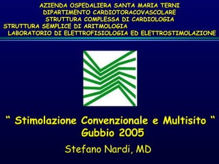
- 1. ““ Stimolazione Convenzionale e Multisito “Stimolazione Convenzionale e Multisito “ Gubbio 2005Gubbio 2005 Stefano Nardi, MD AZIENDA OSPEDALIERA SANTA MARIA TERNIAZIENDA OSPEDALIERA SANTA MARIA TERNI DIPARTIMENTO CARDIOTORACOVASCOLAREDIPARTIMENTO CARDIOTORACOVASCOLARE STRUTTURA COMPLESSA DI CARDIOLOGIASTRUTTURA COMPLESSA DI CARDIOLOGIA STRUTTURA SEMPLICE DI ARITMOLOGIASTRUTTURA SEMPLICE DI ARITMOLOGIA LABORATORIO DI ELETTROFISIOLOGIA ED ELETTROSTIMOLAZIONELABORATORIO DI ELETTROFISIOLOGIA ED ELETTROSTIMOLAZIONE
- 2. Fibrillazione Atriale CronicaFibrillazione Atriale Cronica o Atrio Silenteo Atrio Silente Fibrillazione Atriale CronicaFibrillazione Atriale Cronica o Atrio Silenteo Atrio Silente Tachiaritmie AtrialiTachiaritmie Atriali IntermittentiIntermittenti Tachiaritmie AtrialiTachiaritmie Atriali IntermittentiIntermittenti Pacing VentricolarePacing Ventricolare Normale o bradicardia sinusaleNormale o bradicardia sinusaleNormale o bradicardia sinusaleNormale o bradicardia sinusale La Conduzione AV è adeguata ?La Conduzione AV è adeguata ?La Conduzione AV è adeguata ?La Conduzione AV è adeguata ? DDDRDDDRDDDRDDDR DDDDDDDDDDDD AAIRAAIRAAIRAAIR AAIAAIAAIAAI La Frequenza cardiacaLa Frequenza cardiaca incrementaincrementa adeguatamenteadeguatamente con l’esercizio?con l’esercizio? SincroniaSincronia AVAV SS NN NN SS NN SS AnormaleAnormaleAnormaleAnormale La Conduzione AV è adeguata ?La Conduzione AV è adeguata ?La Conduzione AV è adeguata ?La Conduzione AV è adeguata ? SS NN La Frequenza cardiacaLa Frequenza cardiaca incrementa adeguatamenteincrementa adeguatamente con l’esercizio?con l’esercizio? SS NN DDD(R)*DDD(R)* DDI(R)DDI(R) DDD(R)*DDD(R)* DDI(R)DDI(R) DDDR*DDDR* DDIRDDIR DDDR*DDDR* DDIRDDIR VVI oVVI o VVIRVVIR VVI oVVI o VVIRVVIR La Frequenza cardiacaLa Frequenza cardiaca incrementa adeguatamenteincrementa adeguatamente con l’esercizio?con l’esercizio? Qual è la condizioneQual è la condizione dell’atrio?dell’atrio? * = Con algoritmo di cambio di modo automatico in caso di SVT* = Con algoritmo di cambio di modo automatico in caso di SVT Albero decisionale del modo di stimolazione ottimale
- 3. • Alterata MECCANICA Ventricolo Sinistro • La maggioranza dei pazienti (~77%) con SSS, compresi quelli CHF conduzione AV intatta e QRS stretto (attivazione ventricolare normale).6 • RVA pacing simula LBBB, con un conseguente QRS allargato, desincronizzazione ventricolare, ed alterazione struttura e funzione ventricolare.6,7 • Desincronizzazione “forzata” per RVA pacing aumenta il rischio di FA, CHF & decesso.1,4-6
- 4. Danish II Trial1 AAI(R) vs DDD(R) con AV corto vs DDD(R) con AV lungo CTOPP Trial4 DDD(R) or AAI(R) vs VVI(R) DAVID Trial5 DDD(R) vs VVI ICDs MOST Substudy6 DDDR vs VVIR Ospedaliz. per HF Non specificatamente misurata; lo studio indica che un’alta % di stim.RV riduce la funzione LV Non misurata 1 anno senza eventi (decesso o osped.per HF) era peggiore nel gruppo DDDR con %VP > 40% Incremento di 2.6 volte del rischio quando %VP > 40% (gruppo DDDR) Performance Emodinamica La stim. DDDR a lungo termine provoca dilatazione LA e un’alta % di stim.RV riduce la funzione LV Pazienti con intatta funzione LV, storia negativa per MI o CAD traggono il maggior beneficio dalla stimolazione fisiologica Non misurata Supporta la conclusione che la dissincronia V imposta dalla stimolazione V destra può essere deleteria in pazienti con ridotta funzione del ventricolo sinistroIncidenza di AF L’assenza di AF ai controlli è significativamente migliore con la stimolazione AAIR (p = 0.03); 17% stim. RV nel gruppo La stimolazione fisiologica riduce la probabilità di sviluppare AF cronica Non misurata Rischio aumentato linearmente dell’1% per ogni 1% di aumento della stim.V (fino all’ ~ 85%)
- 5. 0 1 2 3 4 0 20 40 60 80 100 Cum% VP RiskofAFrelativeto DDDRpatientwithCum%VP=0 0 1 2 3 4 0 20 40 60 80 100 Cum % VP RiskofAFrelativeto VVIRpatientwithCum%VP=0 Sweeney MO, et al. Circulation 2003;23:2932-2937 Sottostudio MOSTMOST: la % cumulativa di stimolazione ventricolare predice lo sviluppo di FA • Il rischio di FA aumenta linearmente con la % cumulativa di stim.V, fino a circa 80-85% sia nel gruppo DDDR che in quello VVIR • Il rischio di AF è ridotto dell’ 1% per ogni 1% di diminuzione del Cum %VP nel gruppo DDDR. Rischio Relativo - Studio MOST
- 6. Sottostudio MOST: DDDR vs VVIR, %VP e rischio di prima osped. per HF • Il RISCHIO RELATIVORISCHIO RELATIVO per la prima osped. per HF è sempre maggiore per pazienti VVIR rispetto a DDDR, indipendentemente dalla % di stimolazione 0 2 4 6 8 10 12 0- 20 20- 30 30- 40 > 40 Cum% VP RiskofHFH DDDR VVIR Sweeney MO, et al. Circulation 2003;23:2932-2937 Rischio Relativo - Studio MOST
- 7. 0 1 2 3 4 5 6 7 0 20 40 60 80 100 Cum % VP RiskofHFHrelativeto DDDRpatientwithCum%VP=0 Sweeney MO, et al. Circulation 2003;23:2932-2937 Sottostudio MOST: modo DDDR, % cumulativa di stim.V e rischio di prima ospedalizzazione per CHF. • Il rischio di ospedalizzazione per HF aumenta tra 0% e 40% di Cum VP, ma il rischio si stabilizza sopra il 40% di Cum VP • Il rischio è ridotto a circa il 2% se VP è minimizzata Rischio Relativo - Studio MOST
- 8. 0 1 2 3 4 5 6 7 0 20 40 60 80 100 Cum% VP RiskofHFHrelativeto DDDRpatientwithCum%VP=0 Sweeney MO, et al. Circulation 2003;23:2932-2937 Sottostudio MOSTMOST: modo DDDR, Cum %VP e rischio di prima ospedalizzazione per HF Quando stimolazione V > 40%: Il rischio relativo per i pazienti rimane pressochè costante e aumenta di 2.6 volte rispetto a pazienti con stimolazione V > 40% (es. con stim.V al 45% si ha lo stesso rischio relativo di 65%) • Quando stimolazione V < 40%: Per ogni 10% di riduzione nella % di stimolazione V. c’è una diminuzione relativa del 54% del rischio di prima ospedalizzaz.per HF Rischio Relativo - Studio MOST
- 9. • il modo DDD/R è migliore del VVI/R per gli esiti clinici a lungo termine6 – riduzione del rischio relativo per incidenza di AF – riduzione del rischio relativo per ospedalizzazione per scompenso cardiaco • la stimolazione “fisiologica” AAI/R è migliore della stimolazione DDD/R quando la % di stimolazione è alta.1,6
- 10. Incidence of Heart Failure AnnualRateper1000 30-39 40-49 50-59 60-69 70-79 80-89 Age Ho KL et al. JACC 1993 0 5 10 15 20 25 30 men women Conventional & Multisite PacingConventional & Multisite Pacing
- 11. NYHA CLASS Annualsurvival(%) Hospitalizations/year 100 75 50 25 0 I II III IV 1 10 Survival Hospitalization .1 Hospitalization / NYHA-class Conventional & Multisite PacingConventional & Multisite Pacing
- 12. Stroke Volume Preload Afterload Contractility Cardiac Output Heart Rate • Synergistic LV Contraction • Wall Integrity • Valvular Competence Determinants of Ventricular Function: Review Conventional & Multisite PacingConventional & Multisite Pacing
- 13. VolumeVolume OverloadOverload PressurePressure OverloadOverload Loss ofLoss of MyocardiumMyocardium ImpairedImpaired ContractilityContractility LV Systolic Dysfunction EF < 35% ↓↓ CardiacCardiac OutputOutput HypoperfusionHypoperfusion ↑↑ End Systolic VolumeEnd Systolic Volume ↑↑ End Diastolic VolumeEnd Diastolic Volume Pulmonary CongestionPulmonary Congestion LV Systolic Dysfunction Conventional & Multisite PacingConventional & Multisite Pacing
- 14. 1 Framingham Heart Study (1948 – 1988) in Atlas of Heart Diseases. 2 American Heart Association. Heart Disease and Stroke Statistics—2003 Update. CHF Patients Survival ResultsCHF Patients Survival Results11 100 90 80 70 60 50 40 30 20 10 0 Probabilityofsurvival,% Men (Men (nn = 237)= 237) Women (Women (nn = 230)= 230) Time after CHF diagnosis, years 0 2 4 6 8 10 80% of men and 70% of women who have CHF will die within 8 years.2 80% of men and 70% of women who have CHF will die within 8 years.2 Conventional & Multisite PacingConventional & Multisite Pacing
- 15. • Reduced LVEF remains the single most important risk factor for overall mortality and SCD.1 • Increased risk is measurable at LVEF 30%, but a LVEF < 30% is the single most powerful independent predictor for SCD.2 1 Prior SG, Aliot E, Blonstrom-Lundqvist C, et al. Task Force on Sudden Cardiac Death of the European Society of Cardiology. Eur Heart J, Vol. 22; 16; August 2001. 2 Myerburg RJ, Castellanos A. Cardiac Arrest and Sudden Cardiac Death, in Braunwald E, Zipes DP, Libby P, Heart Disease, A textbook of Cardiovascular Medicine. 6th ed. 2001. W.B. Saunders, Co., p. 895. Relationship of SCDRelationship of SCD and LV Dysfunctionand LV Dysfunction Conventional & Multisite PacingConventional & Multisite Pacing
- 16. Prognosis with Ventricular Dyssynchrony Long-term (45 Mo) Survival 34% 49% QRS < 120 ms QRS > 120 ms Iuliano et al. AHJ 2002 N=669 P < 0.001 1 Year Survival 11% 16% QRS < 120 ms QRS > 120 ms P < 0.001 Baldasseroni S. EHJ 2002 N=5,517 Conventional & Multisite PacingConventional & Multisite Pacing
- 17. Prevalence of VentricularPrevalence of Ventricular Dyssynchrony in HFDyssynchrony in HF Left Bundle Branch Block More Prevalent with Impaired LV Systolic Function 38% 24% 8% Moderate/Severe HF (2) Impaired LVSF (1) Preserved LVSF (1) 1. Masoudi, et al. JACC 2003;41:217-23 2. Aaronson, et al. Circ 1997;95:2660-7
- 18. Deleterious Effects of Ventricular Dyssynchrony on Cardiac Function Reduced diastolic filling time 1 + Weakened contractility 2 + Protracted mitral regurgitation 2 + Post systolic regional contraction 3 = Diminished stroke volume 1. Grines CL, et al Circulation 1989 2. Xiao HB, et al Br Heart J 1991 3. Søgaard P, et al. J Am Coll Cardiol 2002 Courtesy of Ole-A. Breithardt, MD
- 19. PressurePressure mmHgmmHg100100 5050 Ventricular pressure curve VentricularVentricular FillingFilling IVCTIVCTEjectionEjection IVCTIVCT timetime What Happens in the LV?What Happens in the LV?
- 20. timetime PressurePressure mmHgmmHg100100 5050 MitralMitral closureclosure AorticAortic openingopening AorticAortic closureclosure MitralMitral openingopening What Happens with the Valves?What Happens with the Valves? Cardiac Resynchronization TherapyCardiac Resynchronization Therapy the Cardiac Cycle
- 21. the Cardiac Cycle time Pressure mmHg100100 5050 Systolic BP Diastolic BP What Happens in the Aorta? Cardiac Resynchronization TherapyCardiac Resynchronization Therapy
- 22. time PressurePressure mmHgmmHg100100 5050 Left atrium AortaAorta Left ventricleLeft ventricle the Cardiac Cycle Cardiac Resynchronization TherapyCardiac Resynchronization Therapy
- 23. Volume Pressure mmHg a b c d Stroke Volume What is a Pressure-Volume Loop? Cardiac Resynchronization TherapyCardiac Resynchronization Therapy
- 24. LBBB LABB + Incomplete LBBB NO LBBB 25.2% 67.9.% 6.9% 0 2000 4000 6000 8000 5517 3476 1771 TOTAL POPULATION NO LBBB LBBB + LABB +imcomplete LBBB n° Prevalence of wide QRS and LBBB in the Study population (N°=5517) INCHF Cardiac Resynchronization TherapyCardiac Resynchronization Therapy
- 25. % Mortality rate in patients with or without LBBB 0 5 10 15 20 25 30 1 Year-Mortality Total Sudden Death LBBB No LBBB Study population 11.9 16.1 10.5 5.5 4.9 7.3 p<0.001 p<0.001 No LBBB Unadjusted 1 Adjusted 1 RR of Total Death No LBBB Unadjusted 1 RR of Sudden Death Adjusted 1 1.70 (1.34-2.21) 1.36 (1.15-1.61) LBBB 1.58 (1.21-2.06) LBBB 1.34 (1.05-1.42) INCHF Cardiac Resynchronization TherapyCardiac Resynchronization Therapy
- 26. Witch is the prognostic value of QRS width ? • VEST study analysis • NYHA Class II – IV pz • 3,654 ECGs digitally scanned • Age, creatinine, LVEF, heart rate, and QRS duration found to be independent predictors of mortality • Relative risk of widest QRS group 5x greater than narrowest 60% 70% 80% 90% 100% 0 60 120 180 240 300 360 Days in Trial CumulativeSurvival QRS Duration (msec) <90 90-120 120-170 170-220 >220 Adapted from Gottipaty et al. JACC 1999; 33(2):145A (abstract 847-4) Cardiac Resynchronization TherapyCardiac Resynchronization Therapy
- 27. CHF Population 6.5 Mio NYHA III + IV (30 - 35%) 1.95 Mio Wide QRS (10 - 30%) Resynchronization Rx Target Population: 195’000 650’000 Incidence = 580’000 (9.0%) Mortality = 300’000 (4.6%) CHF Population in EuropeCHF Population in Europe Cardiac Resynchronization TherapyCardiac Resynchronization Therapy
- 28. Frank-Starling Mechanism a. At rest, no HF b. HF due to LV systolic dysfunction c. Advanced HF Cardiac Resynchronization TherapyCardiac Resynchronization Therapy
- 29. Pressure-Volume Loop Varying the PreloadPRESSUREPRESSURE VOLUMEVOLUME AA The Frank-Starling law reflects that increased diastolic volume (preload) results in: 1. More tension development 2. Greater stroke volume ESV constantESV constant BB CC Cardiac Resynchronization TherapyCardiac Resynchronization Therapy
- 30. PRESSUREPRESSURE VOLUMEVOLUME 11 22 33 EDV constantEDV constant ESPVRESPVR Pressure-Volume Loop Varying the Afterload Cardiac Resynchronization TherapyCardiac Resynchronization Therapy
- 31. PRESSUREPRESSURE VOLUMEVOLUME ESPVR-2 ESPVR-2 1. Stroke volume increases1. Stroke volume increases 2. End-systolic volume decreases2. End-systolic volume decreases The slope of this almost linearThe slope of this almost linear relation responds to changes ofrelation responds to changes of the cardiac contractile state:the cardiac contractile state: an increase in contractilityan increase in contractility increases the slopeincreases the slope PV Loop Varying the contractility Cardiac Resynchronization TherapyCardiac Resynchronization Therapy
- 32. Poor Quality of Life for HF patients Overall perception of health 36 45 55 48 48 52 56 58 70 Heart Failure NYHA Class IV Heart Failure NYHA Class III Heart Failure NYHA Class II Chronic Bronchitis Valve disease symptomatic AF symptomatic Angina Depression General population Adjusted SF 36 means Hobbs FDR, et al. Eur Heart J 2002 Cardiac Resynchronization TherapyCardiac Resynchronization Therapy
- 33. Sinus node AV node Bundle branch or diffuse block Delayed conduction • Delayed AV sequence • Mitral regurgitation • Decreased filling time Delayed Ventricular ActivationDelayed Ventricular Activation What is abnormal in the HF pts?What is abnormal in the HF pts? Cardiac Resynchronization TherapyCardiac Resynchronization Therapy
- 34. SinusSinus nodenode AVAV nodenode BundleBundle branch orbranch or diffuse blockdiffuse block Delayed conductionDelayed conduction • Abnormal RV-LV sequence • Abnormal LV activation sequence • Segmentary dyskinesia • Aggravation of mitral regurgitation • Disynchrony of RV and LV filling flows Ventricular ContractionVentricular Contraction What is abnormal in the HF pts?What is abnormal in the HF pts? Cardiac Resynchronization TherapyCardiac Resynchronization Therapy
- 35. Sinus node AV node Bundle branch or diffuse block Delayed conduction • Delayed AV sequence • Mitral regurgitation • Decreased filling time Delayed Ventricular ActivationDelayed Ventricular Activation What is abnormal in RVA pacingWhat is abnormal in RVA pacing Cardiac Resynchronization TherapyCardiac Resynchronization Therapy
- 36. SinusSinus nodenode AVAV nodenode BundleBundle branch orbranch or diffuse blockdiffuse block Delayed conductionDelayed conduction • Abnormal RV-LV sequence • Abnormal LV activation sequence • Segmentary dyskinesia • Aggravation of mitral regurgitation • Disynchrony of RV and LV filling flows Ventricular ContractionVentricular Contraction What is abnormal in RVA pacingWhat is abnormal in RVA pacing Cardiac Resynchronization TherapyCardiac Resynchronization Therapy
- 37. • Optimizes AV contraction sequence • Reduces pre-systolic mitral regurgitation • Improves atrial preloading of the ventricle • Increases filling time Mechanism I:Mechanism I: Atrio-Ventricular SynchronyAtrio-Ventricular Synchrony Cardiac Resynchronization TherapyCardiac Resynchronization Therapy What does pacing changeWhat does pacing change??
- 38. OAVD Restores AV Synchrony PP RR INTRINSICINTRINSIC AorticAortic pressurepressure LVLV pressurepressure PPPP PeakPeak atrial systoleatrial systole Start ofStart of LV systoleLV systole Diastolic Mitral Regurgitation Maximum Effective Preload PP VV PACEDPACED PPPP SynchronizedSynchronized LV and atrialLV and atrial systolessystoles Auricchio et al, PACE 1998 Cardiac Resynchronization TherapyCardiac Resynchronization Therapy
- 39. • Optimizes ventricular activation • Increases pumping effectiveness • Reduces regional wall stress • Decreases mitral regurgitation • Resynchronizes ventricular filling flows • Decreases filling pressures Cardiac Resynchronization TherapyCardiac Resynchronization Therapy Mechanism II:Mechanism II: Ventricular CoordinationVentricular Coordination What does pacing changeWhat does pacing change??
- 40. LV Conduction Delay: Fusion of E- and A- Waves Surface ECG Spectral Doppler Fused E & A waves Aortic Flow Fused E & A waves PR LVFTLVFT Aortic Flow PR Cardiac Resynchronization TherapyCardiac Resynchronization Therapy
- 41. Synchronizing the Ventricles: Separation of E- and A- Waves Surface ECG IVRT IVRT A-waveA-wave Aortic Flow E-waveE-wave Spectral Doppler PR PR LVFTLVFT Aortic Flow Cardiac Resynchronization TherapyCardiac Resynchronization Therapy
- 42. • WHO? Only EKG criteria ? • WHEN? Which NYHA class ? • WHERE? RV+LV / LV ? • WHY? Symptoms / Mortality ? VENTRICULAR RESYNCHRONIZATION: KEY QUESTIONS Cardiac Resynchronization TherapyCardiac Resynchronization Therapy
- 43. LV Lead Implant Historical Evolution • Thoracic epicardial LV lead - 1994 1 • RV lead adapted for transvenous LV implant - 1996 2 • CS lead adapted for transvenous LV implant -1997 3 • Special designed transvenous LV lead - 1998 4 • Guiding catheter sheath for LV lead delivery -1998 5 1. Bakker et al. PACE 1994; 2. Cazeau et al. PACE 1996; 3.Daubert et al. PACE 1997; 4. Gras et al. PACE 1998 5. Lurie et al. Circulation 1998 Cardiac Resynchronization TherapyCardiac Resynchronization Therapy
- 44. Blanc et al., Circulation 1997 23 pts mean ± SD 90 100 110 120 130 140 150 SYSTOLICSYSTOLIC Blood PressureBlood Pressure RVARVA LV BVRVORVOBASBAS mmHgmmHg p<.01 p<.03 0 10 20 30 40 Pulmonary CapillaryPulmonary Capillary Wedge PressureWedge Pressure RVARVA LV BVBVRVORVOBASBAS p<.01 p<.01 Acute studies Cardiac Resynchronization TherapyCardiac Resynchronization Therapy
- 45. Kass, Circulation 99 %Change%Change RVSRVS LVSLVS SYSTOLIC PressureSYSTOLIC Pressure 0 2 4 6 8 10 RVSRVS LVSLVS Max LV Dp/DtMax LV Dp/Dt 0 10 20 30 40 meanmean±±SDSD LBBB RBBB p<.05p<.05 p<.01p<.01 p<.01p<.01 nsns Acute studies Cardiac Resynchronization TherapyCardiac Resynchronization Therapy
- 46. Kass et al,Kass et al, Circulation 99Circulation 99 IntrinsicIntrinsic PacedPaced 00 100100 200200 300300 00 4040 8080 120120 RV SeptumRV Septum 00 100100 200200 300300 00 4040 8080 120120 BiventricularBiventricular 00 100100 200200 300300 00 4040 8080 120120 RV ApexRV Apex 00 100100 200200 300300 00 4040 8080 120120 LV FreewallLV Freewall LV VolumeLV Volume (mL)(mL) LVPressureLVPressure (mmHg)(mmHg) LVPressureLVPressure (mmHg)(mmHg) LV VolumeLV Volume (mL)(mL) Acute studies Cardiac Resynchronization TherapyCardiac Resynchronization Therapy
- 47. Cardiac Resynchronization TherapyCardiac Resynchronization Therapy Short term effectsShort term effects RESULTSRESULTS •WIDE variability in a board spectrumWIDE variability in a board spectrum •IMPROVEMENT of Systolic Function IndexIMPROVEMENT of Systolic Function Index •Similar effects on GLOBAL systolic functionSimilar effects on GLOBAL systolic function with LV and BV CONFIGURATIONwith LV and BV CONFIGURATION •BV CONFIGURATION better on regional systolicBV CONFIGURATION better on regional systolic functional index (PW-DTI)functional index (PW-DTI) •NO effects on DIASTOLIC functionNO effects on DIASTOLIC function •Haemodinamic effect related to OAVDHaemodinamic effect related to OAVD •QRS effects not always related toQRS effects not always related to Haemodinamic effectHaemodinamic effect
- 48. InSync Italian Registry QRS duration (msec) 172+32 Ejection fraction (%) 25+7 LV end Diast. Diameter (mm) 71+9 NYHA functional class 3,15+0,61 6 min walking test (m) 269+142 Chronic Atrial Fibrillation 17,4% 190 patients M= 82,8%; Age= 68+ 8 ETIOLOGY: Ischemic 46,6%; Idiopatic 37,9%; Other 15,5% InSync Italian Registry Cardiac Resynchronization TherapyCardiac Resynchronization Therapy
- 49. InSync Italian Registry M. Zardini et al, Eur Hear 2000 LVEF % 0 10 20 30 40 BASELINE FOLLOW-UP % 6m HWT 0 100 200 300 400 500 BASELINE FOLLOW-UP m NYHA class 0 1 2 3 4 BASELINE FOLLOW-UP QOL Score 0 10 20 30 40 50 60 70 80 BASELINE FOLLOW-UP p < .0001 p < .0001 p < .0001 p < .0001 Clinical Outcome Cardiac Resynchronization TherapyCardiac Resynchronization Therapy
- 50. Myocardial Performance Index (MPI)Myocardial Performance Index (MPI) measurementmeasurement EE AA EE AA Mitral flowMitral flow ICTICT IRTIRTETET Aortic flowAortic flow ICT: isovolumetric contraction time ET: ejection time IRT: isovolumetric relaxation time ICT+IRT = MPIICT+IRT = MPI ETET Cardiac Resynchronization TherapyCardiac Resynchronization Therapy
- 51. Echocardiographic evaluation ofEchocardiographic evaluation of the effect of biventricular pacingthe effect of biventricular pacing MC Porciani et al, Eur Heart J Supplements, Vol. 2 (Suppl J) October 2000 LMPILMPI RMPIRMPI Baseline Follow-up p<Baseline Follow-up p< (vs. baseline)(vs. baseline) 1.2 ± 0.671.2 ± 0.67 1.35 ± 0.761.35 ± 0.76 0.8 ± 0.50.8 ± 0.5 0.81 ± 0.390.81 ± 0.39 0.0090.009 0.040.04 InSync Italian Registry
- 52. Auricchio et al., NASPE ‘99 PATH-CHF: Inclusion Criteria (42 pts) • Dilated cardiomyopathy of any etiology • NYHA Class III (> 6 months) or NYHA IV • Optimal individual drug therapy • QRS duration >120 msec • PR Interval >150 msec • Sinus rate > 55 bpm • No conventional pacemaker indication PATHCHF Cardiac Resynchronization TherapyCardiac Resynchronization Therapy
- 53. Cardiac Resynchronization TherapyCardiac Resynchronization Therapy • First controlled, single blinded, randomized and with cross- over technique clinical study 1. best unichamber site (haemodinamic effects) 2. Optimal atrio-ventricular delay (OAVD) 3. Long term clinical efficacy • Three different configuration (A-RVA, A-LV e A-BVP) • “Acute” haemodinamic evalutation of best configuration (unichamber or BVP) PATH-CHF PATHCHF
- 54. 4 weeks 4 weeks One Year 4 weeks Acute Testing at Implant Randomization Prior to Discharge Pre-OP Evaluation Best Unichamber Biventricular No Pace No Pace Biventricular Best Unichamber Best Chronic Pacing Mode FlexStim PATH CHF: Study Design PATHCHF Cardiac Resynchronization TherapyCardiac Resynchronization Therapy
- 55. 325 350 375 400 425 450 475 pre-implant n=20 4 weeks n=20 8 weeks n=20 12 weeks n=20 6 months n=20 12 months n=20 Meters Long Term Benefit: Six-Minute Walk PATHCHF Cardiac Resynchronization TherapyCardiac Resynchronization Therapy Auricchio et al., NASPE ‘99
- 56. 0 10 20 30 40 50 60 pre-implant n=20 4 weeks n=20 8 weeks n=20 12 weeks n=20 6 months n=20 8 months n=20 10 months n=20 12 months n=20 MinnesotaScore Long Term Benefit: Quality of Life PATHCHF Cardiac Resynchronization TherapyCardiac Resynchronization Therapy Auricchio et al., NASPE ‘99
- 57. Long Term Benefit: Peak Oxygen Uptake 0.9 1 1.1 1.2 1.3 1.4 0 1 2 3 4 5 6 7 8 9 10 11 12 Months PeakVO2(l/min) n = 15n = 15 PATHCHF Cardiac Resynchronization TherapyCardiac Resynchronization Therapy Auricchio et al., NASPE ‘99
- 58. Hospitalization for HF MeanMean ±± SEMSEM NN == 1616 0 10 20 30 40 daysofhospitalization 1 Year1 Year Pre-ImplantPre-Implant 1 Year1 Year Post-ImplantPost-Implant PP == .003.003 PATHCHF Cardiac Resynchronization TherapyCardiac Resynchronization Therapy
- 59. MUSTIC: Inclusion Criteria (67 pts) • Dilated cardiomyopathy of any etiology • NYHA Class III • Optimal individual drug therapy • LBBB and QRS duration >150 msec • LVEF<35% and LVEDD>60mm • 6-MWT<450m • SR & no conventional pacemaker indication Cardiac Resynchronization TherapyCardiac Resynchronization Therapy
- 60. MUSTIC study : Results Results Active pacing Inactive pacing p 6-min w (m) 399 ± 100 326 ± 134 .0001 QOL score 29.6 ± 21.3 43.2 ± 22.8 .0002 VO2 (ml/min/Kg) 16.2 ± 4.7 15 ± 4.9 .02 67 pts, mean age 64 yrs, mean LVEF 23%, mean QRS width 177 ms, NYHA III S.Cazeau et al NEJM 2001;344:873-80S.Cazeau et al NEJM 2001;344:873-80 Cardiac Resynchronization TherapyCardiac Resynchronization Therapy
- 61. MIRACLE Inclusion Criteria (571 pts) • Moderate or severe heart failure NYHA Class III or NYHA IV • Stable optimal HF medical therapy regimen for≥1mo – Diuretic (93-94%) – ACE-I or ARB (90-93%), if tolerated – β-blocker (55-62%) - stable regimen for ≥ 3 months • QRS duration ≥130 msec • LVEF ≤ 35% or LVEDD ≥ 55mm (echo measure) • Sinus rate > 55 bpm • 6 MWT<450m Cardiac Resynchronization TherapyCardiac Resynchronization Therapy Abraham WT, Fisher WG, Smith AL, et al. N Engl J Med 2002;346:1845-1853
- 62. Study Design Pre-dischargePre-discharge Random-Random- izationization ControlControl CRTCRT CRTCRTDouble-Double- BlindBlind BaselineBaseline SuccessfulSuccessful ImplantImplant ——6 Months—6 Months— ≤≤ 1 week1 week MIRACLE Cardiac Resynchronization TherapyCardiac Resynchronization Therapy • Primary Efficacy NYHA Functional Class Quality of life (Minnesota Living with HF) 6-minute Walk Distance • Secondary Efficacy Included VO2 peak, Exercise Time, LVEF, LVEDD, MR, QRS Duration, Clinical Composite Response • Other Protocol Specified Endpoints Death or Worsening Heart Failure (Safety) # Days Spent in Hospital (Health Care Utilization) OMTOMT
- 63. Completed 6-MonthCompleted 6-Month Follow-upFollow-up (n = 201)(n = 201) Completed 6-MonthCompleted 6-Month Follow-upFollow-up (n = 215)(n = 215) 16 Death 12 2 Heart transplant 0 1 Infection/explant 1 5 Missed 6M FU 0 ControlControl (n = 225)(n = 225) CRTCRT (n = 228)(n = 228) RandomizedRandomized 6-Month Protocol6-Month Protocol (n = 453)(n = 453) Enrollment and Follow-up Cardiac Resynchronization TherapyCardiac Resynchronization Therapy
- 64. CRT & Submaximal Exercise Distance Walked in 6 MinutesDistance Walked in 6 Minutes Change from Baseline*Change from Baseline* 00 1010 2020 3030 4040 5050 6060 00 33 66 Follow-up Period (Month)Follow-up Period (Month) MetersMeters 11 P=0.004P=0.004 P=0.003P=0.003 P=0.005P=0.005 Baseline (meters)Baseline (meters) 291 ± 101 305 ± 85 CRTCRT ControlControl Cardiac Resynchronization TherapyCardiac Resynchronization Therapy Abraham WT, Fisher WG, Smith AL, et al. N Engl J Med 2002;346:1845-1853
- 65. CRT & QOL Cardiac Resynchronization TherapyCardiac Resynchronization Therapy Change from Baseline*Change from Baseline* 00 55 1010 1515 2020 2525 00 33 66 Follow-up Period (Month)Follow-up Period (Month) ScoreImprovement(points)ScoreImprovement(points) 11 P=0.001P=0.001 P<0.001P<0.001P<0.001P<0.001 CRTCRT ControlControl Minnesota Living with Heart Failure Questionnaire Baseline (score)Baseline (score) 59 ± 21 59 ± 20 Abraham WT, Fisher WG, Smith AL, et al. N Engl J Med 2002;346:1845-1853
- 66. CRT Improves NYHA Functional Class 00 2020 4040 6060 8080 100100 120120 NumberofPatientsNumberofPatients Improved 2 orImproved 2 or more classesmore classes Improved 1Improved 1 classclass No ChangeNo Change WorsenedWorsened ControlControl CRTCRT 6%6% 32%32% 59%59% 4%4% 16%16% 52%52% 30%30% 2%2% P<0.001P<0.001 Cardiac Resynchronization TherapyCardiac Resynchronization Therapy
- 67. Primary Efficacy Results Summary • Pre-specified objective exceeded – P ≤ 0.05 for all three endpoints • Results not influenced by use of beta blockers, heart failure etiology, bundle branch block pattern, QRS duration Control CRT P Value Change in 6-minute walk distance (m) + 10 + 39 0.005 Change in Minnesota LWHFQ Score - 9 - 18 0.001 Change in NYHA Functional Class (% improved) 38% 68% < 0.001 Cardiac Resynchronization TherapyCardiac Resynchronization Therapy
- 68. Paired median change at 6 months from baseline. Error bars are 95% CI. Improvement in Peak VOImprovement in Peak VO22 -0.5-0.5 0.00.0 0.50.5 1.01.0 1.51.5 2.02.0 ControlControl (n=145)(n=145) CRTCRT (n=158)(n=158) ml/kg/minml/kg/min P=0.009P=0.009 Improvement inImprovement in Total Exercise TimeTotal Exercise Time 00 3030 6060 9090 120120 ControlControl (n=146)(n=146) CRTCRT (n=159)(n=159) secondsseconds P=0.001P=0.001 BaselineBaseline (ml/kg/min)(ml/kg/min) 13.7 ± 3.8 14.0 ± 3.5 BaselineBaseline (secondsseconds) 462 ± 217 484 ± 209 CRT Improves Metabolic Exercise Abraham WT, Fisher WG, Smith AL, et al. N Engl J Med 2002;346:1845-1853 Cardiac Resynchronization TherapyCardiac Resynchronization Therapy
- 69. Change in MR Jet AreaChange in MR Jet Area -4-4 -3-3 -2-2 -1-1 00 11 ControlControl (n=118)(n=118) CRTCRT (n=116)(n=116) cmcm22 P<0.001P<0.001 P=0.009P=0.009 Change in LVEDDChange in LVEDD -6-6 -4-4 -2-2 00 22 ControlControl (n=118)(n=118) CRTCRT (n=116)(n=116) mmmm P<0.001P<0.001 Absolute Change in LVEFAbsolute Change in LVEF -2-2 00 22 44 66 88 ControlControl (n=146)(n=146) CRTCRT (n=155)(n=155) %% Baseline (mm)Baseline (mm) 69 ± 10 70 ± 10 Baseline (cmBaseline (cm 2 ) 7.2 ± 4.9 7.6 ± 6.4 Baseline (%)Baseline (%) 22 ± 6 22 ± 6 Paired median change from baseline at 6 months. Error bars are 95% CI. CRT Cardiac Function and Structure Cardiac Resynchronization TherapyCardiac Resynchronization Therapy
- 70. Composite Response 39%39% 34%34% 27%27% 67%67% 17%17% 16%16% 0%0% 20%20% 40%40% 60%60% ImprovedImproved No ChangeNo Change WorsenedWorsened ProportionProportion Control N=225Control N=225 CRT N=228CRT N=228 Chi-square test 83 363 ↓↓ 77%77% ControlControl n=34n=34 CRTCRT n=18n=18 P<0.001P<0.001 Total HF days Hospitalized Cardiac Resynchronization TherapyCardiac Resynchronization Therapy
- 71. Control 225 214 204 197 191 179 70 CRT 228 218 213 209 204 201 99 Patients At RiskPatients At Risk 70%70% 75%75% 80%80% 85%85% 90%90% 95%95% 100%100% 00 11 22 33 44 55 66 Months After RandomizationMonths After Randomization EventFreeEventFreeSurvivalSurvival(%)(%) CRTCRT ControlControl P = 0.033P = 0.033 Relative risk = 0.60;Relative risk = 0.60; 95% CI (0.37, 0.96)95% CI (0.37, 0.96) Time to Death or Worsening HF requiring Hospitalization Cardiac Resynchronization TherapyCardiac Resynchronization Therapy
- 72. – is SAFE and well tolerated – improves QOL, NYHA class & exercise capacity (6 MWT) – improves CARDIAC FUNCTION and STRUCTURE – improves HF composite response – may have a favorable effect on combined measures of morbidity and mortality In NYHA Class III and IV systolic HF patientsIn NYHA Class III and IV systolic HF patients with intraventricular conduction delays, CRT:with intraventricular conduction delays, CRT: conclusions Cardiac Resynchronization TherapyCardiac Resynchronization Therapy MIRACLE Abraham WT, Fisher WG, Smith AL, et al. N Engl J Med 2002;346:1845-1853 First parallele, prospective, double blinded randomized control trial
- 73. • In NYHA class III and IV heart failure patients with ventricular dysynchrony and with or without an ICD indication, CRT significantly improves quality of life, NYHA class, and maximal exercise capacity (peak VO2, exercise time) • Population differences and timing of baseline assessment might explain the discordant 6-minute walk results • CRT adds incremental benefit to the treatment of heart failure conclusionsconclusions Cardiac Resynchronization TherapyCardiac Resynchronization Therapy MIRACLE
- 74. Baseline 1wk 1mo 3mo off-immed off-1wk off-4wk 10 15 20 25 30 35 40 * * † * * * † † Mitralregurgitation(%) MR area Baseline 1wk 1mo 3mo off-immed off-1wk off-4wk 100 125 150 175 200 225 * * * * † * * * † Leftventricularvolume(mL) * LVESV and LVEDV LV Reverse Remodeling after CRT Pacing No pacing N = 25 (Yu CM, et al, Circulation 2002) Cardiac Resynchronization TherapyCardiac Resynchronization Therapy
- 75. Conclusions (1) Severe CHF represent one of the major challenge for cardiologists during the next decades. Pharmacological treatment will certainly remain the baseline procedure but non pharmacological treatments will certainly become an adjunct. Cardiac Resynchronization TherapyCardiac Resynchronization Therapy
- 76. Conclusions (2) The role of pacing has to be determined but the series published up to now are very encouraging and the trials results demonstrate that this procedure does not increase mortality it might become the leader of the non pharmacological procedure for a selected number of patients. Cardiac Resynchronization TherapyCardiac Resynchronization Therapy
- 77. Risk of Sudden Death:Risk of Sudden Death: GISSI-2 TrialGISSI-2 Trial Patients without LV Dysfunction (LVEF >35%) Maggioni AP. Circulation. 1993;87:312-322. Patients with LV Dysfunction (LVEF < 35%) No PVBs 1-10 PVBs/h > 10 PVBs/h 0.86 A 0.88 0.90 0.92 0.94 0.96 0.98 1.00 0 30 60 90 120 150 180 Days Survival p log-rank 0.002 0.88 0.90 0.92 0.94 0.96 0.98 1.00 0 30 60 90 120 150 180 Days Survival B p log-rank 0.0001 0.86 Cardiac Resynchronization TherapyCardiac Resynchronization Therapy
- 78. Challenges in electrical management of HF Ventricular Arrhythmias/SCD: the majority of deaths in Heart Failure are due to SD NYHA II Other 24% CHF 12% Sudden death 64% N=103 NYHA III Sudden death 59% CHF 26% Other 15% N=232 NYHA IV Sudden death 33% CHF 56% Other 11% N=27 N = number of deaths MERIT-HF study Cardiac Resynchronization TherapyCardiac Resynchronization Therapy
- 79. NYHA Class III/IV HF*,NYHA Class III/IV HF*, EFEF ≤≤ 35%, QRS35%, QRS ≥≥ 130 ms,130 ms, Stable HF Medical TherapyStable HF Medical Therapy No indicationNo indication for ICDfor ICD IndicationIndication for ICDfor ICD MIRACLEMIRACLE (InSync)(InSync) MIRACLE ICDMIRACLE ICD (InSync ICD)(InSync ICD) * Separate protocol for MIRACLE ICD Class II* Separate protocol for MIRACLE ICD Class II Inclusion Criteria MIRACLE and MIRACLE ICD Trials W.T. Abraham for MIRACLE and MIRACLE ICD Investigators Cardiac Resynchronization TherapyCardiac Resynchronization Therapy
- 80. BaselineBaseline ImplantImplant AttemptAttempt SuccessfulSuccessful ImplantImplant ControlControl CRTCRT Pre-dischargePre-discharge RandomizationRandomization 1, 3, 61, 3, 6 MonthMonth Follow-upFollow-up 1, 3, 61, 3, 6 MonthMonth Follow-upFollow-up CRTCRT DoubleDouble BlindedBlinded StableStable MedicalMedical TherapyTherapy ≤≤ 11 weekweek CRTCRT Long term follow upLong term follow up every 6 monthsevery 6 months • 369 randomized patients369 randomized patients • 182 CONTROL and 187 CRT182 CONTROL and 187 CRT • Control: No pacingControl: No pacing • Treatment (CRT): atrial synched pacingTreatment (CRT): atrial synched pacing • OMT for HF stability maintainedOMT for HF stability maintained • ICD active in all patients of MIRACLE ICDICD active in all patients of MIRACLE ICD MIRACLE ICD study design W.T. Abraham for MIRACLE and MIRACLE ICD Investigators Cardiac Resynchronization TherapyCardiac Resynchronization Therapy 182182 187187 369369
- 81. Quality of Life 30 35 40 45 50 55 60 Baseline Six Months Score Control n=346 CRT n=375 Treatment Effect, P < 0.001Treatment Effect, P < 0.001 ImprovementImprovement W.T. Abraham for MIRACLE and MIRACLE ICD Investigators Cardiac Resynchronization TherapyCardiac Resynchronization Therapy
- 82. NYHA Functional Class Baseline versus 6 Months 4% 2% 4% 3% 59% 30% 46% 32% 32% 52% 38% 48% 6% 16% 12% 16% 0% 20% 40% 60% 80% 100% MIRACLE Control (n=196) MIRACLE CRT (n=211) MIRACLE ICD Con (n=158) MIRACLE ICD CRT (n=165) Improved 2 or more Improved 1 class No Change Worsening class Treatment Effect, P < 0.001Treatment Effect, P < 0.001 Cardiac Resynchronization TherapyCardiac Resynchronization Therapy
- 83. Peak VO2 12 13 14 15 16 Baseline* Six Months ml/kg/min Control n=263 CRT n=276 Treatment Effect, P = 0.009Treatment Effect, P = 0.009 ** MIRACLE, pre-implant; MIRACLE ICD, post-implantMIRACLE, pre-implant; MIRACLE ICD, post-implant W.T. Abraham for MIRACLE and MIRACLE ICD Investigators Cardiac Resynchronization TherapyCardiac Resynchronization Therapy
- 84. Cardiopulmonary Exercise Time 450 475 500 525 550 575 600 Baseline* Six Months seconds Control n=265 CRT n=277 ** MIRACLEMIRACLE, pre-implant;, pre-implant; MIRACLE ICDMIRACLE ICD, post-implant, post-implant Study Effect, P < 0.001Study Effect, P < 0.001Treatment Effect, P < 0.001;Treatment Effect, P < 0.001; W.T. Abraham for MIRACLE and MIRACLE ICD Investigators Cardiac Resynchronization TherapyCardiac Resynchronization Therapy
- 85. 6 Minute Walk Distance 240 260 280 300 320 340 360 Baseline Six Months DistanceWalked(m) MIRACLE Control N=196 MIRACLE CRT N=211 MIRACLE ICD Control N=158 MIRACLE ICD CRT N=165 Treatment Effect, P = 0.09; Study Effect, P < 0.001Treatment Effect, P = 0.09; Study Effect, P < 0.001 W.T. Abraham for MIRACLE and MIRACLE ICD Investigators Cardiac Resynchronization TherapyCardiac Resynchronization Therapy
- 86. MIRACLE ICD 6 MWT (6 Months vs. 1 Month) 300 310 320 330 340 350 360 One Month Six Months DistanceWalked(m) MIRACLE ICD Control N=143 MIRACLE ICD CRT N=151 Treatment Effect, P = 0.07Treatment Effect, P = 0.07 Cardiac Resynchronization TherapyCardiac Resynchronization Therapy W.T. Abraham for MIRACLE and MIRACLE ICD Investigators
- 87. Survival 80% 85% 90% 95% 100% 0 1 2 3 4 5 6 Months Since Randomization %ofPatientsSurviving Control n=402 CRT n=415 P=0.42 W.T. Abraham for MIRACLE and MIRACLE ICD Investigators Cardiac Resynchronization TherapyCardiac Resynchronization Therapy
- 88. CRT Effect on LV Structure at 6 Months in Moderate to Severe HF -40 -20 0 P<0.001 P=0.06 LVEDV Avg. Change (mL) -6 -4 -2 0 2 MIRACLE MIRACLE ICD Contak CD P<0.05 P=0.81 P=0.001 Data sources: MIRACLE: Circulation 2003 MIRACLE ICD:JAMA 2003 Contak CD: J Am Coll Cardiol 2003 Control CRT LVEDD Avg. Change (mm) NOT REPORTED Cardiac Resynchronization TherapyCardiac Resynchronization Therapy
- 89. CRT Improves Cardiac Function at 6 Mo in Moderate to Severe HF 0 2 4 6 P<0.001 P=0.12 P=0.029 LVEF Avg. Change (Absolute %) -3 -2 -1 0 MIRACLE MIRACLE ICD Contak CD P<0.001 P=0.58 Data sources: MIRACLE: Circulation 2003 MIRACLE ICD:JAMA 2003 Contak CD: J Am Coll Cardiol 2003 Control CRT MR Jet Area Avg. Change (cm2) Not Reported Cardiac Resynchronization TherapyCardiac Resynchronization Therapy
- 90. CRT Improves QOL & Functional Capacity in Moderate to Severe HF -20 -15 -10 -5 0 P<0.001 P=0.02 P=0.017P<0.001 QoL Score (MLWHF) Avg. Change 0% 20% 40% 60% 80% MIRACLE MUSTIC SR MIRACLE ICD Contak CD P<0.001 P=0.006P=0.007 Data sources: MIRACLE: Circulation 2003;107:1985-90 MUSTIC SR: NEJM 2001;344:873-80 MIRACLE ICD:JAMA 2003;289:2685-94 Contak CD: JACC 2003;2003;42:1454-59 Control CRT NYHA Class Proportion Changing 1 or more Classes Improve. ↓ Not Reported Cardiac Resynchronization TherapyCardiac Resynchronization Therapy
- 91. CRT improves Exercise Capacity in Moderate to Severe HF -20 0 20 40 60 P<0.001 P=0.36 P=0.029 P<0.001 6 Min Walk Avg. Change (m) 00 0 1 2 3 MIRACLE MUSTIC SR MIRACLE ICD Contak CD P<0.001 P=0.029 P=0.04 P=0.003 Data sources: MIRACLE: Circulation 2003;107:1985-90 MUSTIC SR: NEJM 2001;344:873-80 MIRACLE ICD:JAMA 2003;289:2685-94 Contak CD: JACC 2003;2003;42:1454-59 Control CRT Peak VO2 Avg. Change (mL/kg/min) Cardiac Resynchronization TherapyCardiac Resynchronization Therapy
- 92. Benefits Sustained Through 2 yr: MIRACLE Study Program 0 100 200 300 400 500 Mean distance walked in 6 minutes (m) 1 2 3 4 0 20 40 60 80 100 6 (N=1124) 12 (N=693) 18 (N=320) 24 (N=68) Months of Active CRT Mean NYHA Functional Class Mean QoL Score Improvement ↓ Baseline Follow-up Paired Data Displayed P<0.001 P<0.001 P<0.001 P=0.01 P<0.001 P<0.001 P<0.001 P<0.001 P<0.001 P<0.001 P<0.001 P<0.001 Source: Abraham, WT et al. AHA 2003 Cardiac Resynchronization TherapyCardiac Resynchronization Therapy
- 93. Cardiac Resynchronization TherapyCardiac Resynchronization Therapy CRT Procedure and Device Related Risks Com plicat ions ( 1) 4,8 3,7 1,5 1,0 1,8 0,3 10,6 10,0 2,3 2,4 1,7 0,3 0 5 10 15 Unsucess. Implant LV Lead Coronary Sinus Infection 30 day mortality Procedure death Percent of Pat ient s MIRACLE+CONTAK CD+MIRACLE ICD InSync III/Attain 4193 Reduced Procedure Time w it h I ncreased Experience (2) 60 120 180 240 300 Up t o first 5 Next 6 t o 10 Next 11 more Cent er-based experience ImplantTime(minutes) P < 0.001 Study Period Attempts Primary LV Lead MIRACLE 11/98 – 12/00 591 Attain 2187 Contak CD 2/98 – 12/00 517 EasyTrak MIRACLE ICD 10/99 – 8/01 636 Attain 4189 InSync III 11/00 – 6/02 334 Attain 4193 1. Greenberg, et al. PACE 2003;26(4p2): 952 (Abstract 93) 2. Unpublished data. Medtronic. Inc.
- 94. Improved Cardiac Function without Oxidative Stress 0,14 0,16 0,18 0,20 0,22 0,24 500 600 700 800 900 dP/ dtmax (mm/ Hg/ s) MVO2/HR(RelativeUnits) Dobutamin LV Pacing P< 0.05 Nelson et al. Circulation 2000 Myocardial Oxidative Metabolism 0 0,02 0,04 0,06 LV RV kmono(min-1 ) p= 0.86 p= 0.62 n=8 Myocardial Efficiency Work Metabolic Index 0 2 4 6 8 10 12 mmHG·L·m-2 Baseline CRT p =0.024 Ukkonen et al. Circulation 2003 n=7 Cardiac Resynchronization TherapyCardiac Resynchronization Therapy
- 95. CRT Does Not Promote Ventricular Arrhythmias • Analyzed 1,044 patients with ICDs from 2 trials: – CONTAK CD – MIRACLE ICD • Odds ratio (CI): 0.92 (0.67 – 1.27) Patients with VT or VF during Follow-up 17,2% 18,4% No CRT CRT Proportion Bradley DJ, et al. JAMA 2003 Cardiac Resynchronization TherapyCardiac Resynchronization Therapy
- 96. Study PopulationStudy Population ∀ ≥ 18 years of age • NYHA Functional Class II • QRS duration ≥ 130 msec • LVEF ≤ 35% • LVEDD ≥ 55 millimeters (echo measure) • Stable optimal HF medical regimen for ≥ 1 mo. • Indication for an ICD Cardiac Resynchronization TherapyCardiac Resynchronization Therapy MIRACLE ICD II
- 97. EndpointsEndpoints Primary EfficacyPrimary Efficacy Peak VO2 Secondary EfficacySecondary Efficacy Cardiopulmonary exercise duration Cardiopulmonary VE/VCO2 NYHA functional class, QOL & 6 M.W. distance Core lab echo-assessed • Left ventricular volumes • LV ejection fraction Clinical composite score √ √ Cardiac Resynchronization TherapyCardiac Resynchronization Therapy MIRACLE ICD II
- 98. InclusionInclusion CriteriaCriteria MIRACLE ICD II MIRACLE ICD JAMA 2003;289:2685 NYHA Functional Class II III/IV Primary Endpoint Peak VO2 QoL, NYHA, 6 MW QRS Duration ≥ 130 msec ≥ 130 msec LVEF ≤ 35% ≤ 35% LVEDD ≥ 55 mm ≥ 55 mm Stable, optimal HF medical regimen Yes Yes Indication for an ICD Yes Yes Device studied InSync ICD (MDT) InSync ICD (MDT) Cardiac Resynchronization TherapyCardiac Resynchronization Therapy MIRACLE ICD II
- 99. Study DesignStudy Design Baseline ex CPX Implant Attempt Successful Implant Control ICD CRT CRT + ICD Pre-discharge Randomization 6 Month Follow-up 6 Month Follow-up CRT Double Blinded Stable Medical Therapy ≤ 1 week • Intent to treat analyses • Comparison between groups • Core labs: metabolic exercise, echocardiography, and neurohormone data CRT Long term follow up every 6 months CPX Cardiac Resynchronization TherapyCardiac Resynchronization Therapy
- 100. Enrollment and FUEnrollment and FU 210 Class II 429 Class III/IV 98 Completed 6M FU 82 Completed 6M FU 2 Death 2 1 Missed 6M FU 1 101 Control (ICD+OPT) 85 CRT (CRT+ICD+OPT) 639 Enrolled and Implant Attempted 19 Unsuccessful 191 (91%) Successful 186 Randomized 5 not randomized - 1 death - 4 LV lead dislodge. Cardiac Resynchronization TherapyCardiac Resynchronization Therapy
- 101. VE/VCO2 Ratio 30 35 40 45 Base 6 Mo P=0.01 Exercise Duration and VE/VCOExercise Duration and VE/VCO22 Cardiopulmonay Exercise Duration 600 660 720 780 s Base 6 Mo P=0.56 • Control (n=79) ♦ CRT (n=66) Cardiac Resynchronization TherapyCardiac Resynchronization Therapy MIRACLE ICD II
- 102. Quality of Life Score (MLWHF) 10 20 30 40 50 Base 6 Mo P=0.49 6 Minute Walk,QoL, NYHA Distance Walked in 6 Minutes 300 350 400 450 m Base 6 Mo P=0.59 • Control (n=96) ♦ CRT (n=81) NYHA Class at 6 Months 0% 10% 20% 30% 40% 50% 60% 70% I II III IV ProportionofPatients P=0.05 Control (n=98) CRT (n=82) Cardiac Resynchronization TherapyCardiac Resynchronization Therapy MIRACLE ICD II
- 103. Left Ventricular End Systolic Diameter 200 250 300 350 400 cm3 Base 6 Mo P=0.01 CRT Promotes Reverse Remodeling in Class II CHF Left Ventricular End Diastolic Diameter 200 250 300 350 400 cm3 Base 6 Mo P=0.04 Left Ventricular Ejection Fraction 20 22 24 26 28 30 % Base 6 Mo P=0.02 • Control (n=85) ♦ CRT (n=69) Cardiac Resynchronization TherapyCardiac Resynchronization Therapy MIRACLE ICD II
- 104. CRT on Composite Response 36% 34% 31% 58% 22% 20% 0% 20% 40% 60% Improved No Change Worsened Proportion Control (n=101) CRT (n=85) Chi-square test P = 0.01 Cardiac Resynchronization TherapyCardiac Resynchronization Therapy MIRACLE ICD II
- 105. CRT Effect on Ventricular Arrhythmias • 25/101 pts assigned to CTR 89 VT or VF events • 18/85 pts assigned to CRT 61 VT/VF events 25% 21% 0% 5% 10% 15% 20% 25% Control CRT p = 0.61 During 6 Month Randomization Period Cardiac Resynchronization TherapyCardiac Resynchronization Therapy MIRACLE ICD II
- 106. Conclusions – Does not alter exercise performance or QOL – Improves measures of CHF disease progression • improves cardiac function and structure • improves heart failure composite response – Neither promotes nor inhibits ventricular arrhythmias In patients with mild systolic HF (NYHA class II) a wide QRS complex, and an indication for an ICD, CRT: Cardiac Resynchronization TherapyCardiac Resynchronization Therapy MIRACLE ICD II
- 107. RandomizeRandomize 1:2:21:2:2 • OptimalOptimal PharmacologicPharmacologic Therapy (OPT)Therapy (OPT) • OPTOPT • BV pacingBV pacing + • OPTOPT • BV pacingBV pacing • DefibrillationDefibrillation + CoComparison ofmparison of MMedical Therapy,edical Therapy, PPacingacing anandd DefibrillatDefibrillationion in Chronic Heart Failurein Chronic Heart Failure ((COMPANIONCOMPANION)) Cardiac Resynchronization TherapyCardiac Resynchronization Therapy
- 108. Cumulative Enrollment in C.R.Cumulative Enrollment in C.R. Randomized TrialsRandomized Trials 0 1000 2000 3000 4000 1999 2001 2003 2005 Results Presented CumulativePatients PATH CHF MUSTIC SR MUSTIC AF MIRACLE CONTAK CD MIRACLE ICD PATH CHF II COMPANION MIRACLE ICD II CARE HF •• Actual ProjectedActual ProjectedDOUG SMITHDOUG SMITH Cardiac Resynchronization TherapyCardiac Resynchronization Therapy
- 109. • Difficult cannulation of coronary sinus • Anatomical abnormalities of coronary venous tree • High thresholds • Phrenic stimulation Biventricular PacingBiventricular Pacing Success rate is conditioned by: Cardiac Resynchronization TherapyCardiac Resynchronization Therapy
- 110. Cardiac Resynchronization TherapyCardiac Resynchronization Therapy 1. Improvemente of Clinical and haemodinamic index (NYHA, LVEF, 6-MWT, V02, QOL). 2. Approved by FDA (class II) for CHF, NYHA III- IV, refrattari a OMT, intra-ventricular contraction delay and IVCD 3. OMOGENEIZZAZIONE tempi di attivazione VSx 4. Reverse remodelling 5. Reduction of mortality (combined with ICD) 6. No effects on metabolic index (Dp/Dt) Long term effects:Long term effects: RESULTSRESULTS
- 111. Cardiac Resynchronization TherapyCardiac Resynchronization Therapy 6. Effetti legati a modifiche del RCIV (TARGET), poi IVCD ed infine OAVD (anche in pz FA) 7. Efficacia INDIPENDENTE dall’etiologia 8. Best response in pts isposta maggiore in classe NYHA III ma anche in classe NYHA II (poco studiata) e IV (casi più gravi) 9. ASSENZA di EFFETTI ARITMOGENI (probabile riduzione effetti pro-aritmici) 10.Riduzione atività ADRENERGICA 11. Riduzione MORTALITA’ EFFETTI CRT A LUNGO TERMINE
- 112. Reduced Mortality in Heart Failure ACE-I & Beta Blockade Reduce Mortality 11,5% 15,6% 12,4% 7,8% 0% 4% 8% 12% 16% SOLVD-T MERIT-HF + CIBIS II 1YearMortality Placebo Treatment Further Reduction with CRT + ICD for Higher Risk Patients HF Mortality Sudden Cardiac Death CRT ICD Cardiac Resynchronization TherapyCardiac Resynchronization Therapy
- 113. Mortality/Morbidity From Published Randomized, Controlled Trials Risk reduction with CRT Study (n random.) FU Mor- tality & Hosp. Mortal. & HF Hosp. Mor- tality HF Mort. HF Hosp. MIRACLE1 (n=453) 6 Mo NR 39%* 27% NR 50%* MIRACLE ICD2 (n=369) 6 Mo 2% 0% 0% NR NR Contak CD3 (n=490) 3-6 Mo NR NR 30% NR 18% Meta-analysis4 (n=1634) 3-6 Mo NR NR 23% 51%* 29%* * P < 0.051. Abraham WT, et al. N Engl J Med 2002 2. Young JB, et al. JAMA 2003 3. Higgins SL, et al. JACC 2003 4. Bradley DJ, et al. JAMA 2003 [Inc. MIRACLE, M.ICD, Contak CD, and MUSTIC] NR = Not reported in publication Individual trials were not powered for mortality or hospitalization
- 114. Weight of Evidence: CRT • More than 4000 patients evaluated in randomized controlled trials • Consistent improvement in QOL, functional status, and exercise capacity • Strong evidence for reverse remodeling – ↓ LV volumes and dimensions ↑ LV ejection fraction – ↓ Mitral regurgitation Courtesy of Dr. Bill Abraham Cardiac Resynchronization TherapyCardiac Resynchronization Therapy
- 115. Cardiac Resynchronization TherapyCardiac Resynchronization Therapy • CRT in NYHA class II ? • Which implication in pts with unstable Haemodinamic profile ? • CRT in chronic Atrial Fibrillation ? • CRT in Right Bundle Branch Block ? • QRS<120ms or QTc dispersion ? • CRT as “Up-grading” in RVA pacing ? Actual Key QuestionsActual Key Questions
- 116. Creating Realistic patients expectations Cardiac Resynchronization TherapyCardiac Resynchronization Therapy • Approximately two-third of patients should experience improvement (responders vs. non-responders)1 • Some patients may not experience immediate improvement
- 117. • Have patients set their own goals of what they would like to do following CRT: Grocery shopping, Decreasing Lasix dose Walking to the mailbox without stopping, Lying flat to sleep • Encourage them to be part of the group that responds to their therapy Cardiac Resynchronization TherapyCardiac Resynchronization Therapy Creating Realistic expectations
- 118. Current treatment of CHF Functional improvement Mortality reduction – pump failure – sudden death DrugsDrugs LV/LV/BiVBiV PacingPacing ? ICDICD DrugsDrugs LV/LV/BiVBiV ICD ?ICD ? ≈ 15% of all conventional ICD could be eligible for BVP Cardiac Resynchronization TherapyCardiac Resynchronization Therapy
- 119. Summary • Large number of patients studied in CRT • Concordant proof that CRT improves QOL, Exercise Capacity, Functional Capacity (Improvements persist through 1 year) • CRT reduces the risk of mortality and HF due to worsening HF • CRT + ICD reduces risk of mortality • CRT improves cardiac function and structure Cardiac Resynchronization TherapyCardiac Resynchronization Therapy
- 120. Relative Cost of CRT Cost per patient $0$20$40$60 CRT+ I CD CRT Hip/ knee replace PTCA CABG Dialysis $ t housands Total Annual Expenditures $0 $5 $10 $15 $20 $ Billions Doug Smith: Doug Smith: Cardiac Resynchronization TherapyCardiac Resynchronization Therapy
- 121. 0 5000 10000 15000 Baseline Post-implant Intensive care Cardiology Others Patient Cost Baseline: 12,784 Euro Patient Cost (Implant included): 12,362 Euro Patient Cost Post-implant: 1,680 Euro Hospital costs per patient Cost Effectiveness Analysis of Biventricular Pacing in HF Curnis A 2001 Cardiac Resynchronization TherapyCardiac Resynchronization Therapy
- 122. Cardiac Resynchronization TherapyCardiac Resynchronization Therapy
- 123. Cardiac Resynchronization TherapyCardiac Resynchronization Therapy
- 129. Cardiac Resynchronization TherapyCardiac Resynchronization Therapy • Percentage of potential RESPONDER • Prognostic value of diagnostic index? • Results of TRIAL • UNIVARIATE/MULTIVARIATE
- 130. Cardiac Resynchronization TherapyCardiac Resynchronization Therapy • PERCENTUALE reale di “responder” (25-30% di “non responder”) • Grado di ACCURATEZZA degli indici predittivi • MORBIDITA’ asociata (BPCO, IRC, CHF, Ipert. Polmonare) • Grado di ASSOCIAZIONE tra ASINERGIE e BBSx • Popolazione “target” CRT? • Severe CHF (NYHA IV) or haemodinamic instability (mortalità 70% a 12M inotropi ev)
- 131. Cardiac Resynchronization TherapyCardiac Resynchronization Therapy • ASSOCIAZIONE QRS>120ms / dissinergie • Pz CMD/HF con BBSx risposta emodinamica più scadente • 15% pop. CMD/HF con QRS>120ms • 1/3 pop. CMD/HF con CHF sistolica presenta QRS>120ms • 30% pop. CMD/HF in classe NYHA III-IV QRS>120ms • 60% pz. CMD/HF alterazioni PQ Witch is the prognostic value of QRS width ?
- 132. Cardiac Resynchronization TherapyCardiac Resynchronization Therapy • Elevata correlazione tra QRS e IVCD • Bassa correlazione tra QRS e RCIV (marker CRT) • QRS parametro specifico ma poco sensibile • Associazione CMD/HF, BBSx ed eventi avversi a distanza (fattore prognostico negativo)
- 133. Cardiac Resynchronization TherapyCardiac Resynchronization Therapy • Fenomeni di ASINERGIA secondari ad attivazione asincrona tra i segmenti miocardici (SIV/PP) • Attivazione SIV quando il restante miocardio è in “compliance” ed attivazione parete L e PL tardiva e connessa a RM tele-diastolico • Creazione di un GRADIENTE ANOMALO VSx/VDx, con progressivo incremento LVEDD e riduzione LVEF regionale, SV/CO, PA media, Dp/Dt• I fenomeni meccanici VSx alterati per ritardati movimenti apertura/chiusura AoV e chiusura di MV • Mancata OMOGENEIZZAZIONE tempi di recupero
- 134. Cardiac Resynchronization TherapyCardiac Resynchronization Therapy • Spostamento del punto di equilibrio ad un livello ancora più critico nei pz CMD/HF • Se PQ lungo, AVD critico e responsabile di un incompleto movimento di chiusura MV (mancanza consecutio onda A/PEP) • Il criterio ECHO (53%) è maggiormente predittivo di ASINERGIA rispetto al criterio ECG (30%) nei pz CHF classe NYHA III-IV
- 135. Cardiac Resynchronization TherapyCardiac Resynchronization Therapy I risultati di uno studio retrospettivo (241 pz CHF) considerano i parametri ECG (BBSx e QTc) nei CHF i migliori indici predittivi negativi all’analisi univariata, mentre i parametri ECHO ed emodinamici i migliori indici predittivi di mortalità (RCIV, VO2 peak, VO2 al raggiungimento della soglia anaerobica, LVEF) al’analisi multivariata (Shanim)
- 136. Cardiac Resynchronization TherapyCardiac Resynchronization Therapy INSYNC (150 centri/5.517 pz) Registro osservazionale (CHF, BBSx e QRS≥140ms) associazione ad un incremento mortalità per ogni causa e ad un incremento frequenza di mortalità per SCD all’analisi univariata. Rischio relativo di mortalità persisteva dopo aggiustamento dei dati (età, patologia cardiaca, altri indicatori di severità HF, prescrizione ACE-I/β-bl.) all’analisi multivariata VEST (3654 ECG pz. CHF NYHA II-IV) Tra i parametri predittivi indipendenti di mortalità c’era la durata del QRS (QRS≥200ms RR>5 volte rispetto a QRS≤90ms)
- 137. Cardiac Resynchronization TherapyCardiac Resynchronization Therapy CONCLUSIONI INSYNC/VEST: ECG basale potente MARKER prognostico nei CHF INSYNC e VEST relazione tra BBSx ed indici di sopravvivenza PRESUPPOSTO di PARTENZA: maggiore QRS, maggiore sarà il grado di asincronia VSx e quindi maggiori saranno i benefici della CRT.
- 138. Cardiac Resynchronization TherapyCardiac Resynchronization Therapy Kass (Circulation '99) Blanc (Circulation '97) Risultati in “ACUTO““ACUTO“ lavori iniziali evidenza di correlazione significativa tra QRS e CRT (valutazione EMODINAMICAEMODINAMICA) Risultati CRT in 22 pz CHF con micromanometro a doppio sensore (LVSP e AoSP) che se l’ASINERGIA rappresenta la chiave predittiva di efficacia, la combinazione QRS≥155ms e Dp/Dt basale≤700mmHg/s eccellente combinazione Nelson
- 139. Cardiac Resynchronization TherapyCardiac Resynchronization Therapy NONNON è un dispositivo di assistenza meccanica NONNON non rappresenta il trapianto cardiaco NONNON non agisce sul Dp/Dt Recupero funzionale di aree asinergiche con CRT Se contrattilità particolarmente depressa risultati meno brillanti fino a diventare nulli
- 140. Cardiac Resynchronization TherapyCardiac Resynchronization Therapy ANALISIANALISI TERMODINAMICATERMODINAMICA le zone con ATTIVAZIONE PRECOCEATTIVAZIONE PRECOCE si contraggono contro un carico di lavoro MINIMOMINIMO (restante miocardio in fase di “compliance”) e quindi l’accorciamento rapido iniziale non può tradursi in un aumento della PRESSIONEPRESSIONE Ao Creazione di un LAVORO NEGATIVOLAVORO NEGATIVO (E pot. che non si trasforma in E. cinetica).
- 141. Cardiac Resynchronization TherapyCardiac Resynchronization Therapy Le zone che si attivano TARDIVAMENTETARDIVAMENTE vengono sottoposte ad un carico lavoro ECCESSIVOECCESSIVO e quindi ad uno elevato STRESS PARIETALE.STRESS PARIETALE. La loro fase di contrazione trova le regioni già attivate in ”COMPLIANCE””COMPLIANCE” esercitando un effetto di stiramento. ANALISIANALISI TERMODINAMICATERMODINAMICA
- 142. Cardiac Resynchronization TherapyCardiac Resynchronization Therapy Lavoro miocardico speso per TRASFERIRETRASFERIRE una certa quantità di E. CINETICAE. CINETICA da una porzione all’altra della camera cardiaca piuttosto che in Ao La contrazione delle porzioni attivate TARDIVAMENTETARDIVAMENTE può avvenire a valvole semilunari chiuse e/o a valvole AV aperte ANALISIANALISI TERMODINAMICATERMODINAMICA
- 143. Cardiac Resynchronization TherapyCardiac Resynchronization Therapy 1.1.AUMENTOAUMENTO del rapporto LVESV/LVEDV 2.2.SPOSTAMENTOSPOSTAMENTO a DESTRA della curva P/V 3.3.ALLUNGAMENTOALLUNGAMENTO PEP>140ms (Q/inizio VTI Ao) 4.4.ACCORCIAMENTOACCORCIAMENTO del rapporto tra Ejection Time/Relaxation Time 5.5.RIDUZIONERIDUZIONE LVEF LAVORO NEGATIVOLAVORO NEGATIVO ANALISIANALISI EMODINAMICAEMODINAMICA
- 144. Cardiac Resynchronization TherapyCardiac Resynchronization Therapy 1.1.ALLUNGAMENTOALLUNGAMENTO IVRT e IVRT 2.2.PROLUNGAMENTOPROLUNGAMENTO temporale del MR 3.3.PEGGIORAMENTOPEGGIORAMENTO del grado di MR 4.4.DELAYDELAY temporale tra eventi P/S (Q-VTI polmonare e Q-VTI aortico >40ms) 5.CONTRAZIONI SEGMENTARIE5.CONTRAZIONI SEGMENTARIE post-sistoliche VSx (M-mode e DTI). C’è meno tempo per riempire e svuotare il VSx. LAVORO NEGATIVOLAVORO NEGATIVO ANALISIANALISI EMODINAMICAEMODINAMICA
- 145. Cardiac Resynchronization TherapyCardiac Resynchronization Therapy (Xiao) CORRELAZIONE durata QRS e ASINERGIA meccanica mediante analisi del Dp/Dt Doppler dal MR Correlazione POSITIVA durata QRS e (1) durata M.R. (2) IVCT e (3) IVRT. Correlazione NEGATIVA tra durata QRS e (1) (+Dp/Dt), (2) intervalli Q-pressione di picco, (3) intervalli Q-Dp/Dt di picco, (4) Tempo di riempimento ventricolare (critico se ≤200ms). ANALISIANALISI ECOCARDIOGRAFICAECOCARDIOGRAFICA
- 146. Cardiac Resynchronization TherapyCardiac Resynchronization Therapy ASINERGIAASINERGIA meccanica VSx/VDx (QRS-VTI AoV (–) QRS-VTI PV) I PARAMETRO di ASINERGIAI PARAMETRO di ASINERGIA IVCDIVCD Elevata CORRELAZIONECORRELAZIONE IVCD / QRS PREVALENZAPREVALENZA ECG dal 25 al 55% ASINERGIAASINERGIA presente nel BBSx e nel PM/RVA INVERSIONEINVERSIONE eventi meccanici VSx/VDx ≈ 10-30ms CONTRAZIONE RITARDATACONTRAZIONE RITARDATA VSx (≈ 85ms) PREVALENZAPREVALENZA ECHO fino al 71%
- 147. Cardiac Resynchronization TherapyCardiac Resynchronization Therapy FASE SISTOLICAFASE SISTOLICA Prolungamento degli intervalli ELETTROMECCANICI (Q/VTI AoV, Q/fine VTI AoV, Q/onda E) FASE DIASTOLICAFASE DIASTOLICA Prolungamento IVRT e IVCT Riduzione del riempimento VSx (VTI MV) Aumento del rapporto VTI-TV/VTI-MV I PARAMETRO di ASINERGIAI PARAMETRO di ASINERGIA IVCDIVCD
- 148. Cardiac Resynchronization TherapyCardiac Resynchronization Therapy IVCD REGIONALEIVCD REGIONALE (1) Q wave/S-wave DTI (lateral wall MV anulus and tricuspidalic) (2) Prolonged MR (non ritardato) (prolungamento IVRT e IVCT) (3) Riduzione del tempo di riempimento VSx (VTI mitralico) IVCD GLOBALEIVCD GLOBALE (1) Q wave/inizio flusso aortico (2) Q wave/inizio flusso polmonare; I PARAMETRO di ASINERGIAI PARAMETRO di ASINERGIA IVCDIVCD
- 149. Cardiac Resynchronization TherapyCardiac Resynchronization Therapy Definizione ECHO: contrazione prolungata PP VSx protratta dopo l’apertura di MV. CHF QRS>120ms (36%-73%) CHF QRS<120ms (26%-51%). DTI (analisi velocità regionali) e sistemi basati su registrazione digitale di “loop” DTI, (“Tissue Tracking”, dato quantitativo dell’ampiezza del movimento longitudinale di ogni segmento miocardico analizzato durante la sistole) e lo “Strain Rate” (discriminazione tra movimenti attivi e passivi) II PARAMETRO di ASINERGIAII PARAMETRO di ASINERGIA RCIVRCIV
- 150. Cardiac Resynchronization TherapyCardiac Resynchronization Therapy Altissimo VALORE PREDITTIVO per CRT. Breithard (JACC ’02), Sogaad (JACC '02) e Pitzalis (JACC '02) con metodica ECHO e valutazioni DTI, “Strain”, “Strain Rate”, “Color Kinesis” ed “Acoustic Quantification”. Marker di ASINERGIA EFFICACE anche in assenza di altri parametri (nessun risultato se manca RCIV anche se QRS>120ms) II PARAMETRO di ASINERGIAII PARAMETRO di ASINERGIA RCIVRCIV
- 151. Cardiac Resynchronization TherapyCardiac Resynchronization Therapy Elevata CORRELAZIONE con: (1) LVEF (2) LVESV (3) LVEDV. (4) Dp/Dt massimo (5) IM moderato-severo (6) RIDUZIONE durata eventi sistolici e diastolici effettivi (7) RITARDATI eventi sistolici e diastolici. Valutazione DTI PP VSx. (regione “target”) II PARAMETRO di ASINERGIAII PARAMETRO di ASINERGIA RCIVRCIV
- 152. Cardiac Resynchronization TherapyCardiac Resynchronization Therapy AVD nei pazienti con PR lungo (1) Prolungamento dell’IVCT (2) Riduzione del riempimento diastolico effettivo (3) Incremento R.M. tele-diastolico (già presente nel RCIV) e ulteriore riduzione SV/COOAVD nei pazienti CMD/HF (1) CORREZIONE FC inappropriatamente basse (2) CORREZIONE AVD patologici (incremento SV/CO) (3) Prima del BVP, CRT era AVD breve e RVSTIM doppio sito (RVA ed RVOT). III PARAMETRO di ASINERGIAIII PARAMETRO di ASINERGIA AVDAVD
- 153. Cardiac Resynchronization TherapyCardiac Resynchronization Therapy • MIGLIORAMENTO indici emodinamici (LVEF/NYHA) in piccola “COORTE” di 16 pazienti CMD/HF con PM- DDD/AVD breve (report ’90) • Dato risultato non riproducibile in studi successivi. • POCHI studi “in acuto” su RVA/RVOT • SINGOLO studio con “TREND” a favore di RVOT (non variazioni SV/CO) • MANCANZA di seguito per dati NON UNIVOCI a favore III PARAMETRO di ASINERGIAIII PARAMETRO di ASINERGIA AVDAVD
- 154. Cardiac Resynchronization TherapyCardiac Resynchronization Therapy AVD espressione della FASE DIASTOLICA (nel BAV inadeguato CRONOTROPISMO) AVD evento finale di una serie di fenomeni legati ad PERFORMANCE legata ad una normale sequenza di attivazione AV (onda A deve precedere PEP) Accoppiamento CRITICO dei due fenomeni (“TIMING” VTI onda A e PEP) SVILUPPO di adeguato aumento del gradiente di pressione AV in fase di PEP, evento temporale che deve precedere la giustapposizione dei lembi mitralici e quindi la fase di contrazione ventricolare effettiva. III PARAMETRO di ASINERGIAIII PARAMETRO di ASINERGIA AVDAVD
- 155. Cardiac Resynchronization TherapyCardiac Resynchronization Therapy CONTRAZIONE ATRIALE PREMATURA (perdita contributo atriale al riempimento) 2. MANCANZA “consecutio” onda A/PEP 3. INCOMPLETA CHIUSURA MV 4. IM TELE-DIASTOLICA (PTDVS > PAS) 5. “TIMING” onda E successiva RITARDATO (progressiva fusione onde E/A per AVD prolungati) 6. RIDUZIONE tempo di riempimento VSx GLOBALE (inizio onda E/fine onda A o VTI mitralico). III PARAMETRO di ASINERGIAIII PARAMETRO di ASINERGIA AVDAVD
- 156. Cardiac Resynchronization TherapyCardiac Resynchronization Therapy Analisi ECHO guidata OAVD specifico per ogni paziente (Karloff, Auricchio) EFFETTI POSITIVI presenti in un ampio SPETTRO. EFFETTI DETRIMENTALI con AVD fuori RANGE (PR<80ms contrazione simultanea ASx/VSx a valvole AV chiuse, con aumento della pressione in ASx e reflusso di sangue nelle VP (riduzione SV/CO). IDENTIFICAZIONE AVD “TARGET” 150ms nel BAV CONGENITO (Karloff) III PARAMETRO di ASINERGIAIII PARAMETRO di ASINERGIA AVDAVD
- 157. Cardiac Resynchronization TherapyCardiac Resynchronization Therapy • TEMPO elettro-meccanico in grado di procurare il MAGGIORE riempimento DIASTOLICO senza interferenze con la fase SISTOLICA • REALE interferenza AVD su SV/CO nel 60% (AMPIA finestra terapeutica di programmazione) • OAVD quel ritardo AV in grado di generare il massimo VTI (Sawhney) OAVD III PARAMETRO di ASINERGIAIII PARAMETRO di ASINERGIA AVDAVD
- 158. Cardiac Resynchronization TherapyCardiac Resynchronization Therapy Numerose PUBBLICAZIONI esprimono il concetto che una OPTIMAL-CRT necessita di un OAVD. (Brecker e Gibson) report (’96) Nesso CAUSALE OAVD/BVP nei pz. CRT/HF, PR lungo, QRS largo e M.R. (A-BVP con AVD breve nei pazienti con PR lungo e MR associata) III PARAMETRO di ASINERGIAIII PARAMETRO di ASINERGIA AVDAVD
- 159. Cardiac Resynchronization TherapyCardiac Resynchronization Therapy Miglioramento INDICI EMODINAMICI (riduzione ASINERGIE REGIONALI e GLOBALI) Miglioramento SINTOMI Miglioramento QOL (Minnesota HF test score) Miglioramento TOLLERANZA allo SFORZO (6-MWT) Miglioramento classe FUNZIONALE (NYHA) EFFETTI A-BVP
- 160. Cardiac Resynchronization TherapyCardiac Resynchronization Therapy Azione OAVD (1) TCIV (2) IVCD (3) tempo di conduzione AV (4) tempo di conduzione intra-atriale ed inter- atriale (5) FC (6) stato adrenergico del paziente.
- 161. Cardiac Resynchronization TherapyCardiac Resynchronization Therapy 1. In ogni paziente una OTTIMIZAZIONEOTTIMIZAZIONE della FUNZIONE SISTOLICA VSx inizia con la programmazione di un OAVD (Auricchio). 2. Concetto di OAVD connesso a quello di RIAG (P>100ms), nel quale si verificano differenti tempi di attivazione AV per il ciclo cardiaco destro e per quello sinistro. 3. RIAG sottraendo al RIAS (“timing” onda P/inizio onda A PW-Doppler MV) il RIAD (“timing” onda P/inizio onda A PW Doppler TV).
- 162. Cardiac Resynchronization TherapyCardiac Resynchronization Therapy (Padeletti) studio condotto sugli Effetti OAVD e concetto di RIAG mediante pacing ADx basso (SIA e triangolo di KOCK) con effetti di RIDUZIONE del RIAG mediante sincronizzazione dell’attività delle due camere atriali, senza interferire con la fase di riempimento diastolico delle due camere ventricolari (Starling, senza modificare SV/CO).
- 163. Cardiac Resynchronization TherapyCardiac Resynchronization Therapy PRESUPPOSTO della CRT: Alterato SINCRONISMO A-BIV ( IVCD/RCIV/AVD) responsabile di un PEGGIORAMENTO emodinamico (Dp/Dt). RAZIONALE della CRT: Possibilità di INVERSIONE delle conseguenze delle asinergie, con risultante miglioramento emodinamico I TRE MECCANISMI di ASINERGIA sono operativi nella maggior parte dei pazienti con BBSx
- 164. Cardiac Resynchronization TherapyCardiac Resynchronization Therapy Studi iniziali con “coil” epicardico Studi successivi con “coil” trans-venosi standard per RVA. Studi successivi con specifici “coil” per il CS
- 165. Cardiac Resynchronization TherapyCardiac Resynchronization Therapy • Segnalazioni di efficacia ANEDOTTICHEANEDOTTICHE • Valutazioni SUCCESSIVE di tipo EMODINAMICAEMODINAMICA • BASSOBASSO valore PREDITTIVOPREDITTIVO studi iniziali • Supposto miglioramento in ACUTOACUTO non confermato in CRONICOCRONICO • DUBBIADUBBIA la natura investigativa e la validità scientifica degli studi (“non controllati” e “non randomizzati”) CONSIDERAZIONICONSIDERAZIONI effetti a breveeffetti a breve terminetermine
- 166. Cardiac Resynchronization TherapyCardiac Resynchronization Therapy • Valutazione in ACUTOACUTO dei parametri EMODINAMICIEMODINAMICI (PA, PP, PCWP, PVC). • Identificazione INIZIALEINIZIALE dei potenziali RESPONDERRESPONDER mediante NYHA, QOL e 6-MWT. • Valutazioni SUCCESSIVESUCCESSIVE della progressione di malattia (MORBIDITA’ e MORTALITA’). CONSIDERAZIONICONSIDERAZIONI effetti a breveeffetti a breve terminetermine
- 167. Cardiac Resynchronization TherapyCardiac Resynchronization Therapy Cazeau (PACE ’94) 8 pz., NYHA IV, LVEF<30% (22±8%). “RANDOM” evaluation of 4 different configuration (RVA, RVOT, RVA/LV,RVOT/LV) BVP improved SV/CO (20-25%), PCWP (19-23%), IVCT/IVRT (13±9%). Bakker (PACE ‘94) A-BVP in 5 pz. CMD/HF, NYHA III-IV, BAV I, epicardial “lead”. 4/5 (80%) improved NYHA, LVEF (m.v.8±2%), SV (12±3 ml), VTI MV, M.R. and OAVD (?) EARLY APPROACH
- 168. Cardiac Resynchronization TherapyCardiac Resynchronization Therapy Leclercq (JACC ’98) 18 pts CHF, NYHA III-IV, QRS>120ms (170±37ms), OMT. Evaluation 4 different PACING configuration AAI, DDD (RVA/RVOT) e BIV (LV/RVA e LV/RVOT) RVA/RVOT META-ANALYSIS: BVP improved in 12/18 pts (66%) CI and PCWP Parametri EMODINIAMICI ed ECG non predittivi della risposta alla CRT (elemento discriminante LVEF basale più bassa nei “responder”)
- 169. Cardiac Resynchronization TherapyCardiac Resynchronization Therapy (Auricchio, Circulation ’99) Studio 27 pz CMD/HF (PATCH-CHF), NYHA III-IV, LVEF ≈20%, RS, QRS>120ms e PR>150ms (“lead” epicardico). Tre diverse configurazioni di pacing (VDx, VSx e BV) Differenti AVD. 22/27 (81%) miglioramento Dp/Dt, PA sistolica e PP. “RESPONDER” identificabili per QRS basale più stretto (128±12 versus 180±22ms). Effetto EMODINAMICO dipendente da OAVD OAVD specifico per ogni paziente Effetti EMODINAMICI ampio “RANGE” (80-200ms)
- 170. Cardiac Resynchronization TherapyCardiac Resynchronization Therapy (Kass, Circulation ’99) 18 pazienti, NYHA III-IV, LVEF 19%, QRS>150ms. Analisi Dp/Dt, PA sistolica e PP Differenti configurazioni di pacing (RVA, RVOT, LV, BV). Correlazione positiva QRS basale e +Dp/Dt Miglioramenti VTI mitralico e Dp/Dt con OAVD (“range” 90-130ms per migliore pre-carico) Effetti DETRIMENTALI AVD<90ms PV “loop” migliore nella configurazione LVP (Yu, AHJ ’03) Configurazione CRT azione sui parametri di fuzione SISTOLICA REGIONALE (DTI) e su MR anche rispetto alla configurazione LVP.
- 171. Cardiac Resynchronization TherapyCardiac Resynchronization Therapy • Risposta individuale VARIABILE • Efficacia su funzione SISTOLICA globale • LVP ≥ BVP su PV “LOOP” • MIGLIORAMENTO funzione SISTOLICA regionale • Effetto combinato con OAVD (fase DIASTOLICA) • OAVD specifico per ogni individuo con effetti positivi in un determinato “RANGE”, al di fuori del quale vengono perduti i benefici emodinamici. • “RESPONDER” QRS basale largo e LVEF più depressa. • EMODINAMICA non sempre correlata all’ECG. EFFETTI CRT A BREVE TERMINE
- 172. Cardiac Resynchronization TherapyCardiac Resynchronization Therapy • Attivazione ventricolare ASINCRONAASINCRONA • REDISTRIBUZIONEREDISTRIBUZIONE carico di lavoro (tensione parietale) regionale (WMSI) con modifica della PERFUSIONE e ripartizione MBF/VO2. • RIDUZIONE MBF/VO2RIDUZIONE MBF/VO2 zone con precoce attivazione • AUMENTO MBFVO2AUMENTO MBFVO2 zone con tardiva attivazione Effetti STIMOLAZIONEEffetti STIMOLAZIONE RVA/RVOTRVA/RVOT
- 173. Cardiac Resynchronization TherapyCardiac Resynchronization Therapy • VARIAZIONIVARIAZIONI IL-GF dei cardiomiociti Effetti STIMOLAZIONEEffetti STIMOLAZIONE RVA/RVOTRVA/RVOT • Progressiva DILATAZIONEDILATAZIONE VSx (LVEDV e LVESV) • Sviluppo di ipertrofia ASIMMETRICAASIMMETRICA (collagene)
- 174. Cardiac Resynchronization TherapyCardiac Resynchronization Therapy • Applicazione “STANDARD”“STANDARD” nella INCOMPETENZAINCOMPETENZA cronotropa (normalizz. FC) • Fenomeni ADATTATIVIADATTATIVI nel BAV completo • Alterata funzione MECCANICA e progressivo sviluppo di IPERTROFIA BVIPERTROFIA BV • Progressiva RIDUZIONERIDUZIONE SV/CO Effetti STIMOLAZIONEEffetti STIMOLAZIONE RVA/RVOTRVA/RVOT
- 175. Cardiac Resynchronization TherapyCardiac Resynchronization Therapy • Relazione NEGATIVANEGATIVA nel rapporto FORZA/FREQUENZAFORZA/FREQUENZA • Modificazioni OMEOSTASIOMEOSTASI Ca++ con prolungamento fase di ripolarizzazione cellulare (QTc lungo) e aumentato rischio di aritmie ventricolari. Effetti STIMOLAZIONEEffetti STIMOLAZIONE RVA/RVOTRVA/RVOT
- 176. Cardiac Resynchronization TherapyCardiac Resynchronization Therapy Induzione BAV completo (13 cani) Valutazioni ECHO per 8 settimane Randomizzazione in doppio cieco a RVP o BVP (VDD e AVD 100ms) Analisi funzionale/morfologica ECHO dopo 8 settimane post BAV completo Durata QRSc e QTc Rapporto indici di MASSA VSx tra SIV e PP Misurazioni PAMc/FC VSx/VDx e P. VSx , mediante elettrodo quadripolare endocardio e misurazione P.VSx (calcolo del Dp/Dt) Effetti STIMOLAZIONEEffetti STIMOLAZIONE RVA/RVOTRVA/RVOT
- 177. Cardiac Resynchronization TherapyCardiac Resynchronization Therapy • Dopo il BAV e prima della RANDOMIZZAZIONE sviluppo ipertrofia ASIMMETRICA VSx (>25%) e incremento LVEDV/LVESV (>45%) • Al termine della RANDOMIZZAZIONE progressiva regressione degli indici di massa (2 settimane/BVP 6 settimane/RVP) • BVP REGRESSIONE dell’ipertrofia più uniforme (rapporto SIV/PP ≈1) • Riduzione LVEDV/LVESV in entrambi i gruppi. • Rimodellamento MECCANICO con normalizzazione del carico di lavoro e miglioramento del sincronismo
- 178. Cardiac Resynchronization TherapyCardiac Resynchronization Therapy Effetti del RVP e BVP nel’incompetenza cronotropa legati ad un RIMODELLAMENTO INVERSO Effetti di tipo CONTRATTILI e STRUTTURALI. Non è possibile il RIMODELLAMENTO ELETTRICO (QTc e QRSc non si normalizzano come la MASSA) Il rimodellamento si associa a variazioni di +Dp/Dt, LVEDV e LVESV VSx. Regressione dell’ipertrofia più rapida ed uniforme con il BVP • Effetti EMODINAMICI del pacing dipendenti dalla configurazione utilizzata (RVP/BVP) • Il grado di IPERTROFIA ASIMMETRICA raggiunto dipende dal SITO di STIMOLAZIONE utilizzato
- 179. Cardiac Resynchronization TherapyCardiac Resynchronization Therapy Risultati CLINICI eterogenei del CRT Percentuale variabile “NON RESPONDER” (25-30%) Criteri di selezione NON UNIFORMI Linee guida AHA/ACC/NASPE continuamente messe in discussione dai risultati di nuovi trial Maggiori effetti della CRT nei pazienti in classe NYHA III ed in fase di stabilità clinica Risultati minori se classe NYHA troppo avanzata (IV), comorbidità associata e/o necessità di inotropi ev
- 180. Cardiac Resynchronization TherapyCardiac Resynchronization Therapy CRITERI condivisi (MIRACLE) CMD/HF primitiva o ischemica Classe NYHA III-IV nonostante OMT Criterio ECG di QRS>120ms Criterio ECHO di LVEDD>55mm ed FE<35%. Recenti META-ANALISI riferite ai principali trial condotti considerano non più necessario il criterio ECG ma sufficiente la diagnosi ECHO di RCIV (IVCD/OAV)
- 181. Cardiac Resynchronization TherapyCardiac Resynchronization Therapy Ridotta efficacia clinica nei pazienti particolarmente compromessi (NYHA IV) ed emodinamicamente instabili (inotropi ev) Eventuale allargamento ai pazienti NYHA II se candidabili all’impianto di un dispositivo DDD/VVI (R) convenzionale e presentano alcuni marker ECHO (LVEDD>55mm, PW restrittivo/pseudo-N, MR) ECG-grafici (VTS/VTNS CRT+ICD) TERAPEUTICI (titolazione non adeguata per AVD)
- 182. Cardiac Resynchronization TherapyCardiac Resynchronization Therapy CRITERIO ECG Relazioni ASINCRONIE/durata QRS HP di partenza: (1) ASINCRONIE scontate se QRS>120ms (2) Correlazione diretta CMD/HF, BBSx e mortalità (3) Criterio ECG specifico ma non sensibile (4) Mancanza di “cut-off” point (5) Ampia variabilità range 120-170ms
- 183. Cardiac Resynchronization TherapyCardiac Resynchronization Therapy • Correlazione INVERSA tra durata QRS e riduzione LVEF • Buona correlazione se QRS>170ms (molto specifico) • Bassa correlazione se QRS<100ms (poco sensibile) • Nessuna correlazione se QRS>100ms e <170ms • Concetto della DISPERSIONE del QRS CORRELAZIONE QRS/LVEF (CMD/HF) Buona correlazione QRS/IVCD Scarsa correlazione QRS/RCIV (marker della CRT)
- 184. Cardiac Resynchronization TherapyCardiac Resynchronization Therapy In alcuni Lavori le percentuali di mortalità sono sovrapponibili se si utilizza il solo parametro QRS ed il “cut-off” point di 120ms) Diversi lavori hanno dimostrato una DISTRIBUZIONE di tipo UNIMODALE della durata QRS nei pazienti CMD/HF, ossia senza “CUT-OFF” point per valori compresi tra 70 a 180ms (Xiao (BHJ ’92):
- 185. Cardiac Resynchronization TherapyCardiac Resynchronization Therapy • Software applicativi (DTI) dimostrano l’ ASSENZAASSENZA di correlazione assoluta QRS/RCIV Metodica ECHOMetodica ECHO • META-ANALISIMETA-ANALISI del PATH-CHFPATH-CHF dimostra come la popolazione arruolata nel trial (QRS>120ms) presentava diversi quadri di RCIV che andavano dalla competa assenza alla presenza di un IVCD.
- 186. Cardiac Resynchronization TherapyCardiac Resynchronization Therapy IMPATTO METODICA ECHO • JACC del ’03 pazienti CMD/HF idiopatica con PREVALENZA del parametro RCIV 92% nel BBSx+EAS, 46% nel BBSx, 78% nel BBSx incompleto, 48% nell’EAS e 41% nel QRS normale. • Efficacia della CRT nei pazienti BBDx (analisi DTI di coesistenti RCIV VSx)
- 187. Cardiac Resynchronization TherapyCardiac Resynchronization Therapy Effetti EMODINAMICI della CRT presenti sia nei pazienti con IVCD che in quelli con QRS<120ms. Estrema variabilità dei valori di “cut off”, alcuni dei quali troppo alti (>150ms). Variabilità dei parametri “TARGET” nel trial PATCH CHF II gli effetti EMODINAMICI (V02 di picco ed alla soglia anaerobica e 6MWT) in ACUTO ed a 3 mesi di FU erano presenti solo nella QRS>150ms Dopo 1 anno di FU effetti presenti in ambedue i gruppi. Un QRS stretto non esclude le ASINERGIE Un QRS largo non garantisce le ASINERGIE
- 188. Cardiac Resynchronization TherapyCardiac Resynchronization Therapy INDICAZIONI AMPIAMENTE CONDIVISE (criteri MIRACLE) CMD/HF di qualsiasi etiologia LVEF<35% (non esistono studi clinici randomizzati con valutato degli effetti della CRT nei pazienti con LVEF>35%). classe NYHA III o IV nonostante OMT. Criteri in parte rivisti e corretti, nel senso che è oramai ampiamente condiviso che il criterio di selezione PRINCIPALE dei pazienti CRT ritiene necessaria e sufficiente la diagnosi ecocardiografica di RCIV e IVCD.
- 189. Cardiac Resynchronization TherapyCardiac Resynchronization Therapy Analisi degli effetti CRT a partenza dai “NON RESPONDER” • Inaccurata SELEZIONE del campione • Presenza di CO-MORBIDITA’ (BPCO, insufficienza renale, ipertensione polmonare con HF Vdx) • Classe NYHA troppo avanzata (IV) • Grave instabilità emodinamica Più utile valutare la presenza di IVCD con metodica ECHO che desumerle sulla base dell’ampiezza del QRS.
- 190. Cardiac Resynchronization TherapyCardiac Resynchronization Therapy Wide variability of QRS duration in a board spectrum (no “cut-of” point) Non Responder Witch is the “target” site for CRT ? Witch is the implication in class NYHA II ? And in class NYHA IV with “unstable” haemodinamic profile ? Is possible CRT in Atrial Fibrillation’s pts ? Is it possible CRT in Right Bundle Branch Block’s patients ? INDICAZIONI DISCUSSE:
- 191. Cardiac Resynchronization TherapyCardiac Resynchronization Therapy • Nei pazienti CMD/HF, il criterio DURATA del QRS presentava una relazione INVERSA con la LVEF. • Un QRS>170ms identifica una condizione di riduzione riduzione della LVEF (Murkofsky) • Il QRS è un criterio molto specifico per ridotta LVEF e IVCD, ma poco sensibile per RCIV
- 192. Cardiac Resynchronization TherapyCardiac Resynchronization Therapy Mancanza di un “cut-off point” standardizzato (1) COMPANION, il CONTAK CD, PATH CHF I e II QRS>120ms (2)BELIVE, INSYNC ICD e MIRACLE (+M-ICD) QRS>130ms (3)INSYNC, MUSTIC e PACMAN QRS>150ms (4)CARE HF QRS>150ms (QRS>120ms se RCIV all’ECHO)
- 193. Cardiac Resynchronization TherapyCardiac Resynchronization Therapy Il limite di alcuni lavori (e mi riferisco non solo a quelli iniziali condotti da Murkofsky) Il criterio QRS largo presenta una notevole correlazione con LVEF per valori >170ms, ma la maggior parte dei lavori pubblicati in letteratura utilizza “cut-off”point anche molto più bassi. Altri lavori includono nella popolazione in esame pazienti con QRS di ampiezza variabile da 80 a 124 ms, valori molto lontani da quelli proposti come indicazione alla CRT.
- 194. Cardiac Resynchronization TherapyCardiac Resynchronization Therapy Un QRS<100ms non esclude una anormale LVEF ed (QRS prolungato nel 43,8% con ridotta LVEF e nel 16% con LVEF normale. La durata del QRS può differire notevolmente nelle diverse derivazioni. Il QRS è più lungo negli uomini che nelle donne. La durata del QRS si correla con le dimensioni del cuore.
- 195. Cardiac Resynchronization TherapyCardiac Resynchronization Therapy Estrema VARIABILITA’ del criterio durata del QRS nei pazienti con CMD/HF anche in ECG seriati. Kervin: analisi dele relazioni esistente tra IVCD e durata del QRS>120ms. Il miglioramento di tale asincronia durante CRT correla un aumento di LVEF in acuto. I pazienti CMD/HF con QRS<120ms presentavano gradi minori di IVCD. AMPIEZZA del QRS non è PREDITTIVA del miglioramento di LVEF dopo CRT.
- 196. Cardiac Resynchronization TherapyCardiac Resynchronization Therapy MECCANISMI di AZIONE della CRT (ECHO) • Contrazione ventricolare più omogenea. • Prolungamento del riempimento DIASTOLICO effettivo • Riduzione delle pressioni di riempimento (PTDVSx). • Riduzione del grado di MR • RIDUZIONE del PEP.
- 197. Cardiac Resynchronization TherapyCardiac Resynchronization Therapy Analisi degli EFFETTI EMODINAMICI della CRT mediante impiego di dispositivi in grado di effettuare un Monitoraggio Emodinamico Continuo (MEC), mediante un sensore di PRESSIONE che sfrutta il principio della CAPACITANZA variabile (inverso della resistenza) per misurare la pressione assoluta. Analisi di PTDVDx, PTSVDx , PAD polmonare (derivata dalla PVDx all’acme del Dp/Dt, ossia al momento dell’apertura della polmonare), il (Dp/Dt), FC, attività adrenergica (HRV) e temperatura.
- 198. Cardiac Resynchronization TherapyCardiac Resynchronization Therapy Le LINEE GUIDA non considerano adeguato proporre ai pazienti NYHA II la CRT. Di contro alcuni lavori documentano risultati positivi di efficacia (classe NYHA e 6-MWT). A tutt’oggi, comunque, appare plausibile proporre la CRT ai pazienti NYHA II se LVEF<35% ed indicazione a PM tradizionale (possibile ulteriore desincronizzazione VSx), oppure se presenti altri marker di rischio (LVEDD>55mm, MR severo, E/A restrittivo o pseudo-N). Risultati di uno studio condotto “in acuto” su 16 pazienti con disturbi della conduzione AV e normale LVEF, nei quali il RVP deprima Dp/Dt rispetto al BVP.
- 199. Cardiac Resynchronization TherapyCardiac Resynchronization Therapy E’ in corso uno studio controllato (PREVENT HF), con lo scopo di valutare l’efficacia della CRT nei pazienti con LVEF ridotta ed indicazione ad impianto di PM tradizionale. Effetti della CRT nei pazienti CMD/HF sotto il profilo funzionale, e quindi come miglioramento del quadro clinico inteso come classe funzionale (NYHA), e della QOL (Minnesota)
- 200. Cardiac Resynchronization TherapyCardiac Resynchronization Therapy PATH CHFPATH CHF significativo miglioramento di QOL (34%) e classe NYHA (26%) nel 63% dei pazienti, con persistenza degli effetti a distanza di 12 mesi di FU. QOL E CLASSE FUNZIONALE: MIRACLEMIRACLE 453 pz, significativo miglioramento di classe NYHA e QOL, dei CRT rispetto ai CTR
- 201. Cardiac Resynchronization TherapyCardiac Resynchronization Therapy MIRACLE-ICD a 6 mesi FU miglioramento classe NYHA del 63% nei pz CRT e del 47% nei pz CRT-off Miglioramento QOL di 19 punti nel gruppo CRT e di 10 punti nel braccio non CRT. CONTAK CD miglioramento di classe NYHA e QOL a 6 mesi di FU nel gruppo pz HF e NYHA III/IV). Il 73% dei pz che ricevevano CRT mostrava un miglioramento di almeno una classe NYHA confrontata con il gruppo non-CRT. La QOL a 6 mesi migliorava di circa 10,1 punti confrontata con i CTR.
- 202. Cardiac Resynchronization TherapyCardiac Resynchronization Therapy Anche se questi studi differiscono nel design (presenza di “crossover” o studio in parallelo) la CRT sembra avere un effetto favorevole su STATO FUNZIONALESTATO FUNZIONALE e QOLQOL. Gli effetti PIU’ MARCATIPIU’ MARCATI nei pazienti CMD/HF con sintomi più importanti (classe NYHA III/IV).
- 203. Cardiac Resynchronization TherapyCardiac Resynchronization Therapy VO2 e CAPACITA’ ALL’ESERCIZIO (6-MWT) Considerati gli iniziali INDICI di efficacia CRT (MIRACLE,MIRACLE ICD e CONTAK CD) 1. 6-MWT (20m40m), parametro influenzabile da effetto placebo, allenamento o risposta familiare. 2. tolleranza all’esercizio (NYHA) 3. Consumo di Ossigeno (VO2) (1-2ml/Kg/min). MIRACLE riduzione pendenza del rapporto VE/VCO2 • Criterio più obiettivo (non è motivazione dip.) • Espressione di una risposta metabolica favorevole • Non differenze significative sulla VO2 alla alla soglia anaerobica. • Associata a miglioramento della capacità esercizio
- 204. Cardiac Resynchronization TherapyCardiac Resynchronization Therapy VALUTAZIONE della PROGRESSIONE di MALATTIA Analisi deL RIMODELLAMENTO INVERSO MORFOLOGICA: dimensioni, forma, volume, massa VSx e dimensioni ASx FUNZIONALE: LVEF, MPI, MR, DT, Qp/Qs, E/A NEURO-ORMONALE DIRETTA: misurazione concentrazioni NE, E, BNF, Dopamina, Grande Endotelina (VIGOR HF primo trial US con valutazione effetti CRT su livelli NE) INDIRETTA: parametri HRV (surrogato)
- 205. Cardiac Resynchronization TherapyCardiac Resynchronization Therapy VIGOR-HF: primo trial negli USA con “lead” epicardico, con 3 mesi di BVP continua, con riduzione dimensioni (1) ASx (70% dei pazienti) e (2) LVEDD e LVESD, LVEDV e LVESV (80% dei pazienti) CONTAK CD: CRT non effetti su variabilità neuro-ormonale ed HRV (>50% pts trattati con β-bloccanti). La sospensione della CRT dopo un mese provocava una inversione degli effetti positivi precedentemente osservati sulle strutture cardiache.
- 206. Cardiac Resynchronization TherapyCardiac Resynchronization Therapy CRT ed indici di sopravvivenza CRT non provoca un incremento della mortalità. End-point primario del CONTAK CD rappresentato dalla progressione dell’HF (insieme di mortalità, ospedalizzazioni ed eventi VT/VF). Studio COMPANION ideato per valutazione effetti su tutte le cause di mortalità. CRT e MORBIDITA’ / MORTALITA’
- 207. Cardiac Resynchronization TherapyCardiac Resynchronization Therapy MODALITA' DI STIMOLAZIONE: BVP vs LVP • I risultati degli studi iniziali (in acuto e non controllati) mostravano una sostanziale equivalenza tra le due tecniche sui parametri EMODINAMICI (Leclercq e Blanc) o effetti superiori del LVP (Cass, Auricchio). • Questi risultati sono stati confermati anche in “FU” a breve termine (Cazeau).
- 208. Cardiac Resynchronization TherapyCardiac Resynchronization Therapy MODALITA' DI STIMOLAZIONE: BVP vs LVP • Altri lavori (Touiza - JACC '01), “non randomizzati” e condotti su piccole “coorti” (33 pts) confermavano una sostanziale equivalenza sui classici “end-point” (NYHA, LVEF, M.R., 6-MWT e VO2 peak), mentre la riduzione del LVESD era più marcata con il BVP. • Anche Garrigue e coll (Heart '02), in uno studio prospettico tra pazienti HF in FA, permetteva di osservare nel confronto una superiorità del BVP.
- 209. Cardiac Resynchronization TherapyCardiac Resynchronization Therapy • La CRT esplica una azione elettro-meccanica a diversi livelli (A-A delay, AV, RCIV, IVCD, V-V delay). • Effetti emodinamici maggiori con CRT del RCIV (LVP e BVP), mentre CRT dell’IVCD possibile solo con BVP. • Yu ed Auricchio (PACE '00) evidenziano come la massima CRT dell’IVCD non si correla con il maggior beneficio emodinamico, mentre CRT del RCIV si correla con l’emodinamica.
- 210. Cardiac Resynchronization TherapyCardiac Resynchronization Therapy • “EBM” supporta uso routinario della CRT quale trattamento aggiuntivo nei pazienti CMD/HF e classe NYHA III-IV nonostante una OMT. • Studi sulla CRT con BVP in pts in R.S. ed FA suggeriscono che la CRT, migliorando entrambi i gruppi, ha un effetto prevalente su RCIV e IVCD rispetto all’OAVD. • Concetto di riserva di CRT.
- 211. Cardiac Resynchronization TherapyCardiac Resynchronization Therapy CRT ed ASSOCIAIZONE con ICD ARITMOGENICITA’ nei PAZIENTI CMD/HF • Potenziale di azione cellulare particolarmente lungo (QTc dispersion), che predispone a post- potenziali (precoci/tardivi) e quindi VT. • Alterazione dei meccanismi di accoppiamento elettrico a livello inter-cellulare, come risultato del rimodellamento delle gap-junction. • Attivazione dei mediatori dell’infiammazione con alterazione dell’interstizio cellulare e comparsa di ritardi nella conduzione dell’impulso elettrico fino alla comparsa di aree di blocco funzionale/anatomico.
- 212. Cardiac Resynchronization TherapyCardiac Resynchronization Therapy • Fenomeni che predispongono a A-A delay, IVCD, RCIV, AVD, V-V delay. • Prolungamento della durata del potenziale di azione e della fase di ripolarizzazione cellulare. • Prolungamento della durata del potenziale di azione con (1) “down-regulation” corrente ripolarizzante di K+, (2) “up-regulation” della corrente depolarizzante del canale ionico legato allo scambiatore Na+/Ca++ e (3) modifiche nell’attivazione dei canali del Na+ voltaggio-dip. CRT ed ASSOCIAIZONE con ICD ARITMOGENICITA’ nei PAZIENTI CMD/HF
- 213. Cardiac Resynchronization TherapyCardiac Resynchronization Therapy • Dispersione della durata del potenziale di azione (differenza tra il potenziale di azione più lungo e quello più corto), fenomeno frequenza- dipendente (a FC basse il QRS dispersion è maggiore). • Modifiche dell’omeostasi del Ca++ • Innesco di depolarizzazione mediata da automaticità triggerata da rientri funzionali. • Modifiche a carico delle proprietà attive della m.p. con fenomeni di rientro, RCIV e IVCD legati a modifiche della cinetica di canali ionici. CRT ed ASSOCIAIZONE con ICD ARITMOGENICITA’ nei PAZIENTI CMD/HF
- 214. Cardiac Resynchronization TherapyCardiac Resynchronization Therapy INDICAZIONI NON UNIVOCHE CRT+ICD DUPLICE PUNTO DI VISTA • Valutazione benefici ICD nei candidati a CRT. Se indicazione di classe I per ICD il problema non sussiste, ma i trial INSYNC ICD e COMPANION suggeriscono l'utilità anche nei pazienti non in classe I ed inoltre i risultati del MADIT II consigliano l'uso di un ICD come “back-up” qualora sia necessaria al CRT nei pazienti CMD post-ischemica. CRT ed ASSOCIAIZONE con ICD ARITMOGENICITA’ nei PAZIENTI CMD/HF
- 215. Cardiac Resynchronization TherapyCardiac Resynchronization Therapy Indicazioni ad eventuale CRT nei candidati ad ICD e senza CHF conclamato. DISCUSSIONE: se si allargano i criteri ai pazienti di classe II e se e, prevedibile un peggioramento clinico a quei pazienti con LVEF depressa ed a quelli con RVP Infatti il MADIT II, nel braccio nel quale i pazienti CAD ed FE,30%, impiantati con ICD, nel follow up presentavano una maggiore incidenza/peggioramento HF. Mortalità HF per (1) deterioramento della funzione di pompa (2) SCD per aritmie letali (causa prevalente di SCD nei pazienti in classe NYHA II/III). ICD quale strumento per ridurre le SCD dovute VT/VF.
- 216. Cardiac Resynchronization TherapyCardiac Resynchronization Therapy • HP a favore dell’ICD per prolungare la sopravvivenza nei pazienti HF e LVEF marcatamente depressa. • HP contro l’ICD per persistenza delle SCD nonostante l’ICD, per dissociazione elettromeccanica. MADIT I, i pts con CAD, LVEF≤35%, VTNS asintomatiche e VTS/VF allo SEE, presentano indici di sopravvivenza migliori con OMT+ICD rispetto ad OMT. MUSTT negli stessi pts (LVEF≤40%) benefici simili con ICD. Nel CABG Patch Trial, studio di prevenzione primaria
- 217. Cardiac Resynchronization TherapyCardiac Resynchronization Therapy MADIT II (pazienti con IMA pregresso, LVEF≤30% ed OMT) ICD riduce la mortalità globale durante un periodo medio di follow-up di 6 mesi (31%). Indicazione ad ICD in una popolazione molto ampia di pazienti con pregresso IMA, non con “screening” elettrofisiologici ma sulla semplice dimostrazione di una LVEF≤30% (espressione di una eventuale instabilità del quadro elettrico). Le implicazioni del MADIT II spingono ad allargare le indicazioni all’ICD. Attualmente non vi sono evidenze sufficienti e conclusive che l’ICD da solo migliori la sopravvivenza a lungo termine dei pazienti
- 218. Cardiac Resynchronization TherapyCardiac Resynchronization Therapy AUMENTO DELLA MORTALITA’: Prolungamento QRS associato ad aumentato rischio di mortalità nei pazienti CMD/HF (INSYNC e VEST) Evidenze sperimentali suggeriscono una aumentata mortalità nei pazienti HF con QRS largo legata ad una aumentata tendenza allo sviluppo di VT, dato supportato da uno studio condotto su pazienti sottoposti a SEE nel quale una durata >120ms del QRS è risultato essere una variabile predittiva indipendente del rischio di induzione di VT sostenute e monomorfe (associazione VT/durata del QRS). QTc dispersion.
- 219. Cardiac Resynchronization TherapyCardiac Resynchronization Therapy Utilizzo LIMITATOLIMITATO a pz. CHF, NYHA III/IV. Potenziale ALLARGAMENTOALLARGAMENTO delle indicazioni ad una fase ancora iniziale della patologia HF. Potenziale effetto di RALLENTAMENTORALLENTAMENTO della progressione di malattia). NYHA II CLASSNYHA II CLASS
- 220. Cardiac Resynchronization TherapyCardiac Resynchronization Therapy • Recente HP del CRT nei pazienti CHF/RBBB (indice predittivo indipendente come LBBB?) • HP di IVCD e RCIV a livello del VSx non evidenziabili all’ECG (RCIV ed IVCD occulti), ma valutabili con altre metodiche (DTI). RBBBRBBB
- 221. Cardiac Resynchronization TherapyCardiac Resynchronization Therapy CRT nei pz. RBBB se associato IVCD. 12 pz. CHF/RBBB e RCIV (DTI) “FU” 1 year 75% (9/12) miglioramento clinico (treadmill test) e riduzione RCIV (DTI). Nei 3/12 (25%) i RCIV (DTI) erano minori. SUBSETS pz dei trial MIRACLE e CONTAK-CD. Pz. RBBB profilo di risposta IDENTICO pz LBBB Garrigue (AHJ ’00)
- 222. Cardiac Resynchronization TherapyCardiac Resynchronization Therapy • Effetti EMODINAMICIEMODINAMICI CRT legati alla ottimizzazione sequenza di attivazione elettrica VSx (DTI) piuttosto che a RIDUZIONE QRS durante BVP Prinzer
- 223. Cardiac Resynchronization TherapyCardiac Resynchronization Therapy Recupero funzionale di aree ASINERGICHE, fenomeno non necessariamente correlato a modifiche della DURATA del QRS ma a modifiche dell’ ATTIVAZIONE ventricolare Risultati in alcuni studi hanno osservato come nei “responder” le variazioni nella RISPOSTA CLINICA si associavano ad un accorciamento SIGNIFICATIVO della durata del QRS dopo CRT. Altri lavori hanno invece supportato l’ipotesi che i pazienti potrebbero presentare un miglioramento emodinamico anche senza sostanziali modifiche della durata del QRS
Editor's Notes
- Selection of the appropriate pacing mode to fit the patient’s electrical and haemodynamic status is usually not difficult. Striving to provide both AV synchrony and rate modulation, whenever possible, assists in the decision-making process. Mode selection dicisions related to electrical considerations take into account three principle issues. These are atrial rhythm status, status of AV conduction, and the presence of chronotropic competence. A mode selection flow chart is shown above.
- Clinical motivations by the device industry to develop unique algorithms (Search AV, AV Hysteresis, etc.) and/or pacing modes (AAISafeR etc.) to actively manage ventricular pacing were brought about by the well-understood relationship between the electrical-mechanical synchrony required by the heart to maintain optimal hemodynamic performance. Specifically, the atrial chamber contractions provide an important filling contribution to ventricular output. And, coordinated pumping between the left and right ventricles is necessary to achieve adequate ejection fractions. When this AV synchrony and ventricular synchronization is lost, patients may suffer from poor systolic function and low cardiac output.
- An avalanche of clinical data is taking shape to support the clinical need of reducing unnecessary RV-pacing and maintaining AV synchrony with physiologic-based pacing modes. Specifically, each of the trials listed on this slide have documented the following: DANISH II Trial – the objective was to compare single chamber atrial and dual chamber pacing in patients with sick sinus syndrome and normal AV conduction. Patients were randomized to one of three rate-adaptive modes (AAIR, DDDR with a short AV or DDDR with a long AV delay). Primary endpoints were left atrial size and left ventricular size and function as measured my M-mode echocardiography. Results showed that in both DDDR groups, LA diameter increased significantly (p&lt;0.01) and in the DDDR-s group, LVFS decreased significantly (p&lt;0.01). Atrial fibrillation was significantly less common in the AAIR group (p=0.03). The study indicates that long-term DDDR pacing induces LA dilation and, in the case of a high proportion of RV pacing, also reduces LV function. Pacing with 90% RV pacing induced changes identical to the changes observed in the VVI group from the DANISH I Trial. [JACC 2003;42:614-23.]1 CTOPP Trial – the objective was to study the effect of physiologic pacing on the development of chronial AF. Patients undergoing first implant were randomized to AAI or DDD. Results showed that physiologic pacing reduced the development of chronic AF by 27.1% (p=0.016) but did not translate into a reduction in the composite primary endpoint of stroke or cardiovascular death. [JACC 2001;38:167-72.]4 DAVID Trial – the objective was to determine the efficacy of dual chamber pacing (DDDR 70 bpm) compared with backup pacing (VVI 40 bpm) in ICD indicated patients who did not require bradycardia pacing. The main outcome to measure was the composite endpoint of time to death or first hospitalization for CHF. Results showed that the VVI group had fewer occurrences of the composite end point (p0.03). However, the component endpoints of death and CHF hospitalization did not individually reach statistical significance. The relationship for %V-pacing in the DDDR group was also studied. Results – patients who survived to the 3-month follow-up had worse 12-month event-free rates when the percentage of right ventricular pacing was 41% to 100% (p=0.09). [JAMA. 2002;288:3115-3123.]5 MOST Trial – this study tested the hypothesis that ventricular desynchronization imposed by right ventricular apical pacing, even when AV synchrony is preserved, increases the risk of heart failure hospitalizations and atrial fibrillation in patients with SND and normal baseline QRS. The trial randomized 2010 patients for 6-year follow-up to DDDR versus VVIR. Results showed that cumalitve time spent in V-pacing was a strong predictor of HF hospitalization in both groups and the riks of AF increased linearly with cumaltive %VP from 0-85%. Additional details on the MOST Trial results follow in subsequent slides. [Circulation. 2003;107:2932-2937.]6
- Sweeney, et al. retrospectively studied the cumulative percent of ventricular pacing and the associated relative risk for developing new onset AF. The best models demonstrated a linearly increasing risk of AF with Cum%VP in DDDR and VVIR modes up to approximately 80-85%. The magnitude of increased risk was approximately 1% for each 1% increase in Cum%VP, and was similar between pacing modes. Key take-away: The rates of AF increased in both the DDDR and VVIR pacing modes. The overall rate of AF was slightly higher in the VVIR group, and the risk of AF increased by 1% for each 1% increase in cumulative %V-pacing (up to 85%) in the DDDR mode.
- The shape of the relationship between Cum%VP and risk of HFH was different between DDDR and VVIR pacing modes. The risk was level above a certain Cum%VP (40%) in the DDDR mode and level below a certain percentage (80%) in the VVIR mode. These results imply that the risk of HFH in the DDDR mode does not increase with further increases in Cum%VP above 40%, but this risk might be reduced to about 2% if ventricular pacing is minimized.
- The focus on this slide is on relative risk for heart failure hospitalization in the DDDR group. Note that relative risk for heart failure hospitalization (HFH) cannot be decreased until the cumulative percent of ventricular pacing falls below the 40% mark. That is, even if a physician is able to reduce %VP from 85% down to 45%, the patients relative risk for heart failure hospitalization would still remain the same. A patient’s risk for HFH is not affected until %VP falls below the 40% mark. Below 40% VP, for each 10% decrease cumulative percent pacing, there is an associated 54% relative decrease in risk in HFH. Bottom line: Novel algorithms or pacing modes that attempt to reduce cumulative %VP, should strive to reduce the percent VP below the 40% mark. If they are unable to show this type of reduction, the relative risk for HFH is unaffected.
- This slide focuses on the VVIR pacing mode. In contrast to the DDDR curve for relative risk of HFH, in the VVIR mode, relative risk cannot be reduced regardless of minimization of ventricular pacing, and this risk is increased by as much as 2.5-fold when Cum%VP exceeds 80%.
- HF is a progressive disease with no therapeutic option to cure the illness. This graph shows the correlation between the severity of HF expressed by the 4 NYHA functional classes and survival as well as hospitalization. You can see a very clear decrease of survival (or in other words: an tremendous increase of mortality) and an increase in the frequency of hospital admissions with increasing NYHA function class.
- Stroke volume is affected by preload, afterload, and contractility. Preload is the amount myocardial stretch at the end of diastole. Afterload is the resistance that needs to be overcome for the heart to eject the blood. There is an inverse relationship between afterload and ventricular function. As the resistance to contraction increases, the force of contraction decreases which results in a decreased stroke volume. Also, as an increase in resistance occurs, there is an increase in myocardial oxygen demand. Remember, afterload affects cardiac performance throughout ventricular ejection, but preload only affects cardiac performance based on filling at the end of diastole. Contractility is the inotropic state of the heart independent of the preload and the afterload. It refers to the intrinsic ability of the right or left ventricles to perform pressure-volume work as a pump. Synergistic LV contraction (ventricular dysynchrony), wall integrity(compliance of the ventricle, infarcted muscle, aneurysm, etc.), and the competence of the valves also affect cardiac output.
- LV dysfunction is defined as an ejection fraction of less than 40%. The number one cause of LV systolic dysfunction is loss of myocardium due to a myocardial infarction ( MI, or heart attack). Pressure overload due to uncontrolled hypertension is another major cause of systolic dysfunction. It is estimated that only 25% of all patients with hypertension are adequately treated. Impaired contractility also contributes to LV dysfunction and is usually the result of drugs such as alcohol or toxins such as chemotherapy. Volume overload from valvular diseases contribute to LV dysfunction. LV dysfunction causes decreased cardiac output , which in turn causes hypoperfusion of the body’s organs. In addition, LV dysfunction causes an increase in the amount of blood left in the ventricle when the heart squeezes and therefore both End Systolic and End Diastolic Volumes are subsequently increased. This increase in volume leads to pulmonary congestion and the patient being short of breath.
- Key message: A wide QRS is associated with a poor prognosis. Additional information: Baldasseroni: Study to determine whether LBBB associated w/ AF had independent, cumulative effect on mortality for CHF. Analysed 1-yr follow-up data for 5517 pts (63+12 yrs) from Italian Network on CHF (IN-CHF; 150 cardiology centers). Of these, 3328 (60.3%) had neither LBBB nor AF (group A), 1206 (2.9%) had isolated complete LBBB (group B), 798 (14.5%) had isolated chronic AF (group C), and 185 (3.3%) had complete LBBB associated w/ chronic AF (group D). Group D presented greater reduction in functional capacity (NYHA) and more significant clinical impairment (higher rate of pts w/ third heart sound, previous hospitalization for CHF, hypotension and cardiac enlargement). In Group D, cause of CHF was dilated cardiomyopathy (38.4%), ischaemic heart disease (35.1%), hypertensive heart disease (17.3%), and other aetiologies (9.2%). LBBB w/ AF (Group D) was associated w/ increased 1-yr mortality from any cause and sudden death and 1-yr hospitalization rate. Synergistic effect remained significant after adjusting for advanced HF clinical variables. LBBB w/ AF identifies CHF specific population w/ high risk of mortality. Iuliano: 669 HF pts (ischemic or nonischemic cardiomyopathy, NYHA II-IV heart failure. Median followup of 45 mo. Prolonged QRS was associated w/ increase in mortality (49.3% vs 34.0%) and sudden death (24.8% vs 17.4%). LBBB was associated w/ worse survival but not sudden death.
- Masoudi and colleagues used retrospective medical chart data of 19,710 pts Medicare beneficiaries hospitalized w/ HF and for whom LV systolic function was confirmed. LBBB present in 8% of those with preserved LV systolic function (diastolic HF) and in 24% of those with EF &lt; 50% (p&lt;0.001). Aaronson developed and validated a multivariable survival model for ambulatory advanced heart failure patients wait listed for a heart transplant. IVCD (QRS &gt; 120 ms) present in 27% of the 268 pts in derivation sample, and in 53% of the 199 pts in validation sample. IVCD identified as contributing risk factor. Other studies have shown that fro the entire HF population about 15% have a wide QRS.
- Diastolic BP is the lowest ventricular pressure reached during any given ventricular cycle.
- LA LINEA AB RAPPRESENTA L’INCREMENTO DI PRESSIONE E VOLUME IN CORRISPONDENZA DEL RIEMPIMENTO DIASTOLICO, SEGUENDO L’APERTURA DELLA VALVOLA MITRALE (PUNTO a). QUANDO LA PRESSIONE VS SUPERA LAPRESSIONE ATRIALE SINISTRA, SI VERIFICA LA CHIUSURA DI MV (PUNTO B). IL TRATTO BC RAPPRESENTALA FASE DI CONTRAZIONE ISOVOLUMETRICA (IVCT) E QUANDO LA PRESSIONE NEL VENTRICOLO SINISTRO SUPERA LA PRESSIONE AORTICA SI VERIFICA APERTURA AoV (PUNTO C) E L’INIZIO DELLA FASE DI EIEZIONE. LA LINEA CD DEFINISCE LA PROGRESSIVA RIDUZIONE DI P/V NEL VENTRICOLO SINISTRO. QUANDO LA PRESSIONE VSX CADE AL DI SOTTO DI QUELLA AORTICA, SI VERIFICA LA CHIUSURA DELLA VALVOLA AORTA (PUNTO D) ED INIZIA LA FASE DI RILASCIAMENTO ISOVOLUMETRICO (IVRT). QUANDO LA PRESSIONE VSX SI RIDUCE AL DI SOTTO DELLA PRESSIONE ATRIALE SX, SI VERIFICA LA RIAPERTURA DELLA MITRALE (PUNTO A). IL PUNTO B DEFINISCE EDV/EDP. IL PUNTO D DEFINISCE ESV/ESP
- The Frank-Starling mechanism plays an important compensatory role in the early stages of HF, which is demonstrated above. On the graph, there are three points, A, B, and C. Point A is a healthy patient where cardiac performance increases as preload increases (the amount of stretch on the ventricle before contraction due to an increase in volume). Point B represents the same individual after developing LV systolic dysfunction. Since the heart is no longer able to contract as effectively as it did, stroke volume falls. As a result, there is a decrease in LV emptying which leads to an elevation of the end-diastolic volume (preload). Since point B is on the ascending portion of the curve, the increased end-diastolic volume initially serves a compensatory role because it leads to a subsequent increase in stroke volume (i.e., more diastolic stretch, the greater the contractility, and the greater the stroke volume...the Frank-Starling mechanism). This however is less than the increase a normal patient would experience. As the patient’s heart failure progresses (represented by point C), which is on the relatively flat portion of the curve, stroke volume only increases slightly relative to further increases in end-diastolic volume (preload). Here the ability of the Frank-Starling mechanism to compensate for worsening LV function is nearly exhausted. In such circumstances, marked elevation of the end-diastolic volume and end-diastolic pressure results in pulmonary congestion, while decreasing cardiac output leads to increasing fatigue and exercise intolerance. The “flat” part of the curve implies that increases in filling no longer lead to an increase in cardiac output. Eventually, the curve starts downward due to decompensation of the heart muscle. When cardiac resynchronization (discussed later) is implemented, the hope is to put the HF patient back on top of the curve rather than on the downward slope.
- The Frank-Starling law results from the length - tension relation. It results in a volume-pressure relationship. Note the relationship when afterload and contractility are held constant. Here, as more volume is added in diastole, the SV increases. The ESV does NOT.
- There is an almost linear relationship between afterload and end-systolic volume, called end-systolic pressure-volume relationship (ESPRV). There is an inverse relationship between afterload and ventricular function. Here, contractility and preload are held constant. If afterload is increased, LV pressure has to increase. More ventricular work is needed to overcome the resistance to ejection. Less fiber shortening takes place. The stroke volume is decreased.
- A positive inotropic agent shifts the ESPVR upward and to the left. This increase in contractility causes an increase in stroke volume and a smaller end-systolic volume.
- Main purpose: Remind all of the poor quality of life that burdens heart failure patients Key messages: Patients with heart failure have statistically significant impairment of all aspects of their quality of life when compared with other chronic disorders. Additional information: From a community screening study involving over 4,000 people in Birmingham, UK. The SF 36 is a standard quality of life instrument that should be familiar to most clinicians. The lower the score, the more significant is the perceived impairment.
- Bakker P, Meijburg H, de Jonge N, van Mechelen R, Wittkampf F, Mower M, Thomas A. Beneficial effects of biventricular pacing in congestive heart failure. PACE 1994;17:820 (abstract 318). CPI study of 5 NYHA Class III/IV pts with DCM, complete LBBB and prolonged PR interval. DDD pacemaker implanted with endocardial RV lead and epicardial LV lead. LV capture lost in 1 pt at 3 months, who had improved initially. 4 pts improved at least 1 NYHA Class at 3 months. Cazeau S, Ritter Ph, Lazarus A, Gras D, Backdach H, Mundler O, Mugica J. Multisite pacing for end-stage heart failure: early experience. PACE 1996;19[II]:1748-1757. Initial experience on 8 pts with wide QRS (mean 200 35 ms) and end-stage HF (NYHA Class IV) with biventricular pacing including 5 pts with transvenous LV system. Daubert JC, Ritter Ph, Gras D, Pavin D, Cazeau S, Mabo Ph. Use of specifically designed coronary sinus leads for permanent left ventricular pacing: preliminary experience. PACE 1997; 20[II]:? (NASPE abstract 17). 15 pts, mean follow-up 6 months (2-10) with either model 2188 or custom 2879 (2188 with different bend) implanted in the LV. Successful implant in 11 pts (73%). 1.3±0.7 V pacing threshold acutely vs. 1.9±1.0 V threshold chronically. No lead dislodgment or other lead-related complication was observed. “In conclusion, this preliminary experience is encouraging in terms of feasibility, safety and long term results. Further improvement in lead configuration is needed to increase implantation success rate.” Gras D, Mabo Ph, Tscheliessnigg KH, Pedersen AK, Tang T. Early results regarding implant procedures of a new biventricular atrial synchronized pacing system. PACE 1998; 21[II]:824 (NASPE Abstract). 18 DCM pts. 16 successful implants (89%), 14 in lateral vein, 2 in GCV. Total procedure duration: 101 35 min. Fluoroscopic time: 24 12 min. Lurie K, , Benditt D, Samiah N, Blanc JJ. A transvenous “Long Guiding Sheath” technique for permanent left ventricular pacing lead implantation in patients with heart failure. Circulation 1998; 98[17]: I-841 (abstract 4414). Report on use of a 45 cm radiopaque peel away introducer sheath by Daig for LV lead placement. The steps: 1) sheath is placed in SVC as introducer; 2) EP catheter (Daig CSL) is put through the sheath and introduced into the CS; 3) A venogram is obtained using the EP catheter; 4) sheath is advanced over the EP catheter into the CS; 5) EP catheter is removed; 6) LV lead (std RV leads used) is placed through the sheath into the CS. 80% success rate with no complications in 20 pts.
- Till now The Registry has enrolled 190 pts, mostly males and aged between 60 and 75. All of them had a history of dilated cardiomiopathy with an average LVDD of 71 mm an an EF of 25%. The average QRS duration 170 msec. The functional capacity was significantly compromised both in terms of NYHA class and of 6min W. Test 17% of the patients were in CAF All of the ethiologies of dilated cardiomyopathy were represented in our population
- Key Point: MIRACLE was designed to assess the treatment effect of cardiac resynchronization by minimizing the bias associated with a device-based therapy trial. Other Information: Following a period of stable medical therapy and a baseline assessment to determine eligibility, patients underwent an implant attempt. Implant included an InSync cardiac resynchronization device Three pacing leads. Standard right atrial and right ventricular; special left ventricular lead implanted transvenously via the coronary sinus Randomization at pre-discharge to Control group (no pacing) and depicted in white, or CRT (atrial-synchronized biventricular pacing mode) depicted in yellow. Randomization occurred in permuted blocks to help ensure balance between groups by center. Patient and heart failure specialist were blinded. HF specialist made decisions about heart failure management. Performed efficacy study evaluations. Electrophysiologist served as an unblinded third party. Evaluations at 1, 3 and 6 months. Crossover to CRT prohibited, except for patients who developed a bradyarrhythmia. Maintenance of background HF medication. Control group patients crossed over to cardiac resynchronization therapy after 6 months. Requirement to follow patients ended with device approval in August 2001. Intention to treat analysis; patients who crossed over were analyzed according to their original treatment assignment. All are inter-group comparisons (Control vs. CRT). To minimize bias, core laboratories assessed neurohormonal (Mayo Clinic), echocardiographic (U of Penn), and cardiopulmonary exercise (U of Cincinnati) test results.
- Key Points: Overall, the attrition rate for this study was very low. Of the 453 patients who participated in the 6 month study protocol, 225 were randomized to control, and 228 to cardiac resynchronization (CRT). Of those, 201 Control group patients and 215 CRT group patients completed their 6 month follow-up visit. Additional information: 7 patients did not maintain their assignment to cardiac resynchronization: 4 due to brady indication, 3 due to worsening HF. Data included based on intention to treat principle, therefore these patients are considered in the Control group for the analysis.
- Key Points: Patients averaged 300 meters at baseline, consistent with NYHA Class III/IV heart failure. There is a favorable treatment effect on this measure of sub-maximal exercise performance as early as one month that is sustained through the 6 month randomization period. Additional Information: Values displayed are paired median changes from baseline with the 95% upper and lower confidence intervals at the specified follow-up periods. Control is represented by the solid white circle, CRT by the solid yellow diamond. All data are paired; data for the same patients are shown for each time point. P-values are based on the comparison of between group changes. All 6 minute walk tests were supervised by a trained clinician who was blinded as to the patient’s randomization group.
- Key Points: Patients averaged 300 meters at baseline, consistent with NYHA Class III/IV heart failure. There is a favorable treatment effect on this measure of sub-maximal exercise performance as early as one month that is sustained through the 6 month randomization period. Additional Information: Values displayed are paired median changes from baseline with the 95% upper and lower confidence intervals at the specified follow-up periods. Control is represented by the solid white circle, CRT by the solid yellow diamond. All data are paired; data for the same patients are shown for each time point. P-values are based on the comparison of between group changes. All 6 minute walk tests were supervised by a trained clinician who was blinded as to the patient’s randomization group.
- Key Points: 68% of CRT patients improved by one or more functional class compared with 38% of Control patients. The difference is highly statistically significant. An improvement in the Control group patients was expected. Drug trials, including the study of carvedilol in moderate to severe heart failure patients, show modest improvement in NYHA functional class for placebo group patients.1 Additional information: The NYHA functional classification was determined by a physician who was blinded to how the patient was randomized. Reference: 1. Packer M, et al for the PRECISE Study Group. Double-Blind, Placebo-Controlled Study of the Effects of Carvedilol in Patients With Moderate to Severe Heart Failure Circulation. 1996;94:2793-2799.
- Key Points: Pre-specified objective met as all primary efficacy endpoints had a P0.05. The magnitude of the effect on the primary efficacy endpoints was not affected by the use of a beta-blocker, by whether the heart failure was of ischemic or non-ischemic origin, whether the intraventricular conduction delay was a left or right bundle branch pattern, or by the duration of the QRS.
- Key Points: (Note – a subset of the 453 patients provide the paired data) Peak VO2 had virtually no change in the Control group of patients compared with a 1.1 ml/kg/min improvement for CRT group patients. The treatment effect was statistically significant. Total exercise time during the cardiopulmonary exercise test showed similar results. A modest average improvement by Control group patients was overshadowed by an increase of over 1 minute by the patients receiving CRT. Other Information: Cardiopulmonary exercise results were assessed by a core laboratory (University of Cincinnati). All data are paired; data for the same patients are shown for each time point.
- Key Points: (Note – a subset of the 453 patients provide the paired data) Measures of both cardiac function and cardiac structure showed improvement with cardiac resynchronization. A 4.6 percentage point improvement in LVEF within the CRT group of patients contrasted significantly with a reduction of 0.2 percentage points in the Control group. Likewise, patients receiving CRT showed a reduction in mitral regurgitation (-2.7 cm2)that was statistically significantly greater than the modest improvement (-0.5 cm2) observed within the Control group of patients. The reduction in left ventricular end diastolic diameter of 3.5 mm for CRT patients was significant compared with no change on average for Control group patients. Other Information: Echocardiographic results are from a single observer at a core laboratory (University of Pennsylvania). All data are paired; data for the same patients are shown for each time point. While ventricular pacing spikes were often observed on the simultaneously recorded ECG, each echo study was blinded with regard to identity and analyzed individually and without reference to echocardiographic images or knowledge of measurements from other studies of the same patient.
- Key Point: 67% of the CRT patients showed improvement in their composite response at 6-months compared to 39% of the Control patients. The difference is statistically significant. Additional Information: Unlike previous endpoints reported that analyze only those patients completing the 6 month randomization period, this measure accounts for the status of all patients randomized. The Composite Response has emerged as an important secondary endpoint of this study. It is the only endpoint, other than mortality, that takes into account all 453 patients. A patient is defined as improved if they improved 1 or more functional classes (by blinded physician), or if the patient indicated a moderate or marked improvement in the patient global assessment score. With the global assessment, the patient answers how they felt since the InSync system was implanted. There are seven possible responses: Markedly Improved, Moderately Improved, Mild Improvement, No Change, Slightly Worse, Moderately Worse, Markedly Worse. A patient is said to have worsened if they died, were hospitalized for worsening heart failure since implant, if they crossover from their assigned group because of worsening heart failure, if they withdraw consent, if they had a worsening of NYHA Functional Class, or if they indicated a moderate/markedly worse ranking on the global assessment. A patient is said to have not changed if the improved or worsened conditions are not met. Reference: Packer M. Proposal for a new clinical endpoint to evaluate the efficacy of drugs and devices in the treatment of chronic heart failure. J Card Fail 2001;7:176-182.
- The MIRACLE study was not powered to study mortality. However, you can conclude from the results here that cardiac resynchronization does not worsen survival.
- Main purpose: Demonstrate evidence of left ventricular reverse remodeling. Key messages: Following 3 months of chronic cardiac resynchronization, LV volumes return slowly to baseline, pre-implant levels after the device is turned off indicating reverse remodeling. On the other hand, mitral regurgitation increases acutely, a finding corroborated by Breithardt and colleagues (J Am Coll Cardiol 2003;41:765–70).
- This slide shows the inclusion criteria of both studies. If the patient had an indication for an ICD, they were enrolled into the MIRACLE ICD trial. In both trials it is important to note that the patients could NOT have a standard indication for pacing. NYHA Class II patients were enrolled into the MIRACLE ICD trial. The data, however, has not yet been reviewed and analyzed.
- An InSync ICD implant was attempted in patients who met inclusion and exclusion criteria within 7 working days of their Baseline evaluation. Randomization was accomplished in blocked groups for each center in order to ensure a 1:1 balance of therapy and control assignments at each participating institution. Randomization/CPX occurred within 7 days of a successful implant. All patients were required to be on stable HF medication that included an ACE inhibitor and a beta blocker if tolerated. Implant success for MIRACLE was 92%, MIRACLE ICD was 89% (MIRACLE trial included the Attain LV Model 2187 and Attain CS Model 2188; MIRACLE ICD trial data included Attain LV Model 2187, Attain CS Model 2188, and Attain SD Model 4189. This percentage of success in the MIRACLE ICD trial did NOT include the Attain OTW Model 4193 lead.) It is thought that the decrease in success rate for the MIRACLE ICD trial is due to the fact that the physicians may not have put forth an equivalent effort to achieve LV lead position because the patient required an ICD implant regardless. (Remember in the MIRACLE trial, if the implant was unsuccessful, the patient did not receive a device.) Note that the Attain SD Model 4189 lead has not been FDA released. Patients were randomized for a period of 6 months to: InSync ICD - control arm with cardiac resynchronization OFF (DDI35), treatment arm with cardiac resynchronization ON (DDD35) InSync – control arm with cardiac resynchronization OFF (VDI30) ,treatment arm with cardiac resynchronization ON (VDD30) The heart failure staff was blinded to the randomization schedule and each patient’s randomization assignment throughout the 6-month follow-up visit. The EP staff were unblinded. Early crossover to ON/CRT was strongly discouraged (8 patients crossed over to ON arm: 4 due to bradycardia and 4 due to worsening HF). The patients were blinded as to their assignment All patients were programmed to ON after completion of the 6-month follow-up visit and testing in their randomized mode. All patients in the MIRACLE ICD trial received an InSync ICD and the ICD was always active.
- Combined Results: When a patient has an improvement in QoL score, the number goes down. Note, here, that the CRT group improved significantly over the control group. Therefore, there was a highly significant treatment affect. The graph here reflects the median at baseline and at 6 months. We expected Control patients to improve as a group. This is true in heart failure drug trials, and one may anticipate that the placebo effect would be magnified with a treatment like cardiac resynchronization that involves a surgical procedure. The MIRACLE study was designed and implemented to assess whether there was in fact a treatment effect associated with cardiac resynchronization. All patients were implanted. Both control and cardiac resynchronization groups followed the same follow-up protocol. There is a treatment effect! The magnitude of that effect on the same endpoints was equal to or greater than that seen with ACE Inhibitors, Carvedilol or digitalis. The improvements with cardiac resynchronization in echocardiographic measures and in cardiopulmonary test results, as well as the reported decrease in hospitalization for worsening heart failure, are consistent with the primary endpoint results.
- For NYHA functional class, we separate the results from the two trials. The patients in both the MIRACLE and MIRACLE ICD improved their NYHA class significantly compared to the control group. In the MIRACLE trial, 68% of patients improved by one functional class or more vs. 38% of control. In the MIRACLE ICD trial, 64% improved by one functional class or more vs. 50% of control. There was a highly significant treatment effect in both studies.
- Combined Results: There was a highly significant treatment effect between the control and the CRT group for patients improving their peak VO2 during exercise. The graph here reflects the median at baseline and at 6 months.
- For cardiopulmonary exercise time, the CRT group improved significantly over the control group. The graph here reflects the median at baseline and at 6 months.
- The 6-min hall walk data presented here reflects the median at baseline and 6 months. Focus first on the solid lines indicating the results of the MIRACLE trial. The patients who received cardiac resynchronization(CR) therapy improved significantly over the control patients. The dashed lines indicate the results of the MIRACLE ICD trial. The patients who received CR therapy did not improve significantly over the control group possibly because these patient’s baseline status was performed prior to having an InSync ICD and the patients knew they were not protected against SCD and this may have limited their exertion. There was a statistical study effect (P &lt;0.001) meaning there was a significant difference in the trials in this measure. Therefore, the data is presented separately.
- Baseline for 6-minute hall walk for the patients in the MIRACLE ICD trial was established at the one month status after the InSync ICD was implanted. At 6 months the two lines are starting to diverge as the CRT patients begin experiencing improvements, and the data is approaching statistical significance. The graph here reflects the median at baseline and 6 months. Results of 1M vs. 6M analysis: Control median paired difference: 5 meters CRT median paired difference: 20 meters
- Combined Results: These studies were NOT powered for survival, therefore we will not make mortality claims for the InSync and InSync ICD devices. Medtronic is conducting a European study in Europe with the primary end points of Mortality and Morbidity, called CARE-HF. It will enroll 800 patients and is randomized 1:1 to optimal drug therapy only, or optimal drug therapy + biventricular pacing. The conclusion one can deduce from this graph is that CRT does not appear to worsen survival.
- Main purpose: Show concordance of proof from randomized controlled trials that CRT improves quality of life and functional status. Key messages: Results from blinded studies that randomized 1,000 NYHA Class III/IV heart failure patients with a wide QRS show that CRT dramatically improves patients’ perceived quality of life and the clinicians’ assessment of functional status. The so-called placebo effect was expected. These studies were designed to assess whether there was a treatment effect, and all consistently demonstrated a positive effect.
- Main purpose: Show concordance of proof from randomized controlled trials that CRT improves exercise capacity. Key messages: From the graph on top, 3 of the 4 randomized trials showed that CRT improves this measure of sub-maximal exercise capacity. All studies showed that CRT improves peak VO2, regarded as a more objective measure of exercise and functional capacity, compared to control. Additional information: The authors of the MIRACLE ICD paper make the following comment on the discrepancy in the 6 minute walk test: “However, the absence of a positive treatment effect on the 6-MW contrasts with these earlier trials, and with the improvements observed in this study with the more objective measurements of peak oxygen consumption and treadmill exercise duration. Whether these discrepancies are due to differences between patient populations, or to the different timing of the 6-MW test (performed before instead of after CRT system implantation) remains uncertain.”
- Main purpose: Show that the improvements observed in quality of life and exercise capacity are sustained through 2 years of CRT. Key messages: The improvements in exercise capacity and quality of life with CRT compared with pre-implant baseline are sustained for at least a year Additional information: This is from a presentation made at AHA 2003. The MIRACLE study program includes the MIRACLE, MIRACLE ICD (Intensive and General phases) and InSync III trials. All of these studies were ended with FDA approval of the specific device under evaluation. The primary reason for the decline in patients is due to the fact that the respective trial ended before the indexed follow-up.
- Main purpose: Explain the risks of a CRT system implant to referral clinicians. Based on Medtronic’s MIRACLE study program and on Guidant’s Contak CD trial. Source of complications is abstract presented at NASPE 2003. Key messages: Each clinical trial utilized a clinical events review committee to evaluate complications, including defined procedure-related mortality. Chiefly due to challenging venous anatomy, implants have been unsuccessful in approximately 10% of patients attempted. Complication rates by category appeared to be reduced with the Medtronic Attain 4193, with an over-the-wire delivery system, used in the InSync III trial. Coronary sinus dissection or perforation generally were resolved without further complication. For comparison, the 30-day mortality in the CABG-PATCH and the AVID trials were 5.4% and 2.4% respectively. Left ventricular lead complications, primarily dislodgements, occurred in 9% of all cases (4% in the InSync II study). There is a learning curve. Implant times came down with increased center-based experience.
- Main purpose: Respond to safety concern that CRT may be an “electronic positive inotrope.” Key messages: CRT improves cardiac function and efficiency without increased myocardial oxygen consumption. Additional information: Graph at left: Comparison of mechanoenergetic responses to LV free wall electrical stimulation vs intravenous dobutamine. Absolute values are shown for dP/dtmax (abscissa) and for heart rate (HR)-adjusted MVO2 index (ordinate). Both sets of studies had similar dP/dtmax baseline and increase because of intervention. However, dobutamine significantly raised MOV2, whereas pacing/stimulation reduced it (P&lt;0.05). Conclusion: unlike inotropes, CRT does not increase myocardial oxygen consumption with improved systolic performance. EF is improved by a more efficient, rather than stronger, contraction. Graph at right: Acute positron emission tomography study in 8 HF pts with wide QRS. Compared atrial pacing to atrial synchronized biventricular pacing (CRT). Stroke volume index increased without increasing LV oxidative metabolism with CRT.
- Main purpose: Address safety concern that CRT is pro-arrhythmic Key messages: No difference between CRT and no CRT in the number of patients with VT/VF events Additional information: These data come from a meta-analysis of CRT. The two studies combined assessed CRT in patients with a pre-existing indication fro an ICD. VT/VF events are collected in the devices’ (InSync ICD, Contak CD) diagnostics.
- Adult, moderate to severe systolic heart failure patients with evidence of ventricular dysynchrony. Stable heart failure medical regimen was required. If on a Beta Blocker, a therapeutic dose for 3-months prior to enrollment was necessary. Patients with an existing indication for a pacemaker or an ICD are excluded. Other exclusion criteria include: Baseline 6 minute walk distance 450 meters Unstable angina, acute MI, coronary artery revascularization/coronary angioplasty within last 3 months Chronic atrial arrhythmias, cardioversion from arrhythmia within previous month, paroxysmal atrial arrhythmia within previous month Supine systolic blood pressure &lt; 80 mm Hg or &gt; 170 mm Hg Supine resting rate &gt; 140 bpm Cerebral vascular event with previous 3 months Cardiac alloograph (patients on heart transplant list not excluded) Serum creatine &gt; 3.0 mg/dL Serum hepatic function 3 times upper limit of normal Primary severe uncorrected valvular disease Enrollment in concurrent study that may confound results
- When the trial started, FDA wanted Peak V02 as primary endpoint. The investigators felt that echo parameters and composite response would be the best parameters of disease progression.
- Backup Only: 7 pts did not maintain treatment assignment 4 due to brady indication, 3 due to worsening HF, data included based on intention to treat principle There were 9 pts who did not have their 6 mo follow up data in the database as of the the database closure date : 5 of 9 Medtronic received follow up form after PMA update data cutoff 1 of 9 died but from had not yet been received 3 of 9 status unknown at time of PMA Update Overall study visit compliance is 98% (2247 due, 2203 completed) 27 of 579 enrolled met inclusion/exclusion criteria (97%)
- VE/VC02 is the metabolic stress test evaluation. It is a better predictor of improvement. It is the respiratory rate divided by the CO2. The higher the number, the worst the situation.
- Class II at baseline had a mean 6 MHW of 370 meters whereas the Class III and IV patients had a mean baseline 6 MHW of approx. 200 meters.
- 63% of the CRT showed improvement at 6-months compared to 38% of the Control patients. The difference is statistically significant using a Chi-square test.
- Main purpose: Show that a large number of patients have been studied in completed and ongoing randomized controlled studies of CRT. Use in conjunction with previous slide. Key messages: Over 3000 patients have been enrolled in randomized controlled clinical trials presented to date. When CARE-HF, another landmark trial assessing mortality and hospitalization, is reported, close to 4,000 patients will have been studied.
- Main purpose: Set up discussion for next slide. Key messages: Despite the significant contributions of ACE inhibitors and beta blockers to help heart failure patients live longer, the annual mortality of heart failure patients remains high. As previously shown, moderate to severe heart failure patients with a wide QRS are at higher risk. Cardiac resynchronization and ICD therapies can help this higher risk group live longer Additional information: SOLVD-T was a landmark trial reported in 1991 that showed ACE inhibitors reduced mortality in symptomatic heart failure patients. The MERIT-HF (metroprolol study in Europe and North America) and the CIBIS II (bucindilol in Europe) studies reported in 1999, demonstrated that the addition of beta blockade to conventional treatment, including ACE-inhibitors, further improved survival. The results from these trials are consistent with those reported from the US cardvedilol trial. As reported in the same review paper, if one extracts NYHA III/IV patients from the combined CIBIS II, MERIT-HF and US carvedilol trials, 1- year mortality in the control and treatment groups are 15.15 and 9.5% respectively.
- Main purpose: Illustrate improvements in mortality and hospitalization with CRT and CRT + ICD on top of optimal medical therapy Key messages: Neither MIRACLE, nor MIRACLE ICD, nor Contak CD were powered to detect differences in mortality or hospitalizations. Additional information: Reprints of MIRACLE: UC200200338EN Reprints of JAMA meta-analysis: UC200303498 EN
- It is very important that a patient set his/her own goals to therapy that are realistic.
- It is very important that a patient set his/her own goals to therapy that are realistic.
- Main purpose: Respond to concern that CRT/CRT + ICD is too expensive. Key messages: Like cardiac resynchronization therapy, a variety of medical procedures exist to help improve patients’ quality of life, clinical status, and survival. Many of these procedures –and their associated costs – are accepted as standard of care. Total annual expenditures for CRT and CRT+ICD remain relatively minor when compared with other standards of care. CRT and CRT+ICD devices last 4 to 6 years Additional information: US data only. Data sources: All except dialysis: Weighted DRG payment for 2003 using the number of discharges in 2000: HCUPnet, Healthcare Cost and Utilization Project. Agency for Healthcare Research and Quality, Rockville, MD. http://www.ahrq.gov/data/hcup/hcupnet.htm. Dialysis: Medicare 2000 payment per patient: The United States Renal Data System (USRDS), 2002. www.usrds.org +This cost comparison is meant for illustrative purposes only; it is not intended as a therapeutic comparison.
- References: Hunnicutt D, Smith A, Leon AR, Langberg JL. Experience with CRT in patients requiring inotropic support. Accepted for presentation at: North American Society of Pacing and Electrophysiology 24th Annual Scientific Sessions; May 14-17, 2003; Washington, DC. DeLurgio DB, Chinchoy E, Ransom SA, et al. Stability of the optimal right ventricular to left ventricular timing sequence during biventricular pacing for congestive heart failure as determined by Doppler-derived stroke volume. Presented at: North American Society of Pacing and Electrophysiology 23rd Annual Scientific Sessions; May 8-11, 2002; San Diego, CA. Moss AJ, Zareba W, Hall J, et al for the Multicenter Automatic Defibrillator Implantation Trial II Investigators. Prophylactic implantation of a defibrillator in patients with myocardial infarction and reduced ejection fraction.N Engl J Med 2002;346:877-883.
- So we naturally headed towards an endocardial approach. Nowdays Using coronary venous system we try to reach the best pacing site on the base of Auricchio’s work in 1998.
- Bakker P, Meijburg H, de Jonge N, van Mechelen R, Wittkampf F, Mower M, Thomas A. Beneficial effects of biventricular pacing in congestive heart failure. PACE 1994;17:820 (abstract 318). CPI study of 5 NYHA Class III/IV pts with DCM, complete LBBB and prolonged PR interval. DDD pacemaker implanted with endocardial RV lead and epicardial LV lead. LV capture lost in 1 pt at 3 months, who had improved initially. 4 pts improved at least 1 NYHA Class at 3 months. Cazeau S, Ritter Ph, Lazarus A, Gras D, Backdach H, Mundler O, Mugica J. Multisite pacing for end-stage heart failure: early experience. PACE 1996;19[II]:1748-1757. Initial experience on 8 pts with wide QRS (mean 200 35 ms) and end-stage HF (NYHA Class IV) with biventricular pacing including 5 pts with transvenous LV system. Daubert JC, Ritter Ph, Gras D, Pavin D, Cazeau S, Mabo Ph. Use of specifically designed coronary sinus leads for permanent left ventricular pacing: preliminary experience. PACE 1997; 20[II]:? (NASPE abstract 17). 15 pts, mean follow-up 6 months (2-10) with either model 2188 or custom 2879 (2188 with different bend) implanted in the LV. Successful implant in 11 pts (73%). 1.3±0.7 V pacing threshold acutely vs. 1.9±1.0 V threshold chronically. No lead dislodgment or other lead-related complication was observed. “In conclusion, this preliminary experience is encouraging in terms of feasibility, safety and long term results. Further improvement in lead configuration is needed to increase implantation success rate.” Gras D, Mabo Ph, Tscheliessnigg KH, Pedersen AK, Tang T. Early results regarding implant procedures of a new biventricular atrial synchronized pacing system. PACE 1998; 21[II]:824 (NASPE Abstract). 18 DCM pts. 16 successful implants (89%), 14 in lateral vein, 2 in GCV. Total procedure duration: 101 35 min. Fluoroscopic time: 24 12 min. Lurie K, , Benditt D, Samiah N, Blanc JJ. A transvenous “Long Guiding Sheath” technique for permanent left ventricular pacing lead implantation in patients with heart failure. Circulation 1998; 98[17]: I-841 (abstract 4414). Report on use of a 45 cm radiopaque peel away introducer sheath by Daig for LV lead placement. The steps: 1) sheath is placed in SVC as introducer; 2) EP catheter (Daig CSL) is put through the sheath and introduced into the CS; 3) A venogram is obtained using the EP catheter; 4) sheath is advanced over the EP catheter into the CS; 5) EP catheter is removed; 6) LV lead (std RV leads used) is placed through the sheath into the CS. 80% success rate with no complications in 20 pts.
- Main purpose: Demonstrate evidence of left ventricular reverse remodeling. Key messages: Following 3 months of chronic cardiac resynchronization, LV volumes return slowly to baseline, pre-implant levels after the device is turned off indicating reverse remodeling. On the other hand, mitral regurgitation increases acutely, a finding corroborated by Breithardt and colleagues (J Am Coll Cardiol 2003;41:765–70).
- Key Points: (Note – a subset of the 453 patients provide the paired data) Measures of both cardiac function and cardiac structure showed improvement with cardiac resynchronization. A 4.6 percentage point improvement in LVEF within the CRT group of patients contrasted significantly with a reduction of 0.2 percentage points in the Control group. Likewise, patients receiving CRT showed a reduction in mitral regurgitation (-2.7 cm2)that was statistically significantly greater than the modest improvement (-0.5 cm2) observed within the Control group of patients. The reduction in left ventricular end diastolic diameter of 3.5 mm for CRT patients was significant compared with no change on average for Control group patients. Other Information: Echocardiographic results are from a single observer at a core laboratory (University of Pennsylvania). All data are paired; data for the same patients are shown for each time point. While ventricular pacing spikes were often observed on the simultaneously recorded ECG, each echo study was blinded with regard to identity and analyzed individually and without reference to echocardiographic images or knowledge of measurements from other studies of the same patient.
- Key Points: (Note – a subset of the 453 patients provide the paired data) Peak VO2 had virtually no change in the Control group of patients compared with a 1.1 ml/kg/min improvement for CRT group patients. The treatment effect was statistically significant. Total exercise time during the cardiopulmonary exercise test showed similar results. A modest average improvement by Control group patients was overshadowed by an increase of over 1 minute by the patients receiving CRT. Other Information: Cardiopulmonary exercise results were assessed by a core laboratory (University of Cincinnati). All data are paired; data for the same patients are shown for each time point.
- The MIRACLE study was not powered to study mortality. However, you can conclude from the results here that cardiac resynchronization does not worsen survival.
- Combined Results: These studies were NOT powered for survival, therefore we will not make mortality claims for the InSync and InSync ICD devices. Medtronic is conducting a European study in Europe with the primary end points of Mortality and Morbidity, called CARE-HF. It will enroll 800 patients and is randomized 1:1 to optimal drug therapy only, or optimal drug therapy + biventricular pacing. The conclusion one can deduce from this graph is that CRT does not appear to worsen survival.
- Main purpose: Show concordance of proof from randomized controlled trials that CRT improves quality of life and functional status. Key messages: Results from blinded studies that randomized 1,000 NYHA Class III/IV heart failure patients with a wide QRS show that CRT dramatically improves patients’ perceived quality of life and the clinicians’ assessment of functional status. The so-called placebo effect was expected. These studies were designed to assess whether there was a treatment effect, and all consistently demonstrated a positive effect.
- Main purpose: Show concordance of proof from randomized controlled trials that CRT improves exercise capacity. Key messages: From the graph on top, 3 of the 4 randomized trials showed that CRT improves this measure of sub-maximal exercise capacity. All studies showed that CRT improves peak VO2, regarded as a more objective measure of exercise and functional capacity, compared to control. Additional information: The authors of the MIRACLE ICD paper make the following comment on the discrepancy in the 6 minute walk test: “However, the absence of a positive treatment effect on the 6-MW contrasts with these earlier trials, and with the improvements observed in this study with the more objective measurements of peak oxygen consumption and treadmill exercise duration. Whether these discrepancies are due to differences between patient populations, or to the different timing of the 6-MW test (performed before instead of after CRT system implantation) remains uncertain.”
- Main purpose: Illustrate improvements in mortality and hospitalization with CRT and CRT + ICD on top of optimal medical therapy Key messages: Neither MIRACLE, nor MIRACLE ICD, nor Contak CD were powered to detect differences in mortality or hospitalizations. Additional information: Reprints of MIRACLE: UC200200338EN Reprints of JAMA meta-analysis: UC200303498 EN
- Main purpose: Respond to safety concern that CRT may be an “electronic positive inotrope.” Key messages: CRT improves cardiac function and efficiency without increased myocardial oxygen consumption. Additional information: Graph at left: Comparison of mechanoenergetic responses to LV free wall electrical stimulation vs intravenous dobutamine. Absolute values are shown for dP/dtmax (abscissa) and for heart rate (HR)-adjusted MVO2 index (ordinate). Both sets of studies had similar dP/dtmax baseline and increase because of intervention. However, dobutamine significantly raised MOV2, whereas pacing/stimulation reduced it (P&lt;0.05). Conclusion: unlike inotropes, CRT does not increase myocardial oxygen consumption with improved systolic performance. EF is improved by a more efficient, rather than stronger, contraction. Graph at right: Acute positron emission tomography study in 8 HF pts with wide QRS. Compared atrial pacing to atrial synchronized biventricular pacing (CRT). Stroke volume index increased without increasing LV oxidative metabolism with CRT.
- Main purpose: Address safety concern that CRT is pro-arrhythmic Key messages: No difference between CRT and no CRT in the number of patients with VT/VF events Additional information: These data come from a meta-analysis of CRT. The two studies combined assessed CRT in patients with a pre-existing indication fro an ICD. VT/VF events are collected in the devices’ (InSync ICD, Contak CD) diagnostics.
- Backup Only: 7 pts did not maintain treatment assignment 4 due to brady indication, 3 due to worsening HF, data included based on intention to treat principle There were 9 pts who did not have their 6 mo follow up data in the database as of the the database closure date : 5 of 9 Medtronic received follow up form after PMA update data cutoff 1 of 9 died but from had not yet been received 3 of 9 status unknown at time of PMA Update Overall study visit compliance is 98% (2247 due, 2203 completed) 27 of 579 enrolled met inclusion/exclusion criteria (97%)
- Main purpose: Explain the risks of a CRT system implant to referral clinicians. Based on Medtronic’s MIRACLE study program and on Guidant’s Contak CD trial. Source of complications is abstract presented at NASPE 2003. Key messages: Each clinical trial utilized a clinical events review committee to evaluate complications, including defined procedure-related mortality. Chiefly due to challenging venous anatomy, implants have been unsuccessful in approximately 10% of patients attempted. Complication rates by category appeared to be reduced with the Medtronic Attain 4193, with an over-the-wire delivery system, used in the InSync III trial. Coronary sinus dissection or perforation generally were resolved without further complication. For comparison, the 30-day mortality in the CABG-PATCH and the AVID trials were 5.4% and 2.4% respectively. Left ventricular lead complications, primarily dislodgements, occurred in 9% of all cases (4% in the InSync II study). There is a learning curve. Implant times came down with increased center-based experience.
- Main purpose: Show that a large number of patients have been studied in completed and ongoing randomized controlled studies of CRT. Use in conjunction with previous slide. Key messages: Over 3000 patients have been enrolled in randomized controlled clinical trials presented to date. When CARE-HF, another landmark trial assessing mortality and hospitalization, is reported, close to 4,000 patients will have been studied.
- Main purpose: Respond to concern that CRT/CRT + ICD is too expensive. Key messages: Like cardiac resynchronization therapy, a variety of medical procedures exist to help improve patients’ quality of life, clinical status, and survival. Many of these procedures –and their associated costs – are accepted as standard of care. Total annual expenditures for CRT and CRT+ICD remain relatively minor when compared with other standards of care. CRT and CRT+ICD devices last 4 to 6 years Additional information: US data only. Data sources: All except dialysis: Weighted DRG payment for 2003 using the number of discharges in 2000: HCUPnet, Healthcare Cost and Utilization Project. Agency for Healthcare Research and Quality, Rockville, MD. http://www.ahrq.gov/data/hcup/hcupnet.htm. Dialysis: Medicare 2000 payment per patient: The United States Renal Data System (USRDS), 2002. www.usrds.org +This cost comparison is meant for illustrative purposes only; it is not intended as a therapeutic comparison.
- Main purpose: Set up discussion for next slide. Key messages: Despite the significant contributions of ACE inhibitors and beta blockers to help heart failure patients live longer, the annual mortality of heart failure patients remains high. As previously shown, moderate to severe heart failure patients with a wide QRS are at higher risk. Cardiac resynchronization and ICD therapies can help this higher risk group live longer Additional information: SOLVD-T was a landmark trial reported in 1991 that showed ACE inhibitors reduced mortality in symptomatic heart failure patients. The MERIT-HF (metroprolol study in Europe and North America) and the CIBIS II (bucindilol in Europe) studies reported in 1999, demonstrated that the addition of beta blockade to conventional treatment, including ACE-inhibitors, further improved survival. The results from these trials are consistent with those reported from the US cardvedilol trial. As reported in the same review paper, if one extracts NYHA III/IV patients from the combined CIBIS II, MERIT-HF and US carvedilol trials, 1- year mortality in the control and treatment groups are 15.15 and 9.5% respectively.
- It is very important that a patient set his/her own goals to therapy that are realistic.