Download to read offline
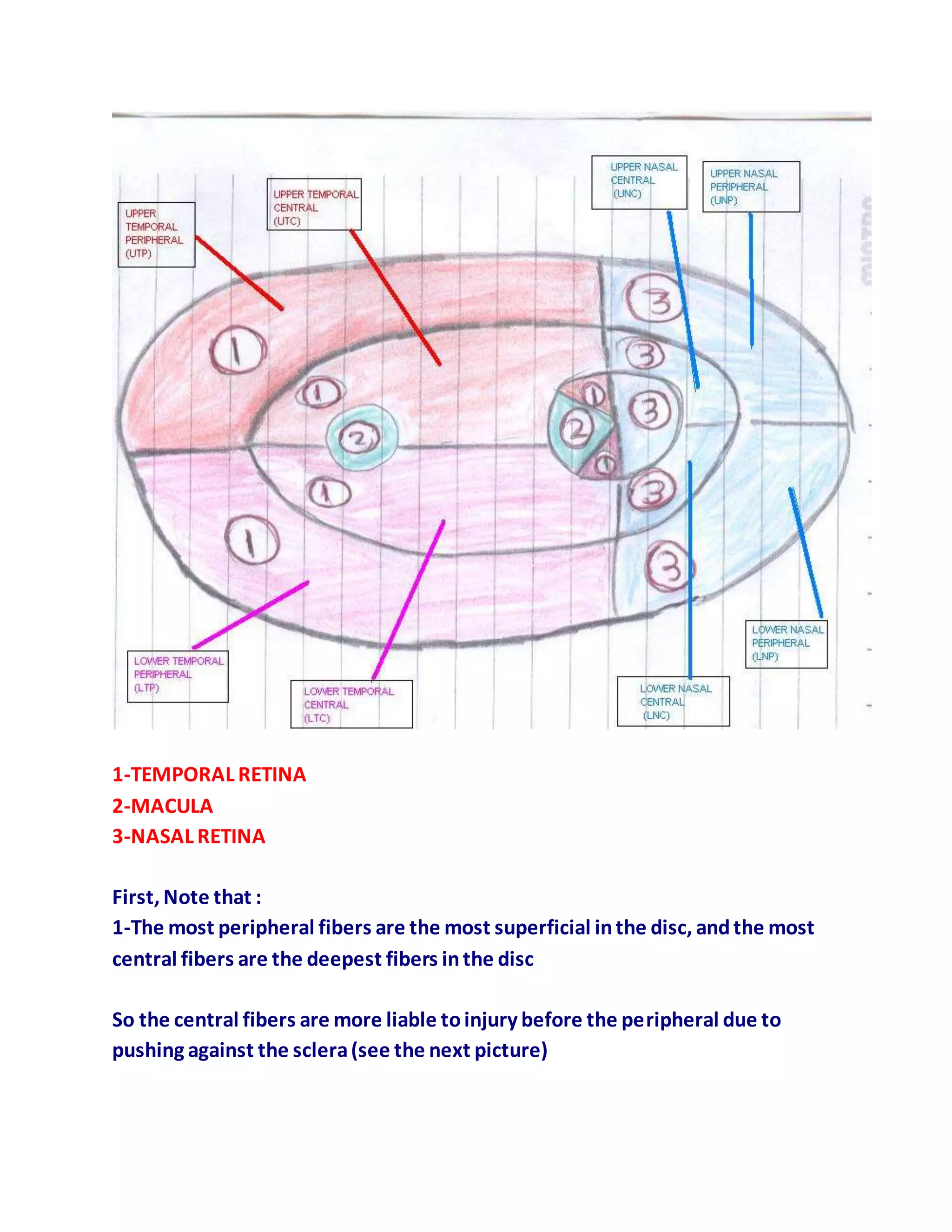

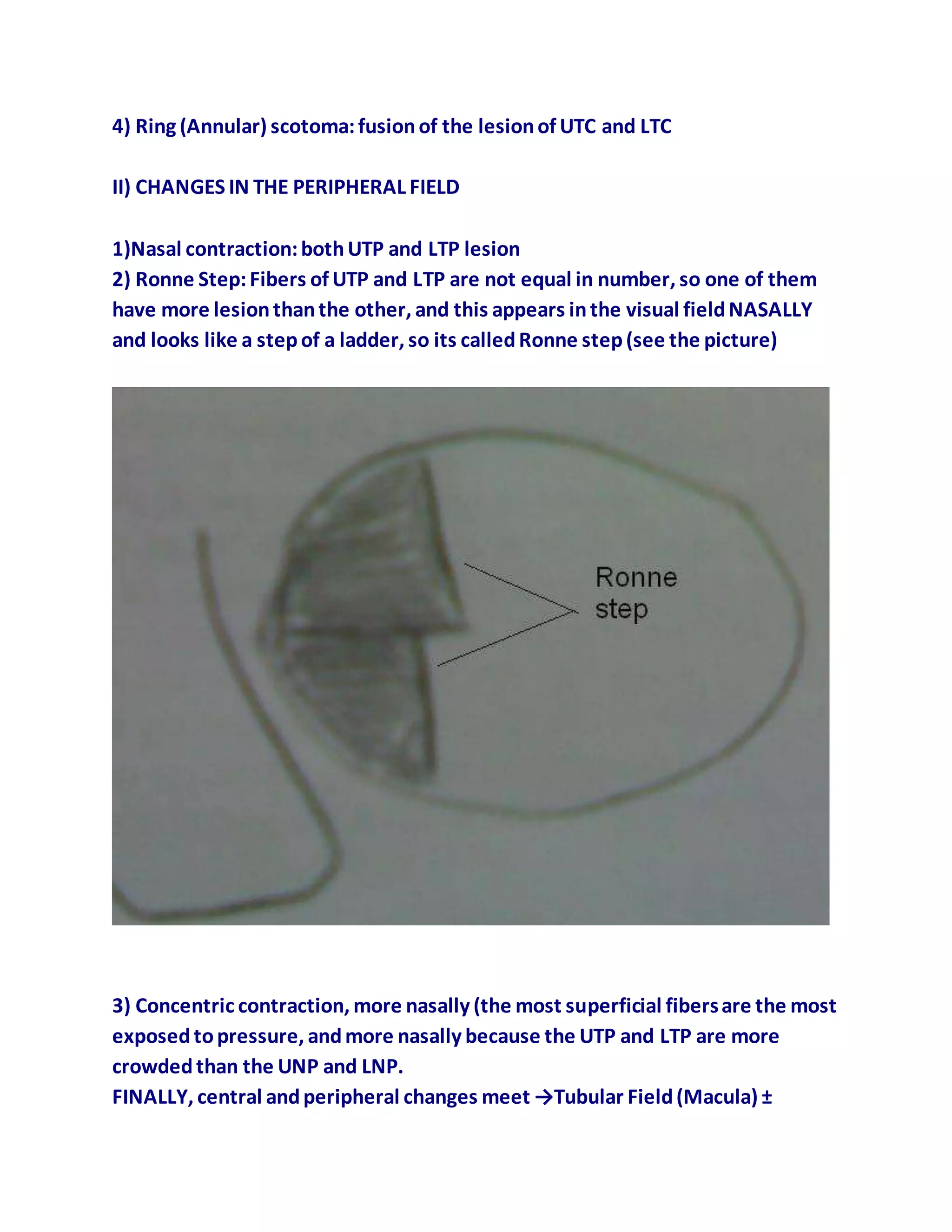
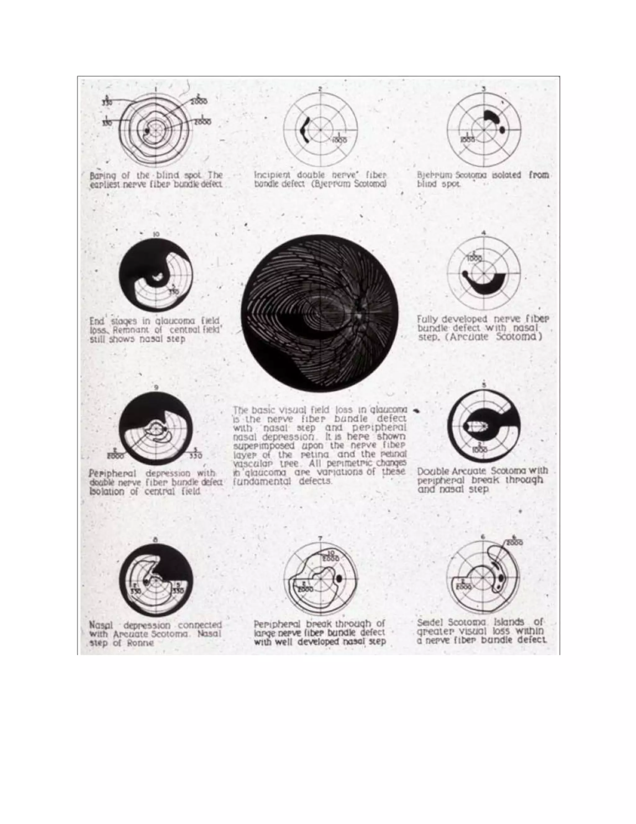
1. The central retinal fibers at the optic disc are deeper and more susceptible to injury than peripheral fibers due to pressure against the sclera. 2. Lesions in different retinal areas will affect the contralateral visual field in the same location. The most crowded fibers are in the temporal retina, then nasal, then macula. 3. Visual field changes progress from paracentral scotomas to arcuate scotomas to ring scotomas centrally, and nasal contraction to Ronne step to concentric contraction peripherally, eventually meeting in the macula to form a tubular visual field.


