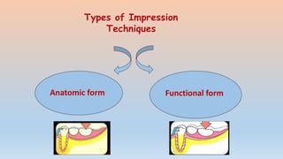
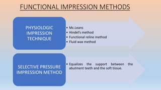
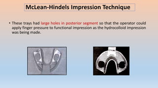
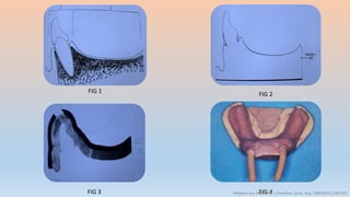
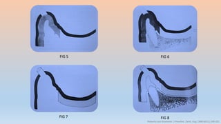
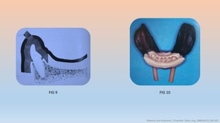
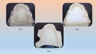
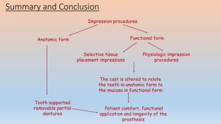
Impression Procedures in Removable Partial
Denture
Guided by:
Dr. U.M. Radke Dr. N.A. Pande Dr. S Deshmukh
HOD & Guide Professor Reader
Dr. T.K. Mowade Dr. R. Banerjee DR. A. Chandak
Reader Reader READER
Presented by:-
Dr. Richa Sahai
II MDS
Contents • Introduction
• Definitions
• Rpd impression vs Complete denture impression
• Impression techniques
• Anatomic form impression
• Functional impression techniques
• Review of literature
• Recent Advances
• Summary and Conclusion
• References
• A widely accepted axiom of removable partial prosthodontics holds that
it is fully as important that  the denture be designed and constructed
in such a way as best to preserve the oral structures as it is to restore
function.
• The principle of functional basing is employed to create conditions which
favor maximum longevity of the remaining structures.
INTRODUCTION
Impression
A negative likeness or copy in reverse of the surface of an object ; imprint of
teeth and adjacent structures for use in dentistry. GPT – 9
Partial Denture Impression
A negative likeness of a part or all of a partially edentulous arch. GPT – 9
Impression trays
A receptacle in to which suitable impression material is placed to make
negative likeness
OR
A device that is used to carry, confine and control impression material while
making an impression.
Impression trays can be classified broadly into :
Stock trays and Custom trays
1. By the manner in which they Harden or Set :
• Impression plaster
• ZOE
• Alginate
• Elastomers
Chemical / Irreversible
• Thermoplastic
• Impression compound
• Waxes
• Non-thermoplastic
• Agar
Reversible
Phillips-science of dental material,11th edition
Classification of impression materials
Non-elastic
Elastic
Aqueous
Hydrocolloid
Non-aqueous
Elastomers
Polysulfide
Silicones
Polyether
Condensation
Addition
Agar
(reversible)
Alginate
(irreversible)
Plaster
Compound
ZnO - Eugenol
Waxes
2. According to elasticity :
3.According to the use of the materials in dentistry :
A) Materials used for obtaining impression of Dentulous mouth
• Alginate
• Agar
• Non-aqueous Elastomers
B) Materials used for obtaining Impression of Edentulous mouth:
• Impression Compound
• Impression Plaster
• Zinc Oxide eugenol
• Wax
• Impression plaster
• Agar
• Alginate
Mucostatic
• Impression Compound
• Putty elastomersMuco-compressive
4. According to the viscosity or tissue displacement:
RPD IMPRESSION
• Records not only relative
soft yielding tissues as
well as a hard unyielding
substance .
CD Impression
• Records the edentulous
mucosa with underlying
bone only.
To Start :
1. PRIMARY IMPRESSION; Study casts
2. Checking Maxillary Tray and Mandibular tray For Correct Size.
3. Control of Gagging
4. Control of Saliva
1. Bubbles or voids
in and around rest
preparations.
2. Contact of
cusp with the
tray.
3. Show through
between teeth and
modeling plastic or
modeling plastic and
hard palate
4. Voids or
bubbles in
palatal vault.
5.Peripheral under
extension.
6. Interproximal
tearing of the
impression
material.
7. Lack of detail on
the impression
surface.
8. Any doubt as
to the accuracy
of the
impression.
Types of Impression
Techniques
Anatomic form Functional form
• The proponents of functional impression put great emphasis on the tissue
compression to get a registration of “Functional ridge form” in order to place
load properly on ridge and there by minimize cantilever action on abutment
teeth.
• Two requirements must be satisfied:
– That it records and relates the supporting soft tissue under some loading.
– It distributes load over a maximum area and establish precisely accurate
functional borders for the denture.
FUNCTIONAL IMPRESSION METHODS
• Mc.Leans
• Hindel’s method
• Functional reline method
• Fluid wax method
PHYSIOLOGIC
IMPRESSION
TECHNIQUE
• Equalizes the support between the
abutment teeth and the soft tissue.SELECTIVE PRESSURE
IMPRESSION METHOD
McLean’s Physiologic Impression
1. Fabrication of the custom-made impression tray
over the edentulous areas of the preliminary cast.
2. Making of the occlusal rim on the custom tray.
3. Impression material loaded on the tray is inserted
into the patient’s mouth.
4. An alginate over-impression is made by using a
large stock tray.
• The functional master impression will come along when the over-impression is
removed and it is called as ‘pick up’ impression.
McLean’s Physiologic Impression
Disadvantage:
• The finger pressure had to be applied on the hydrocolloid impression tray
during the ‘over-impression’.
• This finger pressure could only be approximated to the occlusal load applied
earlier.
McLean-Hindels Impression Technique
• These trays had large holes in posterior segment so that the operator could
apply finger pressure to functional impression as the hydrocolloid impression
was being made.
1. Fabrication of the occlusal rim in a special tray using the
primary cast.
2. The supporting tissues under rest – anatomical
impression is recorded by special tray.
3. Large holes with over impression is made by a special
stock tray.
4. A pseudo-functional stress is gave by pushes the
edentulous ridge by using the finger pressure on the
special tray.
The chief modifications were:
Impression of the
edentulous ridge was
made in the anatomic
form made with zinc
oxide eugenol and
not under finger
pressure.
However, when the
over impression was
being made with the
hydrocolloid, finger
pressure was applied
through the modified
tray to the anatomic
impression.
The pressure had to
be maintained till the
alginate sets.
The finished
impression was a
reproduction of the
anatomic surface of
ridge and the surfaces
of teeth but the two
were related to each
other as if the
masticating forces
were taking place on
the denture base.
Disadvantages:-
• If the action of the retentive clasps of RPD is sufficient in order to maintain
the denture base in soft tissue displace, blood vessels interruption will occur
with worst soft tissue destruction and bone resorption.
• If the action of the retentive clasps of RPD is not sufficient in order to
maintain the denture base in soft tissue displace, it will result in artificial
teeth premature contact.
Functional Reline Method
• It consists of adding of a new surface to the tissue side of the denture base.
• The procedures of this technique should be done before the insertion of
RPD.
• It also can be done later in future if the denture base having loose or no
longer fits to the ridge due to bone resorption. (Steffel, 1954)
Procedure :
1. Partial denture frame work is constructed on cast made from single
impression. (anatomic form)
2. To allow room for impression material between the denture base and the
ridge, a uniform space is provided by adapting a metal spacer over ridge of
cast before processing the denture base.
3. After processing the denture base, metal spacer is removed and uniform
space is achieved.
4. Low fusing modeling plastic is applied onto the tissue surface of denture
base, tampered and placed in patient’s mouth.
• To provide space for impression material 1mm of modeling plastic is
removed and impression is made with free flowing zinc oxide- euqenol
paste or light bodied polysulfide rubber base.
Advantages:
• The amount of soft tissue
displacement can be controlled
by the amount of relief given to
the modeling plastic before the
final impression is being made.
Disadvantages:
• Failures to maintain correct
relationship between framework
and abutment teeth, during
impression procedure
• Failure to achieve correct
occlusal contact following reline.
• The patient must keep the mouth
partially open because the
border tissues, cheek and tongue
are thus best controlled
• The relationship between partial
denture framework and teeth
must be observed
Fluid Wax Functional Technique
• Objective  Obtaining maximum denture base peripheral borders extension
which it will not interfered with the movable border tissues function.
 It also used in ridge stress-bearing areas recording for functional
form and recording of non pressure- bearing areas in anatomic form.
• The borders made should be short for all movable tissues which is not more
than 2 mm short. This is due to the insufficient strength of fluid wax to support
itself beyond the distance.
OBJECTIVE
To obtain maximum
extension of
peripheral borders
while not interfering
with function of
movable border
tissues.
To record stress
bearing areas of
the ridge in their
functional form.
To record non
pressure bearing
areas in their
anatomic form.
1. A container of wax is placed in water bath
maintained at 51°C-54°C.
2. Wax is painted onto impression surface with
brush.
3. The tray is then seated in the mouth and the patient
must hold his mouth half open for about 5 min; the
impression is left in the mouth for 12 min.
The new cast need to be poured as soon as possible after the impression is
completed. This due the wax fragile and might be distortion.
Disadvantages
1. Time consuming.
2. Accurately followed timing of procedures.
3. The excessive tissue displacement might result to the impression if the
timing period is not followed properly as mentioned.
Selective Tissue Placement Impression method
OBJECTIVES
1. This attempts to direct more force to those portions of the ridge that are able
to absorb stress without adverse response.
2. Protect the areas of the ridge which is least able to absorb force.
• Buccal shelf is primary stress bearing area, as a result only sight relief is
provided.
• The crest of the ridge is not stress bearing area and hence the surface of the
tray is relieved down to metal retention using laboratory bur.
1. fabrication of the special tray on the master cast from
the anatomical impression.
2. Trimming of the special tray tissue surface by using
burs for adequate relief.
3. prepared special tray is loaded by impression
material, such as zinc oxide eugenol and inserted into
the patients’ mouth.
The framework is placed in the mouth and great care is to be taken ensuring that it is
seated on the teeth by pressure on the occlusal rests & indirect retainers only - no
finger pressure is applied to the base area. Once the framework is fully seated,
border moulding is carried out.
Altered Cast Technique
FIG 1 Fig 2
Contemp Clin Dent. 2010 Apr-Jun; 1(2): 103–1
Objective  To reduce the support differential for a free-end saddle by
obtaining a compressive impression of the edentulous area under conditions
which mimic functional loading.
FIG 3 FIG 4
Contemp Clin Dent. 2010 Apr-Jun; 1(2): 103–1
FIG 5 FIG 6
FIG 7 Fig 8
Fig 9
FIG 10 FIG 11
FIG 12 FIG 13
John B. Holmes* conducted a study to determine the “Influence of impression
procedures and occlusal loading on partial denture movement”
He concluded that:
1. The movement of partial dentures from occlusal loading is related to the
impression technique and material used.
2. The altered cast technique provided the least amount of movement from
occlusal loading at the time of insertion .
3. Partial dentures made with Korecta wax 4 (altered cast) resulted in least
amount of movement. *J PROSTHET. DENT. May 1965:15(3);474-481
REVIEW OF LITERATURE
Modification of Altered Cast Technique
Technique :
Richard Bauman & James- JPD 1982(47) 2,212-213
FIG 3 FIG 4
FIG 5
FIG 6
Richard Bauman & James- JPD 1982(47) 2,212-213
• Pour a completely new master cast into the combined impression.
• After the stone has set, remove the metal tray and irreversible hydrocolloid
impression material.
• Mount the new master cast on the articulator by means of the interocclusal
record.
• Arrange the teeth and process the denture base.
Richard Bauman & James- JPD 1982(47) 2,212-213
Technique for semi-precision
and precision partial dentures
*Louis Blatterfien et al. J Prosthet. Dent, 1980:43 (1);9-14
FIG 1 Fig 2
FIG 3 FIG 4
FIG 5 FIG 6
FIG 7 FIG 8*Louis Blatterfien et al. J Prosthet. Dent, 1980:43 (1);9-14
FIG 9
FIG 10
FIG 11
*Louis Blatterfien et al. J Prosthet. Dent, 1980:43 (1);9-14
Roberto von Krammer. J Prosthet. Dent, Aug 1988:60(1);199-201
FIG 1 FIG 2
FIG 3 FIG 4Roberto von Krammer. J Prosthet. Dent, Aug 1988:60(1);199-201
FIG 5 FIG 6
FIG 7 FIG 8
Roberto von Krammer. J Prosthet. Dent, Aug 1988:60(1);199-201
FIG 9 FIG 10
Roberto von Krammer. J Prosthet. Dent, Aug 1988:60(1);199-201
Impression technique for maxillary
removable partial dentures
- C D. LEACH & T E. DONOVAN JPD 1983 (50)2,283-285
Fig 1 Fig 2
Fig 3
Fig 4 Fig 5
Fig 6
Single tray dual impression for
distal extension partial dentures
JOSEPH A. R- JPD 1970(24,1,41-46)
Joseph A. Rapuano. J Prosthet Dent, July 1970:24(1);41-46
Fig 1 Fig 2
Fig 3 Fig 4
Fig 5 Fig 6
Fig 7
Fig 8
Joseph A. Rapuano. J Prosthet Dent, July 1970:24(1);41-46
Fig 9 Fig 10
Joseph A. Rapuano. J Prosthet Dent, July 1970:24(1);41-46
Summary and Conclusion
Impression procedures
Anatomic form Functional form
Physiologic impression
procedures
Selective tissue
placement impressions
The cast is altered to relate
the teeth in anatomic form to
the mucosa in functional form
Patient comfort, functional
application and longevity of the
prosthesis
Tooth supported
removable partial
dentures
“Unless a partial denture is made with adequate
abutment support, optimal base support and with
harmonious and functional occlusion, it should be
clear to all concerned that such a denture should
be considered only a temporary treatment”
1. Glossary of Prosthodontic Terms -8 th Edn, 2005.
2. Stewart, Rudd, Kuebker : Clinical Removable Partial Prosthodontics.
3. McGivney GP, Alan B Carr David T Brown : McCracken’s Removable Partial
Dentures-11 th Edn.
4. Joseph E. Grasso, Ernest L. Miller : Removable Partial Prosthodontics.
5. Alan A. Grant, Wesley Johnson : Removable Partial Dentures.
6. APPLEGATE OC. An evaluation of the support for removable partial
denture. J PROSTHET DENT 1960; 10:112-23.
References
7. HINDEL GW. Load distribution in extension saddle denture. J PROSTHET DENT
1952; 22:92-100.
8. HINDEL GW. Stress analysis in distal extension partial. J PROSTHET DENT
1957; 7:197-205.
9. HOLMES JB. Influence of impression procedures and occlusal loading on
partial denture movement. J PROSTHET DENT, 1965; 15:474-81.
10. F. James Kratochvil : Partial Removable Prosthodontics.
11. Robert P. Renner, Louis J. Boucher : Removable Partial Dentures.
12. Kenneth D Rudd, Morrow: Dental Lab, Procedure for Removable Partial
Dentures.
impression techniques in Removable Partial Denture

impression techniques in Removable Partial Denture

  • 1.
    Impression Procedures inRemovable Partial Denture Guided by: Dr. U.M. Radke Dr. N.A. Pande Dr. S Deshmukh HOD & Guide Professor Reader Dr. T.K. Mowade Dr. R. Banerjee DR. A. Chandak Reader Reader READER Presented by:- Dr. Richa Sahai II MDS
  • 2.
    Contents • Introduction •Definitions • Rpd impression vs Complete denture impression • Impression techniques • Anatomic form impression • Functional impression techniques • Review of literature • Recent Advances • Summary and Conclusion • References
  • 3.
    • A widelyaccepted axiom of removable partial prosthodontics holds that it is fully as important that  the denture be designed and constructed in such a way as best to preserve the oral structures as it is to restore function. • The principle of functional basing is employed to create conditions which favor maximum longevity of the remaining structures. INTRODUCTION
  • 4.
    Impression A negative likenessor copy in reverse of the surface of an object ; imprint of teeth and adjacent structures for use in dentistry. GPT – 9 Partial Denture Impression A negative likeness of a part or all of a partially edentulous arch. GPT – 9
  • 5.
    Impression trays A receptaclein to which suitable impression material is placed to make negative likeness OR A device that is used to carry, confine and control impression material while making an impression.
  • 6.
    Impression trays canbe classified broadly into : Stock trays and Custom trays
  • 7.
    1. By themanner in which they Harden or Set : • Impression plaster • ZOE • Alginate • Elastomers Chemical / Irreversible • Thermoplastic • Impression compound • Waxes • Non-thermoplastic • Agar Reversible Phillips-science of dental material,11th edition Classification of impression materials
  • 8.
  • 9.
    3.According to theuse of the materials in dentistry : A) Materials used for obtaining impression of Dentulous mouth • Alginate • Agar • Non-aqueous Elastomers B) Materials used for obtaining Impression of Edentulous mouth: • Impression Compound • Impression Plaster • Zinc Oxide eugenol • Wax
  • 10.
    • Impression plaster •Agar • Alginate Mucostatic • Impression Compound • Putty elastomersMuco-compressive 4. According to the viscosity or tissue displacement:
  • 11.
    RPD IMPRESSION • Recordsnot only relative soft yielding tissues as well as a hard unyielding substance . CD Impression • Records the edentulous mucosa with underlying bone only.
  • 12.
    To Start : 1.PRIMARY IMPRESSION; Study casts 2. Checking Maxillary Tray and Mandibular tray For Correct Size. 3. Control of Gagging 4. Control of Saliva
  • 13.
    1. Bubbles orvoids in and around rest preparations. 2. Contact of cusp with the tray. 3. Show through between teeth and modeling plastic or modeling plastic and hard palate 4. Voids or bubbles in palatal vault. 5.Peripheral under extension. 6. Interproximal tearing of the impression material. 7. Lack of detail on the impression surface. 8. Any doubt as to the accuracy of the impression.
  • 14.
  • 15.
    • The proponentsof functional impression put great emphasis on the tissue compression to get a registration of “Functional ridge form” in order to place load properly on ridge and there by minimize cantilever action on abutment teeth. • Two requirements must be satisfied: – That it records and relates the supporting soft tissue under some loading. – It distributes load over a maximum area and establish precisely accurate functional borders for the denture.
  • 16.
    FUNCTIONAL IMPRESSION METHODS •Mc.Leans • Hindel’s method • Functional reline method • Fluid wax method PHYSIOLOGIC IMPRESSION TECHNIQUE • Equalizes the support between the abutment teeth and the soft tissue.SELECTIVE PRESSURE IMPRESSION METHOD
  • 17.
  • 18.
    1. Fabrication ofthe custom-made impression tray over the edentulous areas of the preliminary cast. 2. Making of the occlusal rim on the custom tray. 3. Impression material loaded on the tray is inserted into the patient’s mouth. 4. An alginate over-impression is made by using a large stock tray. • The functional master impression will come along when the over-impression is removed and it is called as ‘pick up’ impression.
  • 19.
  • 20.
    Disadvantage: • The fingerpressure had to be applied on the hydrocolloid impression tray during the ‘over-impression’. • This finger pressure could only be approximated to the occlusal load applied earlier.
  • 21.
    McLean-Hindels Impression Technique •These trays had large holes in posterior segment so that the operator could apply finger pressure to functional impression as the hydrocolloid impression was being made.
  • 22.
    1. Fabrication ofthe occlusal rim in a special tray using the primary cast. 2. The supporting tissues under rest – anatomical impression is recorded by special tray. 3. Large holes with over impression is made by a special stock tray. 4. A pseudo-functional stress is gave by pushes the edentulous ridge by using the finger pressure on the special tray.
  • 24.
    The chief modificationswere: Impression of the edentulous ridge was made in the anatomic form made with zinc oxide eugenol and not under finger pressure. However, when the over impression was being made with the hydrocolloid, finger pressure was applied through the modified tray to the anatomic impression. The pressure had to be maintained till the alginate sets. The finished impression was a reproduction of the anatomic surface of ridge and the surfaces of teeth but the two were related to each other as if the masticating forces were taking place on the denture base.
  • 25.
    Disadvantages:- • If theaction of the retentive clasps of RPD is sufficient in order to maintain the denture base in soft tissue displace, blood vessels interruption will occur with worst soft tissue destruction and bone resorption. • If the action of the retentive clasps of RPD is not sufficient in order to maintain the denture base in soft tissue displace, it will result in artificial teeth premature contact.
  • 26.
    Functional Reline Method •It consists of adding of a new surface to the tissue side of the denture base. • The procedures of this technique should be done before the insertion of RPD. • It also can be done later in future if the denture base having loose or no longer fits to the ridge due to bone resorption. (Steffel, 1954)
  • 27.
    Procedure : 1. Partialdenture frame work is constructed on cast made from single impression. (anatomic form) 2. To allow room for impression material between the denture base and the ridge, a uniform space is provided by adapting a metal spacer over ridge of cast before processing the denture base.
  • 28.
    3. After processingthe denture base, metal spacer is removed and uniform space is achieved. 4. Low fusing modeling plastic is applied onto the tissue surface of denture base, tampered and placed in patient’s mouth.
  • 29.
    • To providespace for impression material 1mm of modeling plastic is removed and impression is made with free flowing zinc oxide- euqenol paste or light bodied polysulfide rubber base.
  • 30.
    Advantages: • The amountof soft tissue displacement can be controlled by the amount of relief given to the modeling plastic before the final impression is being made. Disadvantages: • Failures to maintain correct relationship between framework and abutment teeth, during impression procedure • Failure to achieve correct occlusal contact following reline. • The patient must keep the mouth partially open because the border tissues, cheek and tongue are thus best controlled • The relationship between partial denture framework and teeth must be observed
  • 31.
    Fluid Wax FunctionalTechnique • Objective  Obtaining maximum denture base peripheral borders extension which it will not interfered with the movable border tissues function.  It also used in ridge stress-bearing areas recording for functional form and recording of non pressure- bearing areas in anatomic form. • The borders made should be short for all movable tissues which is not more than 2 mm short. This is due to the insufficient strength of fluid wax to support itself beyond the distance.
  • 32.
    OBJECTIVE To obtain maximum extensionof peripheral borders while not interfering with function of movable border tissues. To record stress bearing areas of the ridge in their functional form. To record non pressure bearing areas in their anatomic form.
  • 33.
    1. A containerof wax is placed in water bath maintained at 51°C-54°C. 2. Wax is painted onto impression surface with brush. 3. The tray is then seated in the mouth and the patient must hold his mouth half open for about 5 min; the impression is left in the mouth for 12 min. The new cast need to be poured as soon as possible after the impression is completed. This due the wax fragile and might be distortion.
  • 34.
    Disadvantages 1. Time consuming. 2.Accurately followed timing of procedures. 3. The excessive tissue displacement might result to the impression if the timing period is not followed properly as mentioned.
  • 35.
    Selective Tissue PlacementImpression method OBJECTIVES 1. This attempts to direct more force to those portions of the ridge that are able to absorb stress without adverse response. 2. Protect the areas of the ridge which is least able to absorb force. • Buccal shelf is primary stress bearing area, as a result only sight relief is provided. • The crest of the ridge is not stress bearing area and hence the surface of the tray is relieved down to metal retention using laboratory bur.
  • 36.
    1. fabrication ofthe special tray on the master cast from the anatomical impression. 2. Trimming of the special tray tissue surface by using burs for adequate relief. 3. prepared special tray is loaded by impression material, such as zinc oxide eugenol and inserted into the patients’ mouth.
  • 39.
    The framework isplaced in the mouth and great care is to be taken ensuring that it is seated on the teeth by pressure on the occlusal rests & indirect retainers only - no finger pressure is applied to the base area. Once the framework is fully seated, border moulding is carried out.
  • 40.
    Altered Cast Technique FIG1 Fig 2 Contemp Clin Dent. 2010 Apr-Jun; 1(2): 103–1 Objective  To reduce the support differential for a free-end saddle by obtaining a compressive impression of the edentulous area under conditions which mimic functional loading.
  • 41.
    FIG 3 FIG4 Contemp Clin Dent. 2010 Apr-Jun; 1(2): 103–1
  • 42.
    FIG 5 FIG6 FIG 7 Fig 8
  • 43.
  • 44.
  • 45.
  • 46.
    John B. Holmes*conducted a study to determine the “Influence of impression procedures and occlusal loading on partial denture movement” He concluded that: 1. The movement of partial dentures from occlusal loading is related to the impression technique and material used. 2. The altered cast technique provided the least amount of movement from occlusal loading at the time of insertion . 3. Partial dentures made with Korecta wax 4 (altered cast) resulted in least amount of movement. *J PROSTHET. DENT. May 1965:15(3);474-481
  • 47.
  • 48.
    Modification of AlteredCast Technique Technique : Richard Bauman & James- JPD 1982(47) 2,212-213
  • 49.
    FIG 3 FIG4 FIG 5 FIG 6 Richard Bauman & James- JPD 1982(47) 2,212-213
  • 50.
    • Pour acompletely new master cast into the combined impression. • After the stone has set, remove the metal tray and irreversible hydrocolloid impression material. • Mount the new master cast on the articulator by means of the interocclusal record. • Arrange the teeth and process the denture base. Richard Bauman & James- JPD 1982(47) 2,212-213
  • 51.
    Technique for semi-precision andprecision partial dentures
  • 52.
    *Louis Blatterfien etal. J Prosthet. Dent, 1980:43 (1);9-14 FIG 1 Fig 2 FIG 3 FIG 4
  • 53.
    FIG 5 FIG6 FIG 7 FIG 8*Louis Blatterfien et al. J Prosthet. Dent, 1980:43 (1);9-14
  • 54.
    FIG 9 FIG 10 FIG11 *Louis Blatterfien et al. J Prosthet. Dent, 1980:43 (1);9-14
  • 55.
    Roberto von Krammer.J Prosthet. Dent, Aug 1988:60(1);199-201
  • 56.
    FIG 1 FIG2 FIG 3 FIG 4Roberto von Krammer. J Prosthet. Dent, Aug 1988:60(1);199-201
  • 57.
    FIG 5 FIG6 FIG 7 FIG 8 Roberto von Krammer. J Prosthet. Dent, Aug 1988:60(1);199-201
  • 58.
    FIG 9 FIG10 Roberto von Krammer. J Prosthet. Dent, Aug 1988:60(1);199-201
  • 59.
    Impression technique formaxillary removable partial dentures - C D. LEACH & T E. DONOVAN JPD 1983 (50)2,283-285
  • 60.
    Fig 1 Fig2 Fig 3
  • 61.
    Fig 4 Fig5 Fig 6
  • 62.
    Single tray dualimpression for distal extension partial dentures JOSEPH A. R- JPD 1970(24,1,41-46)
  • 63.
    Joseph A. Rapuano.J Prosthet Dent, July 1970:24(1);41-46 Fig 1 Fig 2 Fig 3 Fig 4
  • 64.
    Fig 5 Fig6 Fig 7 Fig 8 Joseph A. Rapuano. J Prosthet Dent, July 1970:24(1);41-46
  • 65.
    Fig 9 Fig10 Joseph A. Rapuano. J Prosthet Dent, July 1970:24(1);41-46
  • 67.
    Summary and Conclusion Impressionprocedures Anatomic form Functional form Physiologic impression procedures Selective tissue placement impressions The cast is altered to relate the teeth in anatomic form to the mucosa in functional form Patient comfort, functional application and longevity of the prosthesis Tooth supported removable partial dentures
  • 68.
    “Unless a partialdenture is made with adequate abutment support, optimal base support and with harmonious and functional occlusion, it should be clear to all concerned that such a denture should be considered only a temporary treatment”
  • 69.
    1. Glossary ofProsthodontic Terms -8 th Edn, 2005. 2. Stewart, Rudd, Kuebker : Clinical Removable Partial Prosthodontics. 3. McGivney GP, Alan B Carr David T Brown : McCracken’s Removable Partial Dentures-11 th Edn. 4. Joseph E. Grasso, Ernest L. Miller : Removable Partial Prosthodontics. 5. Alan A. Grant, Wesley Johnson : Removable Partial Dentures. 6. APPLEGATE OC. An evaluation of the support for removable partial denture. J PROSTHET DENT 1960; 10:112-23. References
  • 70.
    7. HINDEL GW.Load distribution in extension saddle denture. J PROSTHET DENT 1952; 22:92-100. 8. HINDEL GW. Stress analysis in distal extension partial. J PROSTHET DENT 1957; 7:197-205. 9. HOLMES JB. Influence of impression procedures and occlusal loading on partial denture movement. J PROSTHET DENT, 1965; 15:474-81. 10. F. James Kratochvil : Partial Removable Prosthodontics. 11. Robert P. Renner, Louis J. Boucher : Removable Partial Dentures. 12. Kenneth D Rudd, Morrow: Dental Lab, Procedure for Removable Partial Dentures.

Editor's Notes

  • #7 1-- Stock trays for partially edentulous patients may be perforated to retain the impression material or they may be constructed with a rim-lock for this purpose. 2--Another type of stock tray designed for the reversible type of hydrocolloid are water cooled trays. It contains tubes through which water can be circulated for purpose of cooling the tray.
  • #9 Factors that influence the selection of impression materials are: Convenience of use Time of manipulation and set Cost Need for special trays Operator training and preference
  • #13 1 RPD - Partial denture impression Records not only relative soft yielding tissues (the oral mucosa) as well as a hard unyielding substance (the remaining teeth). Removable partial denture impression need to record the teeth that are irregular in contour as well as varying in their vertical relations to occlusal plane 1 CD - The complete denture impression records the edentulous mucosa with underlying bone only.
  • #14 Objectives: To obtain an impression of all the standing teeth and denture - supporting tissues of each jaw from which study casts may be prepared. The purpose of the study casts are: To enable special trays and occlusion rims to be constructed if necessary. To examine the occlusion in detail on an articulator. By use of a surveyor, to plan the path of insertion of the proposed denture, arrive at a tentative design and plan any mouth preparation. 4 – gauze , antisialagogues Diag -- RPD framework drawn on the study cast 
  • #15 Reasons for Rejecting Impression .
  • #16 Anatomic -- It represents all the hard and soft tissues at rest. The surface of the residual ridge at rest and it is the shape of the ridge before functional load is applied . Functional form-- Recording the mucosa in a compressed or a displaced state by one impression and then relating it to the abutment teeth by means of secondary impression. The shape of the ridge after functiona load is applied .
  • #17 After 1 -- The functional method selected is greatly determined by the support potential of the residual ridge. Last – Need of functional impression – the displaceability of the mucosa or residual ridge is not uniform. , in cases of distal extension based partial dentures and shoet span distal extension bases
  • #20 1- No need for adaptation of spacer. 2- Occlusal rim is used to make sure patients are bite while making the impression. 3- after 3  Patient needs to be instructed to bite on the occlusal rim. Very imp --  Custom tray should not be removed from patient’s mouth after impression making 4 -
  • #23 Start -- Hindel and others developed/modified impression tray for second impression procedures.
  • #24 1. In order to avoid excessive pressure on the tissues, the stoppers need to be placed on the tray that extend over the stress-bearing areas 3.  Steady constant pressure should be applied on the occlusal rim during the procedure. Till the alginate is set, the pressure should be held 4.  A pseudo-functional is same to the functional impression. End -- 5.  Excessive tissue compression can be avoided by using the stopper on the custom stray.
  • #27 After 2 last -- This premature contact may become objectionable for almost all patients.
  • #30 After 4 -- Sequence is repeated until accurate impression is obtained.
  • #32 Adv last -- Greater the relief lesser will be the tissue displacement.
  • #33  start -- The fluid wax are waxes that firm at room temperature but it have an ability to flow when put in mouth temperature. There are two types of fluid waxes used in RPD, which are: Iowa Wax and Korrecta wax. After 2 --  In fluid wax impression technique, the key of using it are space and time.
  • #35 2. Tray borders should not more than 2mm short because the fluid wax does not have sufficient strength beyond that distance. 3. When tissue contact is present, wax will be glossy and where tissue contact is not present it will be dull. 12 mins -- This is to ensure that wax had flown completely. After 3..  All the steps need to be repeated till the impression is completed, and need to be carefully handle.
  • #37 Start -- The selective tissue placement impression method is based on these clinical observations, the histological nature of tissue that covers the residual alveolar bone, the nature of the residual ridge bone, and its positional relationship to the direction of stresses that will be placed on it.
  • #38 The fabrication of tray is without a wax spacer. After 3 --  The patients need to open their mouth and the finger pressure is used to record the impression. Make sure that only the stress bearing areas are compressed during the procedures.
  • #39 A secondary impression for the distal extension mandibular rpd is made in individual trays attatched to the denture framework. The framework has been tried in the patient mouth and fits the mouth and the master cast is planned the outline of acrylic resin trays is penciled on the cast A single sheet of baseplate wax relief provided with a window for the posterior tissue stop and anterior tissue index The framework is warmed and pressed to position on cast. All regions of cast that will be contacted by autopolymerizing acrylic resin or VLC resin are painted with tinfoil substitute (Alcote) or model release agent (MRA).
  • #40 5. A sheet of VLC resin material is adapted to cast and over the framework with finger pressure. Excess material over borders of cast is removed with sharp knife while material is still soft. 6. The cured acrylic resin trays with framework are removed from cast, and trays are trimmed to outline of wax spacer. 7. Borders of trays will be adjusted to extend 2 mm short of tissue reflections. Holes will be placed in trays corresponding to crest of residual ridge and retromolar pads to allow escape of excess impression material when impression is made.
  • #42 Objective – end – The distribution of load from the denture to the residual ridge is thus improved and the denture is more stable. 1 -- Maxillary edentulous arch with partial missing teeth , missing 13,15,16, 24, 27, 36, 37, 46, 47 and mandibular anterior 4 unit fixed partial denture . 2-- Mandibular edentulous arch with distal extension LAST -- Maxillary preliminary impressions were made with irreversible hydrocolloid and study casts were obtained. study casts were placed on a surveyor for examination and design of the cast framework. Mouth preparation was done and final impression of both arch were made with medium bodied elastomeric impression material. Secondary casts obtained were placed on a surveyor for examination and design of the cast framework
  • #43 Start -- Master casts were duplicated, refractory cast were obtained, and the design was transferred from the master cast to the refractory cast. Investing and casting was completed. The completed framework is examined to ensure that it fits the cast accurately Fig 3-- Maxillary metal framework Fig 4 -- Mandibular metal framework
  • #44 Fig 5 --Maxillary metal try in FIG 6-- Mandibular metal try in the cast framework was verified for acceptable fit orally An acrylic resin custom tray was attached to the mandibular metal framework and the tray was then border molded in the usual fashion for the desired extension, the fit of the metal framework to the teeth and soft tissues were checked . FIG 7 -- An acrylic resin custom tray attached to the mandibular metal framework Fig 8 -- Border molded tray for the desired extension
  • #45 The final impression was made using zinc oxide eugenol impression paste. While making impression, finger pressure was applied only to the parts of the framework that comes in contact with the teeth. fig 9-- Final impression made using zinc oxide eugenol. impression paste
  • #46 The cast was altered in the laboratory. Fig 10 Cast with two saw cuts perpendicular to each other on either side of saddle The second cut was made parallel and medial to the edentulous ridge, extending from the most posterior aspect of the cast to the most medial aspect of the first cut. FIG 11 The cut surface of the cast was then grooves placed to aid in the retention of the newly poured stone
  • #47 Fig 12 -- Seating of the framework on the cast with sticky wax Fig 13 -- Final impression with beading and boxing poured in usual manner in die stone Fig 14 -- Working altered cast after beading and boxing was removed and usual remaining steps in the fabrication of removable partial dentures were carried out (jaw relation and try in procedures), denture delivery was done and post delivery instructions were given *A favorably extended base will provide stimulation to the underlying bone and distribute forces uniformly. The altered cast technique allows the ridge, recorded in functional form, to be related to the teeth so that when the prosthesis is seated, it derives support simultaneously from the teeth and the denture base*
  • #53 disadvantage of the altered cast procedure is -- difficult laboratory phase, which involves sectioning the master cast, attaching the framework to the dentulous portion of the cast, and keeping the framework in position while new stone is poured in the edentulous parts. Another disadvantage is that occlusal records cannot normally be made at the same visit at which the altered cast impression is made. This modified technique accomplishes the objectives of cast correction while eliminating these disadvantages. Fig 1 -- Attach an acrylic resin tray to the framework of the partial denture in the edentulous area, and add a wax occlusion rim to the tray . Fig 2 -- Functionally border mold the tray using green stick modeling compound.
  • #54 Fig 3 -- Make a corrective wash impression of the edentulous ridge. ( Elastic impression materials or fluid wax may be used according to the preferences of the dentist.) Care must be taken to ensure that all occlusal rests are firmly seated while the corrective impression is being made. Reduce the occlusion rim which is attached to the resin tray after the impression material has set so that the rim is short of contact with the opposing teeth. FIG 4 -- Add green stick modeling compound to the rim and make a centric jaw relation record Fig 5 -- Thoroughly chill the occlusal record and replace the prosthesis in the mouth FIG 6 -- Select a tray and make an irreversible hydrocol-loid impression over the prosthesis and remaining Teeth.
  • #55 AFTER 2 -- Care must be taken to avoid damaging the jaw relation record which is embedded in the irreversible hydrocolloid impression.
  • #57 Preliminary hydrocolloid impression Cast poured with impression Base plate wax adapted over the teeth A second layer of base plate adapted over the complete cast
  • #58 Windows cut to provide stops for impression tray Acrylic tray Inner surface of tray with the stops Border molding done
  • #59 Impression made in polyether gel Borders of impression are rimmed and boxed Final cast with abutments related to the ridge in the loaded form
  • #61 1.Impression area divided into 2 zones 2. Undercuts are blocked out 3. Spacer adapted and custom tray fabricated 4. Tubes for injection of the impression material
  • #62 Spacer removed from zone I Impression made of zone I Spacer removed from zone II Tray with impression of zone I is replaced and located correctly
  • #63 Impression material injected for zone II Completed impression Zone I : ZOE Zone II : Irreversible hydrocolloid
  • #65 1. Outline for resin tray 2. Tray completed and perforated 3. Impression of the palatal tissues
  • #66 1. Wax stop for over impression 2. Syringe Impression material injected around the special tray 3. Over Impression is made
  • #68 1. Spacer adapted over the teeth 2. Special tray 3. Opening made over teeth region 4. Tray in patients mouth
  • #69 1. Border molding of the edentulous segment 2. Impression being made. 3. Final impression of the residual ridge 4. Occlusal rim being fabricated over the tray
  • #70 Irreversible hydrocolloid is forced through the anterior opening Final impression completed
  • #72  Various techniques used for the construction of removable partial dentures are based on the characteristics and behavior of hard and soft tissues. The prosthesis thus designed should be constructed to preserve the oral structures as well as restore function