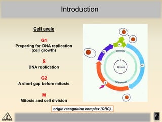
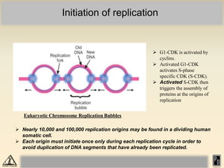
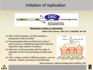
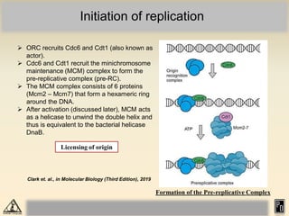
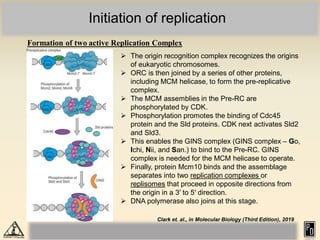
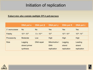
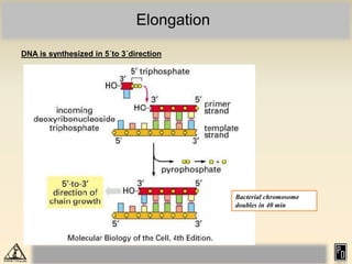
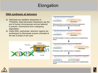
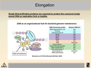
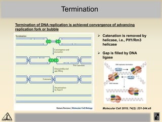
M.Sc. Zoology Semester I Molecular Biology
DNA replication in eukaryotes
Praveen Deepak
Assistant Professor
Department of Zoology
S. S. College, Jehanabad
Introduction
 DNA replication is the process of producing two identical copies from one
original DNA molecules.
 It is the basis of inheritance and therefore it occurs in all living organisms.
 Fundamental mechanism of DNA replication is same, however there are
some variation due to difference in molecular properties.
 DNA replication in eukaryotes (karyobionta) is more complex than the DNA
replication in prokaryotes (akaryobionta).
 DNA in eukaryotes are highly condensed nucleosomal structure which
needs to be unwound before replication.
 Majority of DNA replication (synthesis) occurs during S phase of the cell
cycle (telomere is synthesized in late S phase).
 Due to complex nature of eukaryotic chromosomes, replication is not as well
understood as that in prokaryotes.
 Replication is bidirectional as in the prokaryotes.
Introduction
Cell cycle
G1
Preparing for DNA replication
(cell growth)
S
DNA replication
G2
A short gap before mitosis
M
Mitosis and cell division
origin recognition complex (ORC)
Prokaryotic and eukaryotic DNA supercoils
 Most prokaryotic genomes are less
than 5 Mb in size (Bacillus
megaterium has 30 Mb), while in
eukaryotes, ranges from 10 Mb in
some fungi to > 100,000 Mb in
certain plants, salamanders and
lungfishes).
 Prokaryotic chromosomes are
circular, while eukaryotic one is
linear.
 Chromatin is found in the
eukaryotic chromosome.
 Opening DNA in prokaryotes is
fast, while fork formation and
movement is slow in eukaryotes.
DNA replication at heterochromatin region and telomeric region is slow, while at
euchromatin and centromeric region, replication is faster.
Nucleosome formation
Structure of a nucleosome
The positive charge of
histones (lysine and
arginine amino acids), is
a major feature of the
histone molecules
enabling them to bind
the negatively charged
phosphate backbones of
DNA. Pairs of four
different histones (H2A,
H2B, H3, and H4)
combine to form an
eight-protein bead
around which DNA is
wound (nucleosome)
Bhagavan & Ha, in Essentials of Medical Biochemistry (Second Edition), 2015
Initiation of replication
Eukaryotic Chromosome Replication Bubbles
 Nearly 10,000 and 100,000 replication origins may be found in a dividing human
somatic cell.
 Each origin must initiate once only during each replication cycle in order to
avoid duplication of DNA segments that have already been replicated.
 G1-CDK is activated by
cyclins.
 Activated G1-CDK
activates S-phase
specific CDK (S-CDK).
 Activated S-CDK then
triggers the assembly of
proteins at the origins of
replication
Initiation of replication
Replication origins in eukaryotes.
Gilbert D.M. Science. 2001 Oct 5; 294(5540): 96–100.
 After S-CDK activation, S-CDK transfers a
phosphate to Sld2 and Sld3.
 Phosphorylated Sld2 and Sld3 bind to Dbp11,
which acts as a scaffolding protein that holds the
replication origin proteins in position.
 After that, cdc45 associates with the origin of
replication to form the pre-loading complex (pre-
LC).
 Then, a large number of different proteins, such as
helicase, initiates unwinding of the DNA helix.
Clark & Pazdernik, in Molecular Biology (Second Edition), 2013
Initiation of replication
Formation of the Pre-replicative Complex
 ORC recruits Cdc6 and Cdt1 (also known as
actor).
 Cdc6 and Cdt1 recruit the minichromosome
maintenance (MCM) complex to form the
pre-replicative complex (pre-RC).
 The MCM complex consists of 6 proteins
(Mcm2 – Mcm7) that form a hexameric ring
around the DNA.
 After activation (discussed later), MCM acts
as a helicase to unwind the double helix and
thus is equivalent to the bacterial helicase
DnaB.
Licensing of origin
Clark et. al., in Molecular Biology (Third Edition), 2019
Initiation of replication
Formation of two active Replication Complex
 The origin recognition complex recognizes the origins
of eukaryotic chromosomes.
 ORC is then joined by a series of other proteins,
including MCM helicase, to form the pre-replicative
complex.
 The MCM assemblies in the Pre-RC are
phosphorylated by CDK.
 Phosphorylation promotes the binding of Cdc45
protein and the Sld proteins. CDK next activates Sld2
and Sld3.
 This enables the GINS complex (GINS complex – Go,
Ichi, Nii, and San.) to bind to the Pre-RC. GINS
complex is needed for the MCM helicase to operate.
 Finally, protein Mcm10 binds and the assemblage
separates into two replication complexes or
replisomes that proceed in opposite directions from
the origin in a 3′ to 5′ direction.
 DNA polymerase also joins at this stage.
Clark et. al., in Molecular Biology (Third Edition), 2019
Initiation of replication
Eukaryotes also contain multiple DNA polymerases
DNA pol α DNA pol β DNA pol γ DNA pol δ DNA pol ε
3´ exonucease No No Yes Yes Yes
Fidelity 10-4- 10-5 5 × 10⁻⁴ 10-5 10-5- 10-6 10-6- 10-7
Processivity Moderate Low High High High
Role Lagging
strand primer
synthesis
DNA repair Mitochodria I
DNA
replication
Lagging
strand
replication
Leading
strand
replication
Elongation
DNA polymerase starts synthesizing after
its recruitment in Pre-Replicative complex
(Pre-RC).
Histones are synthesized only during S
phase and are added as replication
proceeds.
Some histone parts are “inherited” and
some are new.
The spacing of histones every 200 nt
might be the reason for the shorter
Okazaki fragments in eukaryotes and the
slower speed of replication.
Elongation
 DNA polymerase adds DNA nucleotides to the 3′ end of the newly synthesized polynucleotide
strand or primer synthesized by DNA primase (RNA polymerase).
 Proliferating cell nuclear antigen (PCNA) acts as DNA clamp and processivity factor for DNA
polymerase δ.
 ATR is a serine/threonine kinase which maintains genome integrity by stabilizing replication
forks.
Elongation
Replication complex and its moving on
DNA Clamp
Molecular Cell 2006; 23(2): 155-160.
Elongation
DNA is synthesized in 5´to 3´direction
Bacterial chromosome
doubles in 40 min
Elongation
DNA synthesis at telomere
 Telomeres are repetitive sequences of
TTAGGGs, these telomeres (Hexamers) cap the
end of human chromosomes and are believed
to prevent chromosome from undergoing
degradation.
 Unlike DNA polymerase, telomeric regions are
synthesized by telomerase enzyme activated at
the late S phase of cell cycle.
Elongation
Single Strand Binding proteins are required to protect the unwound single
strand DNA at replication fork or bubble.
Termination
Termination of DNA replication is achieved convergence of advancing
replication fork or bubble
 Catenation is removed by
helicase, i.e., Plf1/Rm3
helicase
 Gap is filled by DNA
ligase
Molecular Cell 2019; 74(2): 231-244.e9
Further reading
 Watson J.D., Baker T.A., Bell S.P., Gann A., Levine M., Losick R. 2013. Molecular Biology of
the Gene, 7th Edition. Pearson education, London, UK.
 Alberts B., Johnson A., Lewis J., Raff M., Roberts K., Walter P. 2002. Molecular Biology of
the Cells. Garland Science, New York, USA.
 Lodish H., Berk A., Lawrence Zipursky S., Matsudaira P., Baltimore D., Darnell J.E.. 2000.
Molecular Cell Biology. W. H. Freeman and Company, New York, USA.
 Krebs J.E., Goldstein E.S., Kilpatrick S.T. 2017. Lewin’s Genes XII. Jones and Bartlett
Publishers, Inc., Burlington, MA, USA.
 Hartt D.L. & Jones E.W. 2001. Genetics – Analysis of genes and genomes. Jones & Barlett
Publishers.
 Miglani G.S. 2002. Advance Genetics, Narosa Publications. P520.
 Van Steensel B. 2011. Chromatin constructing the big picture. The EMBO Journal.

1768816628DNA replication in eukaryotes.pdf

  • 1.
    M.Sc. Zoology SemesterI Molecular Biology DNA replication in eukaryotes Praveen Deepak Assistant Professor Department of Zoology S. S. College, Jehanabad
  • 2.
    Introduction  DNA replicationis the process of producing two identical copies from one original DNA molecules.  It is the basis of inheritance and therefore it occurs in all living organisms.  Fundamental mechanism of DNA replication is same, however there are some variation due to difference in molecular properties.  DNA replication in eukaryotes (karyobionta) is more complex than the DNA replication in prokaryotes (akaryobionta).  DNA in eukaryotes are highly condensed nucleosomal structure which needs to be unwound before replication.  Majority of DNA replication (synthesis) occurs during S phase of the cell cycle (telomere is synthesized in late S phase).  Due to complex nature of eukaryotic chromosomes, replication is not as well understood as that in prokaryotes.  Replication is bidirectional as in the prokaryotes.
  • 3.
    Introduction Cell cycle G1 Preparing forDNA replication (cell growth) S DNA replication G2 A short gap before mitosis M Mitosis and cell division origin recognition complex (ORC)
  • 4.
    Prokaryotic and eukaryoticDNA supercoils  Most prokaryotic genomes are less than 5 Mb in size (Bacillus megaterium has 30 Mb), while in eukaryotes, ranges from 10 Mb in some fungi to > 100,000 Mb in certain plants, salamanders and lungfishes).  Prokaryotic chromosomes are circular, while eukaryotic one is linear.  Chromatin is found in the eukaryotic chromosome.  Opening DNA in prokaryotes is fast, while fork formation and movement is slow in eukaryotes. DNA replication at heterochromatin region and telomeric region is slow, while at euchromatin and centromeric region, replication is faster.
  • 5.
    Nucleosome formation Structure ofa nucleosome The positive charge of histones (lysine and arginine amino acids), is a major feature of the histone molecules enabling them to bind the negatively charged phosphate backbones of DNA. Pairs of four different histones (H2A, H2B, H3, and H4) combine to form an eight-protein bead around which DNA is wound (nucleosome) Bhagavan & Ha, in Essentials of Medical Biochemistry (Second Edition), 2015
  • 6.
    Initiation of replication EukaryoticChromosome Replication Bubbles  Nearly 10,000 and 100,000 replication origins may be found in a dividing human somatic cell.  Each origin must initiate once only during each replication cycle in order to avoid duplication of DNA segments that have already been replicated.  G1-CDK is activated by cyclins.  Activated G1-CDK activates S-phase specific CDK (S-CDK).  Activated S-CDK then triggers the assembly of proteins at the origins of replication
  • 7.
    Initiation of replication Replicationorigins in eukaryotes. Gilbert D.M. Science. 2001 Oct 5; 294(5540): 96–100.  After S-CDK activation, S-CDK transfers a phosphate to Sld2 and Sld3.  Phosphorylated Sld2 and Sld3 bind to Dbp11, which acts as a scaffolding protein that holds the replication origin proteins in position.  After that, cdc45 associates with the origin of replication to form the pre-loading complex (pre- LC).  Then, a large number of different proteins, such as helicase, initiates unwinding of the DNA helix. Clark & Pazdernik, in Molecular Biology (Second Edition), 2013
  • 8.
    Initiation of replication Formationof the Pre-replicative Complex  ORC recruits Cdc6 and Cdt1 (also known as actor).  Cdc6 and Cdt1 recruit the minichromosome maintenance (MCM) complex to form the pre-replicative complex (pre-RC).  The MCM complex consists of 6 proteins (Mcm2 – Mcm7) that form a hexameric ring around the DNA.  After activation (discussed later), MCM acts as a helicase to unwind the double helix and thus is equivalent to the bacterial helicase DnaB. Licensing of origin Clark et. al., in Molecular Biology (Third Edition), 2019
  • 9.
    Initiation of replication Formationof two active Replication Complex  The origin recognition complex recognizes the origins of eukaryotic chromosomes.  ORC is then joined by a series of other proteins, including MCM helicase, to form the pre-replicative complex.  The MCM assemblies in the Pre-RC are phosphorylated by CDK.  Phosphorylation promotes the binding of Cdc45 protein and the Sld proteins. CDK next activates Sld2 and Sld3.  This enables the GINS complex (GINS complex – Go, Ichi, Nii, and San.) to bind to the Pre-RC. GINS complex is needed for the MCM helicase to operate.  Finally, protein Mcm10 binds and the assemblage separates into two replication complexes or replisomes that proceed in opposite directions from the origin in a 3′ to 5′ direction.  DNA polymerase also joins at this stage. Clark et. al., in Molecular Biology (Third Edition), 2019
  • 10.
    Initiation of replication Eukaryotesalso contain multiple DNA polymerases DNA pol α DNA pol β DNA pol γ DNA pol δ DNA pol ε 3´ exonucease No No Yes Yes Yes Fidelity 10-4- 10-5 5 × 10⁻⁴ 10-5 10-5- 10-6 10-6- 10-7 Processivity Moderate Low High High High Role Lagging strand primer synthesis DNA repair Mitochodria I DNA replication Lagging strand replication Leading strand replication
  • 11.
    Elongation DNA polymerase startssynthesizing after its recruitment in Pre-Replicative complex (Pre-RC). Histones are synthesized only during S phase and are added as replication proceeds. Some histone parts are “inherited” and some are new. The spacing of histones every 200 nt might be the reason for the shorter Okazaki fragments in eukaryotes and the slower speed of replication.
  • 12.
    Elongation  DNA polymeraseadds DNA nucleotides to the 3′ end of the newly synthesized polynucleotide strand or primer synthesized by DNA primase (RNA polymerase).  Proliferating cell nuclear antigen (PCNA) acts as DNA clamp and processivity factor for DNA polymerase δ.  ATR is a serine/threonine kinase which maintains genome integrity by stabilizing replication forks.
  • 13.
    Elongation Replication complex andits moving on DNA Clamp Molecular Cell 2006; 23(2): 155-160.
  • 14.
    Elongation DNA is synthesizedin 5´to 3´direction Bacterial chromosome doubles in 40 min
  • 15.
    Elongation DNA synthesis attelomere  Telomeres are repetitive sequences of TTAGGGs, these telomeres (Hexamers) cap the end of human chromosomes and are believed to prevent chromosome from undergoing degradation.  Unlike DNA polymerase, telomeric regions are synthesized by telomerase enzyme activated at the late S phase of cell cycle.
  • 16.
    Elongation Single Strand Bindingproteins are required to protect the unwound single strand DNA at replication fork or bubble.
  • 17.
    Termination Termination of DNAreplication is achieved convergence of advancing replication fork or bubble  Catenation is removed by helicase, i.e., Plf1/Rm3 helicase  Gap is filled by DNA ligase Molecular Cell 2019; 74(2): 231-244.e9
  • 18.
    Further reading  WatsonJ.D., Baker T.A., Bell S.P., Gann A., Levine M., Losick R. 2013. Molecular Biology of the Gene, 7th Edition. Pearson education, London, UK.  Alberts B., Johnson A., Lewis J., Raff M., Roberts K., Walter P. 2002. Molecular Biology of the Cells. Garland Science, New York, USA.  Lodish H., Berk A., Lawrence Zipursky S., Matsudaira P., Baltimore D., Darnell J.E.. 2000. Molecular Cell Biology. W. H. Freeman and Company, New York, USA.  Krebs J.E., Goldstein E.S., Kilpatrick S.T. 2017. Lewin’s Genes XII. Jones and Bartlett Publishers, Inc., Burlington, MA, USA.  Hartt D.L. & Jones E.W. 2001. Genetics – Analysis of genes and genomes. Jones & Barlett Publishers.  Miglani G.S. 2002. Advance Genetics, Narosa Publications. P520.  Van Steensel B. 2011. Chromatin constructing the big picture. The EMBO Journal.