- Cell biology began with ancient Greek philosophers observing common structures in plants and animals in the 4th century BC.
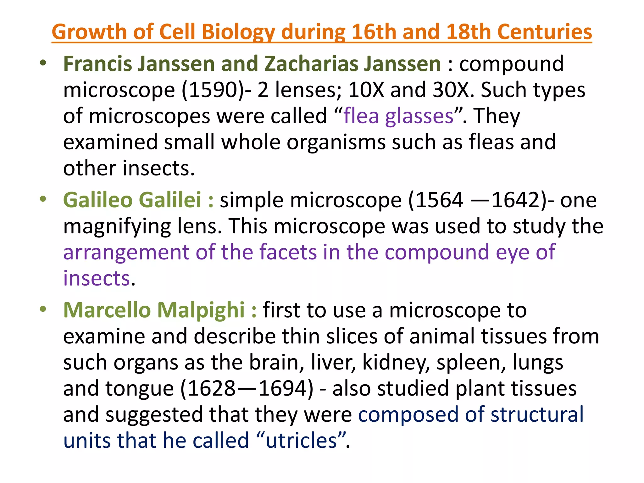
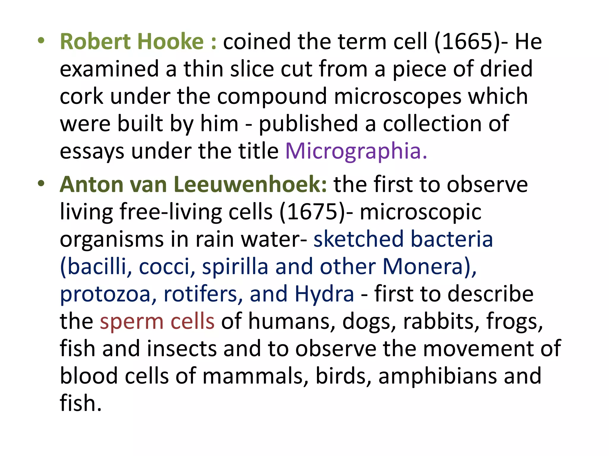
- Advances in microscope technology in the 16th-18th centuries allowed scientists like Hooke, van Leeuwenhoek, and Malpighi to observe cells directly for the first time.
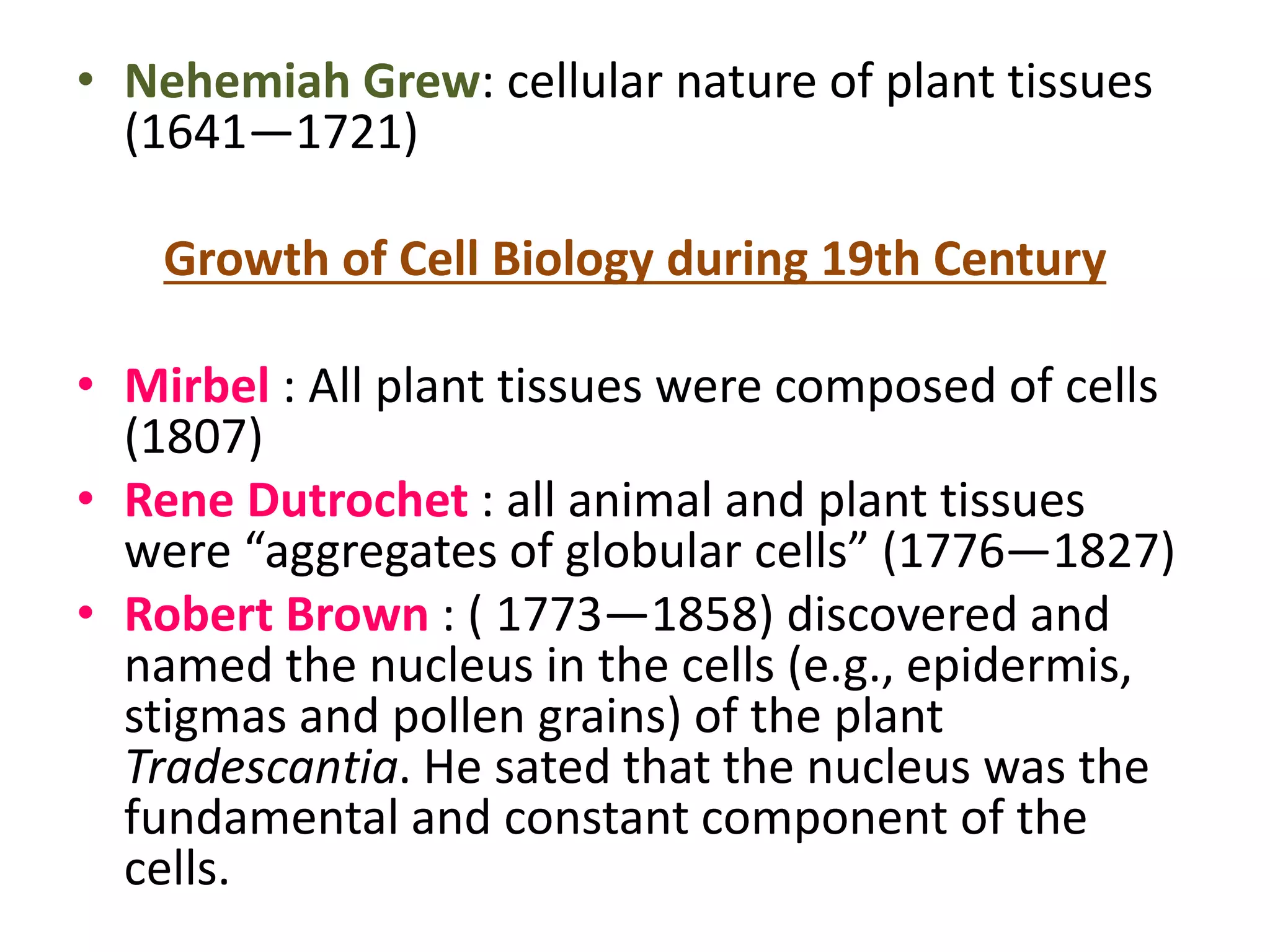
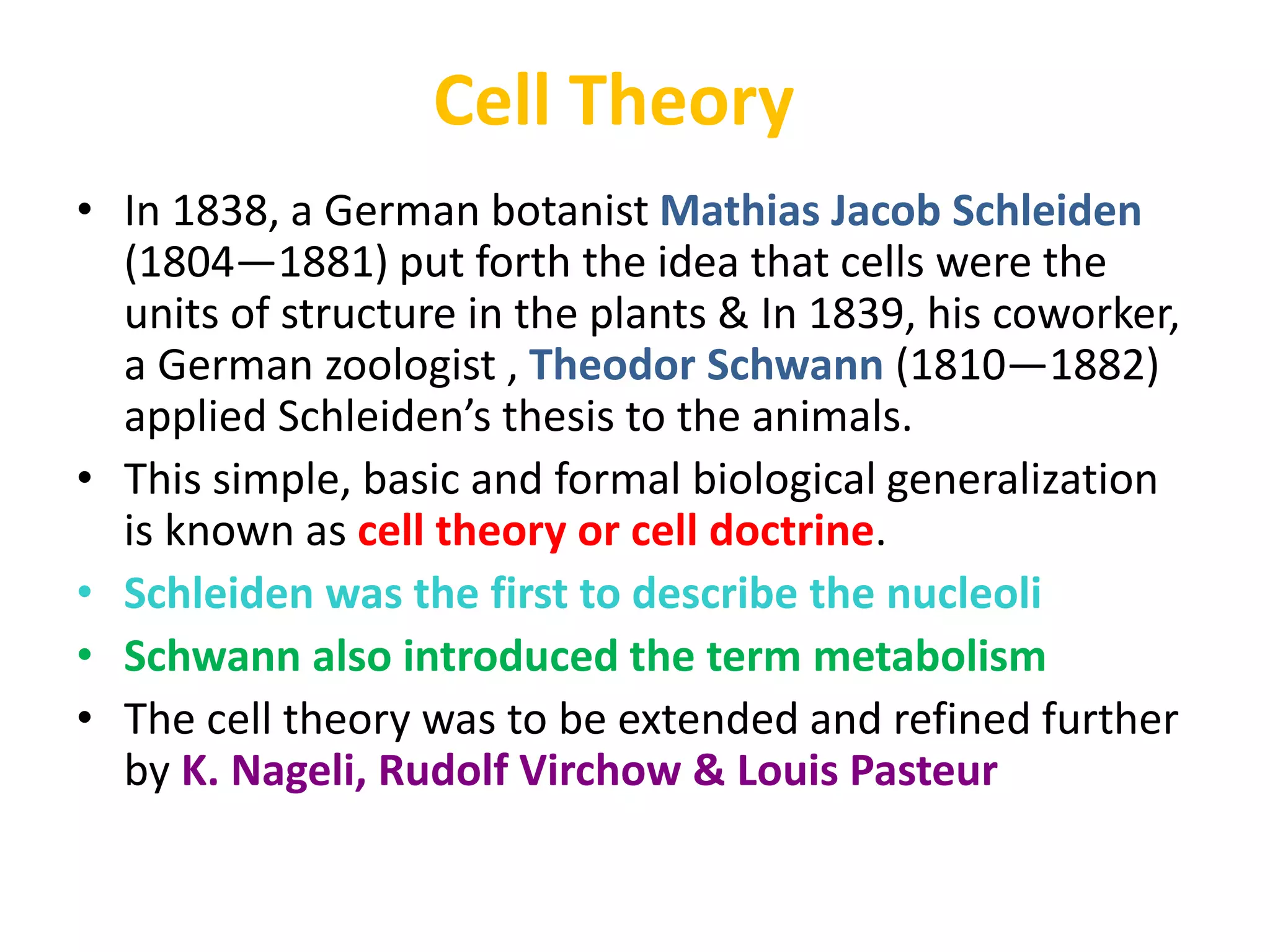
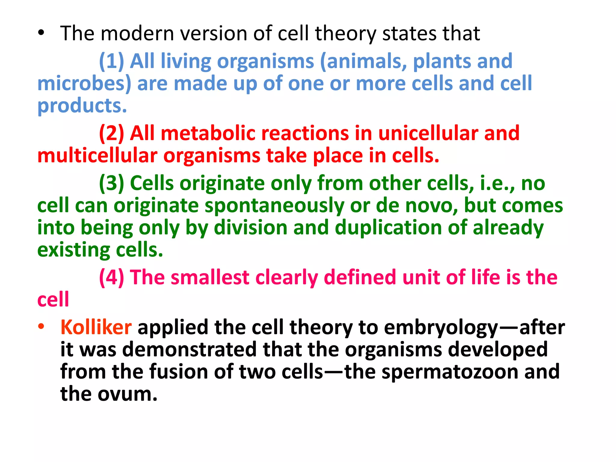
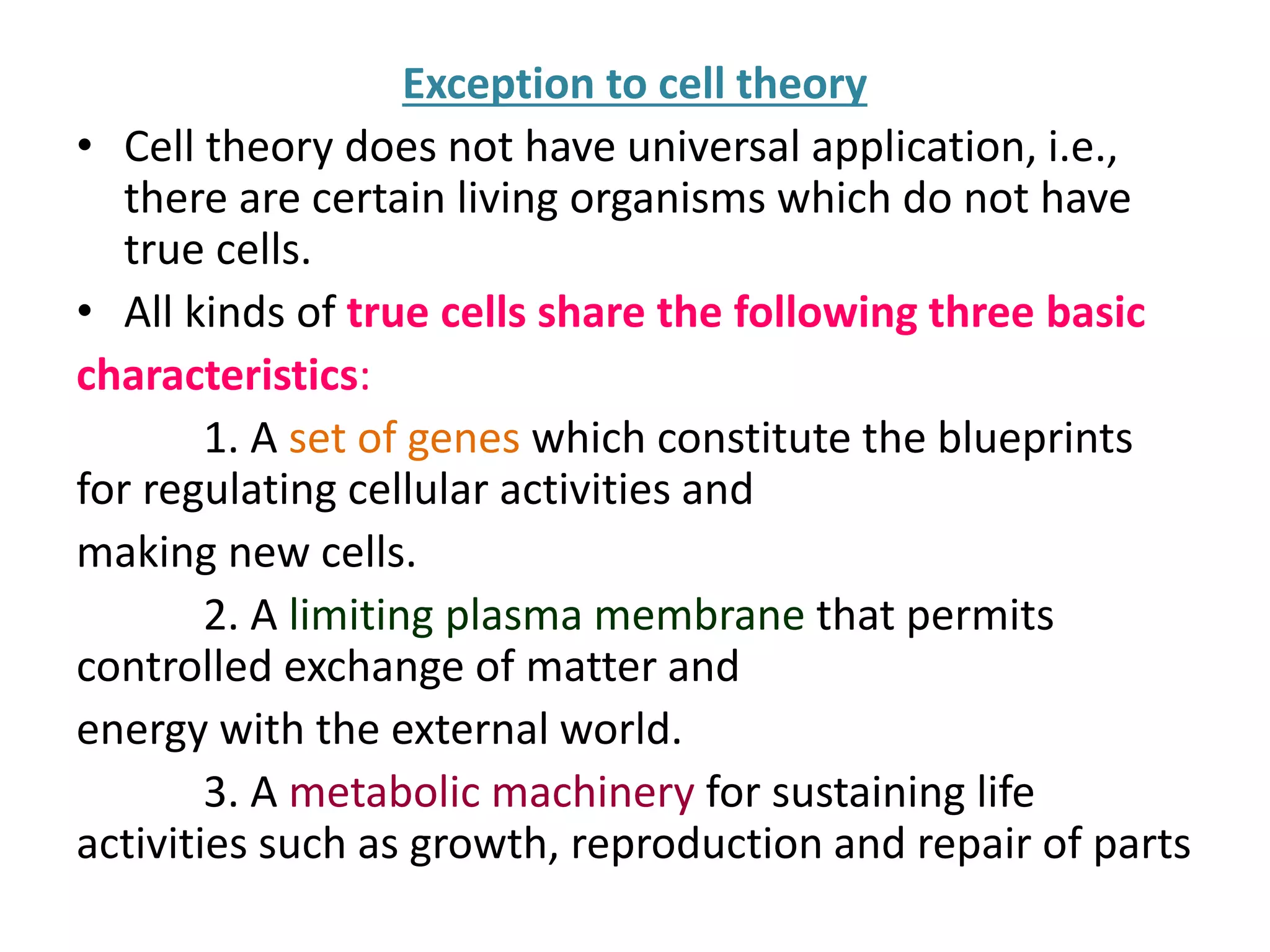
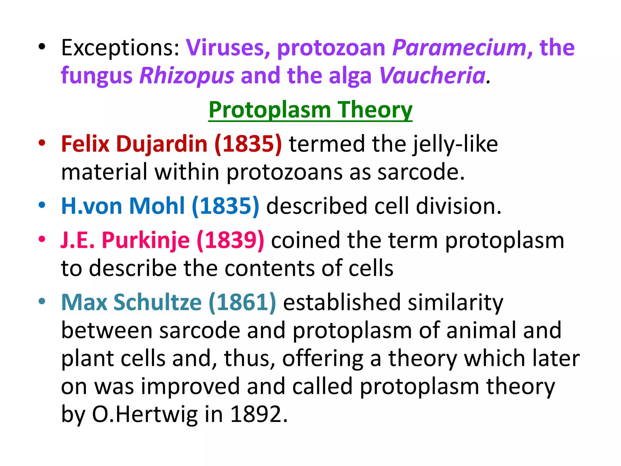
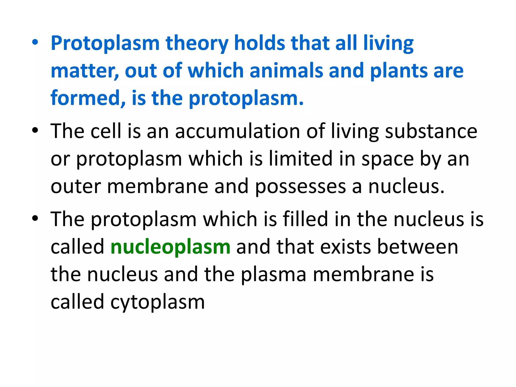
- In 1838, Schleiden proposed that plants are composed of cells, and in 1839, Schwann extended this to animals, establishing the foundations of the cell theory.
INTRODUCTION TO CELLS
INTRODUCTION TO CELL THEORY
HISTORY
FORMULATION OF CELL THEORY
CLASSICAL CELL THEORY
DRAWBACKS OF CLASSICAL THEORY
MORDEN CELL THEORY
EXCEPTION OF CELL THEORY
SIGNIFICANCE OF CELL THEORY
HOW HAS THE CELL THEORY BEEN USED
CONCLUSION
INTRODUCTION TO CELLS
INTRODUCTION TO CELL THEORY
HISTORY
FORMULATION OF CELL THEORY
CLASSICAL CELL THEORY
DRAWBACKS OF CLASSICAL THEORY
MORDEN CELL THEORY
EXCEPTION OF CELL THEORY
SIGNIFICANCE OF CELL THEORY
HOW HAS THE CELL THEORY BEEN USED
CONCLUSION
This power point presentation is an attempt to present some direct and some indirect evidences in favour of DNA as genetic material. Very few organisms have RNA as genetic material for example plant virus and some bacteriophages
ultra structure of Ribosome, Prokaryotic Ribosome, Eukaryotic Ribosome, Svedberg unit, Centrifugal force, assembly of Ribosome, functions of Ribosome, models of Ribosomes, fine structure of Ribosome, Discovery of Ribosome,
The word cell is derived from the Latin word “cellula” which means “a little room”
It was the British botanist Robert Hooke who, in 1664, while examining a slice of bottle cork under a microscope, found its structure resembling the box-like living quarters of the monks in a monastery, and coined the word “cells”
Diversity of cell size & shape By KK Sahu SirKAUSHAL SAHU
SYNOPSIS
Introduction to cell
Historical Aspects
Cell Diversity
Types Of Cell Diversity
Cell Diversity In Origin
Cell Diversity In size
Cell Diversity In Shape
Some Other Types
5) Differentiation And Specialisation Of Cell Diversity
6) Conclusion
7) References
History of Genetics - Pre-Mendelian GeneticsAsad Afridi
this presentation is about history of genetics. all theory suggested and proposed after Mendel are discussed in this presentation. such as fluid theories, preformation theories and particulate theories
INTRODUCTION
HISTORY
ORIGIN OF LIFE
THEORY OF ETERNITY
THEORY OF SPECIAL CREATION
THEORY OF SPONTANEOUS GENERATION
CELL THOERY
MODERN CELL THEORY
EXCEPTION OF CELL THEORY
CONCLUSION
REFERANCES
This presentation you will get how the cell theory developed.
Robert Hooke observed cells in cork and coined the term "cells”.
Anton Van Leeuwenhoek observed first living cells under the simple microscope.
Matthias Schleiden (1838) German lawyer turned botanist, concluded that, despite differences in the structure of various tissues, plants were made of cells and that the plant embryo arose from a single cell.
In 1839, Theodor Schwann, a German zoologist and colleague of Schleiden’s, published a comprehensive report on the cellular basis of animal life. Schwann concluded that the cells of plants and animals are similar structures.
By 1855, Rudolf Virchow, a German pathologist concluded that
“Omnis cellula e cellula”- new cells are formed only from pre-existing cells.
All organisms are composed of one or more cells.
2) The cell is the structural unit of life.
3) Cells can arise only by division from a pre-existing cell
the smallest structural and functional unit of an organism, typically microscopic and consisting of cytoplasm and a nucleus enclosed in a membrane. Microscopic organisms typically consist of a single cell, which is either eukaryotic or prokaryotic.
Seminar of U.V. Spectroscopy by SAMIR PANDASAMIR PANDA
Spectroscopy is a branch of science dealing the study of interaction of electromagnetic radiation with matter.
Ultraviolet-visible spectroscopy refers to absorption spectroscopy or reflect spectroscopy in the UV-VIS spectral region.
Ultraviolet-visible spectroscopy is an analytical method that can measure the amount of light received by the analyte.
(May 29th, 2024) Advancements in Intravital Microscopy- Insights for Preclini...Scintica Instrumentation
Intravital microscopy (IVM) is a powerful tool utilized to study cellular behavior over time and space in vivo. Much of our understanding of cell biology has been accomplished using various in vitro and ex vivo methods; however, these studies do not necessarily reflect the natural dynamics of biological processes. Unlike traditional cell culture or fixed tissue imaging, IVM allows for the ultra-fast high-resolution imaging of cellular processes over time and space and were studied in its natural environment. Real-time visualization of biological processes in the context of an intact organism helps maintain physiological relevance and provide insights into the progression of disease, response to treatments or developmental processes.
In this webinar we give an overview of advanced applications of the IVM system in preclinical research. IVIM technology is a provider of all-in-one intravital microscopy systems and solutions optimized for in vivo imaging of live animal models at sub-micron resolution. The system’s unique features and user-friendly software enables researchers to probe fast dynamic biological processes such as immune cell tracking, cell-cell interaction as well as vascularization and tumor metastasis with exceptional detail. This webinar will also give an overview of IVM being utilized in drug development, offering a view into the intricate interaction between drugs/nanoparticles and tissues in vivo and allows for the evaluation of therapeutic intervention in a variety of tissues and organs. This interdisciplinary collaboration continues to drive the advancements of novel therapeutic strategies.
Slide 1: Title Slide
Extrachromosomal Inheritance
Slide 2: Introduction to Extrachromosomal Inheritance
Definition: Extrachromosomal inheritance refers to the transmission of genetic material that is not found within the nucleus.
Key Components: Involves genes located in mitochondria, chloroplasts, and plasmids.
Slide 3: Mitochondrial Inheritance
Mitochondria: Organelles responsible for energy production.
Mitochondrial DNA (mtDNA): Circular DNA molecule found in mitochondria.
Inheritance Pattern: Maternally inherited, meaning it is passed from mothers to all their offspring.
Diseases: Examples include Leber’s hereditary optic neuropathy (LHON) and mitochondrial myopathy.
Slide 4: Chloroplast Inheritance
Chloroplasts: Organelles responsible for photosynthesis in plants.
Chloroplast DNA (cpDNA): Circular DNA molecule found in chloroplasts.
Inheritance Pattern: Often maternally inherited in most plants, but can vary in some species.
Examples: Variegation in plants, where leaf color patterns are determined by chloroplast DNA.
Slide 5: Plasmid Inheritance
Plasmids: Small, circular DNA molecules found in bacteria and some eukaryotes.
Features: Can carry antibiotic resistance genes and can be transferred between cells through processes like conjugation.
Significance: Important in biotechnology for gene cloning and genetic engineering.
Slide 6: Mechanisms of Extrachromosomal Inheritance
Non-Mendelian Patterns: Do not follow Mendel’s laws of inheritance.
Cytoplasmic Segregation: During cell division, organelles like mitochondria and chloroplasts are randomly distributed to daughter cells.
Heteroplasmy: Presence of more than one type of organellar genome within a cell, leading to variation in expression.
Slide 7: Examples of Extrachromosomal Inheritance
Four O’clock Plant (Mirabilis jalapa): Shows variegated leaves due to different cpDNA in leaf cells.
Petite Mutants in Yeast: Result from mutations in mitochondrial DNA affecting respiration.
Slide 8: Importance of Extrachromosomal Inheritance
Evolution: Provides insight into the evolution of eukaryotic cells.
Medicine: Understanding mitochondrial inheritance helps in diagnosing and treating mitochondrial diseases.
Agriculture: Chloroplast inheritance can be used in plant breeding and genetic modification.
Slide 9: Recent Research and Advances
Gene Editing: Techniques like CRISPR-Cas9 are being used to edit mitochondrial and chloroplast DNA.
Therapies: Development of mitochondrial replacement therapy (MRT) for preventing mitochondrial diseases.
Slide 10: Conclusion
Summary: Extrachromosomal inheritance involves the transmission of genetic material outside the nucleus and plays a crucial role in genetics, medicine, and biotechnology.
Future Directions: Continued research and technological advancements hold promise for new treatments and applications.
Slide 11: Questions and Discussion
Invite Audience: Open the floor for any questions or further discussion on the topic.
The increased availability of biomedical data, particularly in the public domain, offers the opportunity to better understand human health and to develop effective therapeutics for a wide range of unmet medical needs. However, data scientists remain stymied by the fact that data remain hard to find and to productively reuse because data and their metadata i) are wholly inaccessible, ii) are in non-standard or incompatible representations, iii) do not conform to community standards, and iv) have unclear or highly restricted terms and conditions that preclude legitimate reuse. These limitations require a rethink on data can be made machine and AI-ready - the key motivation behind the FAIR Guiding Principles. Concurrently, while recent efforts have explored the use of deep learning to fuse disparate data into predictive models for a wide range of biomedical applications, these models often fail even when the correct answer is already known, and fail to explain individual predictions in terms that data scientists can appreciate. These limitations suggest that new methods to produce practical artificial intelligence are still needed.
In this talk, I will discuss our work in (1) building an integrative knowledge infrastructure to prepare FAIR and "AI-ready" data and services along with (2) neurosymbolic AI methods to improve the quality of predictions and to generate plausible explanations. Attention is given to standards, platforms, and methods to wrangle knowledge into simple, but effective semantic and latent representations, and to make these available into standards-compliant and discoverable interfaces that can be used in model building, validation, and explanation. Our work, and those of others in the field, creates a baseline for building trustworthy and easy to deploy AI models in biomedicine.
Bio
Dr. Michel Dumontier is the Distinguished Professor of Data Science at Maastricht University, founder and executive director of the Institute of Data Science, and co-founder of the FAIR (Findable, Accessible, Interoperable and Reusable) data principles. His research explores socio-technological approaches for responsible discovery science, which includes collaborative multi-modal knowledge graphs, privacy-preserving distributed data mining, and AI methods for drug discovery and personalized medicine. His work is supported through the Dutch National Research Agenda, the Netherlands Organisation for Scientific Research, Horizon Europe, the European Open Science Cloud, the US National Institutes of Health, and a Marie-Curie Innovative Training Network. He is the editor-in-chief for the journal Data Science and is internationally recognized for his contributions in bioinformatics, biomedical informatics, and semantic technologies including ontologies and linked data.
Multi-source connectivity as the driver of solar wind variability in the heli...Sérgio Sacani
The ambient solar wind that flls the heliosphere originates from multiple
sources in the solar corona and is highly structured. It is often described
as high-speed, relatively homogeneous, plasma streams from coronal
holes and slow-speed, highly variable, streams whose source regions are
under debate. A key goal of ESA/NASA’s Solar Orbiter mission is to identify
solar wind sources and understand what drives the complexity seen in the
heliosphere. By combining magnetic feld modelling and spectroscopic
techniques with high-resolution observations and measurements, we show
that the solar wind variability detected in situ by Solar Orbiter in March
2022 is driven by spatio-temporal changes in the magnetic connectivity to
multiple sources in the solar atmosphere. The magnetic feld footpoints
connected to the spacecraft moved from the boundaries of a coronal hole
to one active region (12961) and then across to another region (12957). This
is refected in the in situ measurements, which show the transition from fast
to highly Alfvénic then to slow solar wind that is disrupted by the arrival of
a coronal mass ejection. Our results describe solar wind variability at 0.5 au
but are applicable to near-Earth observatories.
Professional air quality monitoring systems provide immediate, on-site data for analysis, compliance, and decision-making.
Monitor common gases, weather parameters, particulates.
2. • 384 —322 B.C.: Ancient Greek philosophers such
as Aristotle and Paracelsus concluded that “all
animals and plants, however, complicated, are
constituted of a few elements which are repeated
in each of them.”
• They were referring to the macroscopic
structures of an organism such as roots, leaves
and flowers common to different plants, or
segments and organs that are repeated in the
animal kingdom.
• 1485 : Da Vinci recommended the uses of lenses
in viewing small objects.
• 1558 : Conrad Gesner published his on the
structure of a group of protists called
foraminifera.
3. Growth of Cell Biology during 16th and 18th Centuries
• Francis Janssen and Zacharias Janssen : compound
microscope (1590)- 2 lenses; 10X and 30X. Such types
of microscopes were called “flea glasses”. They
examined small whole organisms such as fleas and
other insects.
• Galileo Galilei : simple microscope (1564 —1642)- one
magnifying lens. This microscope was used to study the
arrangement of the facets in the compound eye of
insects.
• Marcello Malpighi : first to use a microscope to
examine and describe thin slices of animal tissues from
such organs as the brain, liver, kidney, spleen, lungs
and tongue (1628—1694) - also studied plant tissues
and suggested that they were composed of structural
units that he called “utricles”.
4. • Robert Hooke : coined the term cell (1665)- He
examined a thin slice cut from a piece of dried
cork under the compound microscopes which
were built by him - published a collection of
essays under the title Micrographia.
• Anton van Leeuwenhoek: the first to observe
living free-living cells (1675)- microscopic
organisms in rain water- sketched bacteria
(bacilli, cocci, spirilla and other Monera),
protozoa, rotifers, and Hydra - first to describe
the sperm cells of humans, dogs, rabbits, frogs,
fish and insects and to observe the movement of
blood cells of mammals, birds, amphibians and
fish.
5. • Nehemiah Grew: cellular nature of plant tissues
(1641—1721)
Growth of Cell Biology during 19th Century
• Mirbel : All plant tissues were composed of cells
(1807)
• Rene Dutrochet : all animal and plant tissues
were “aggregates of globular cells” (1776—1827)
• Robert Brown : ( 1773—1858) discovered and
named the nucleus in the cells (e.g., epidermis,
stigmas and pollen grains) of the plant
Tradescantia. He sated that the nucleus was the
fundamental and constant component of the
cells.
6. Cell Theory
• In 1838, a German botanist Mathias Jacob Schleiden
(1804—1881) put forth the idea that cells were the
units of structure in the plants & In 1839, his coworker,
a German zoologist , Theodor Schwann (1810—1882)
applied Schleiden’s thesis to the animals.
• This simple, basic and formal biological generalization
is known as cell theory or cell doctrine.
• Schleiden was the first to describe the nucleoli
• Schwann also introduced the term metabolism
• The cell theory was to be extended and refined further
by K. Nageli, Rudolf Virchow & Louis Pasteur
7. • The modern version of cell theory states that
(1) All living organisms (animals, plants and
microbes) are made up of one or more cells and cell
products.
(2) All metabolic reactions in unicellular and
multicellular organisms take place in cells.
(3) Cells originate only from other cells, i.e., no
cell can originate spontaneously or de novo, but comes
into being only by division and duplication of already
existing cells.
(4) The smallest clearly defined unit of life is the
cell
• Kolliker applied the cell theory to embryology—after
it was demonstrated that the organisms developed
from the fusion of two cells—the spermatozoon and
the ovum.
8. Exception to cell theory
• Cell theory does not have universal application, i.e.,
there are certain living organisms which do not have
true cells.
• All kinds of true cells share the following three basic
characteristics:
1. A set of genes which constitute the blueprints
for regulating cellular activities and
making new cells.
2. A limiting plasma membrane that permits
controlled exchange of matter and
energy with the external world.
3. A metabolic machinery for sustaining life
activities such as growth, reproduction and repair of parts
9. • Exceptions: Viruses, protozoan Paramecium, the
fungus Rhizopus and the alga Vaucheria.
Protoplasm Theory
• Felix Dujardin (1835) termed the jelly-like
material within protozoans as sarcode.
• H.von Mohl (1835) described cell division.
• J.E. Purkinje (1839) coined the term protoplasm
to describe the contents of cells
• Max Schultze (1861) established similarity
between sarcode and protoplasm of animal and
plant cells and, thus, offering a theory which later
on was improved and called protoplasm theory
by O.Hertwig in 1892.
10. • Protoplasm theory holds that all living
matter, out of which animals and plants are
formed, is the protoplasm.
• The cell is an accumulation of living substance
or protoplasm which is limited in space by an
outer membrane and possesses a nucleus.
• The protoplasm which is filled in the nucleus is
called nucleoplasm and that exists between
the nucleus and the plasma membrane is
called cytoplasm
14. Organismal Theory
• The body of all multicellular organisms is a
continuous mass of protoplasm which remains
divided incompletely into small centres, the cells, for
the various biological activities.
• Thus, a multicellular organism is a highly differentiated
protoplasmic individual, differing with a unicellular
Protozoa only in size and degree of differentiation of
the protoplasm.
• The differentiation involves separation of the
protoplasm into subordinate semi-independent
compartments, the so-called cells.
• Organismal theory too fails to ascertain the position of
viruses