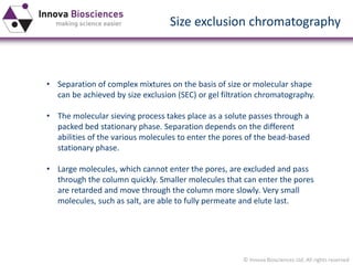
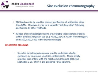
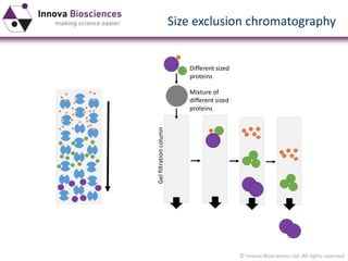
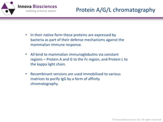
Download as PDF, PPTX

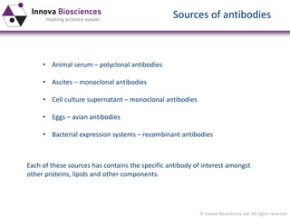

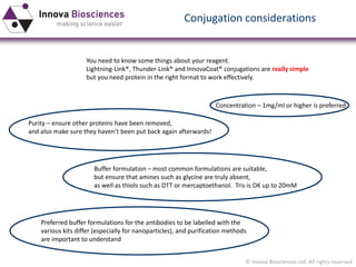
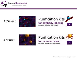

The document is a comprehensive overview of antibody purification methods, including sources of antibodies, various purification techniques, and factors affecting the quality of purified antibodies. It discusses methods such as ion exchange chromatography, size exclusion chromatography, and antigen affinity chromatography, explaining their advantages and limitations. Additionally, it highlights the considerations necessary for effective antibody use, such as buffer components and potential contaminants.


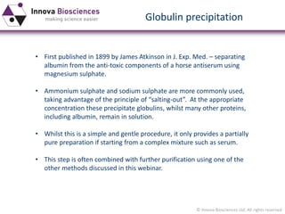
![Polymer [ बहुलक ] Chemistry Notes PDF - Irfanullah Mehar - JJ Sir Chemistry.pdf](https://cdn.slidesharecdn.com/ss_thumbnails/polymerchemistrynotespdf-irfanullahmehar-jjsirchemistry-260210172118-3f9b37f7-thumbnail.jpg?width=640&height=640&fit=bounds)


