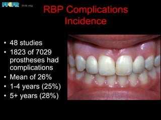
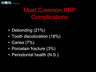
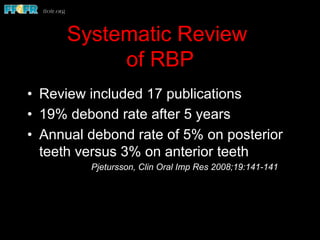
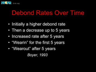
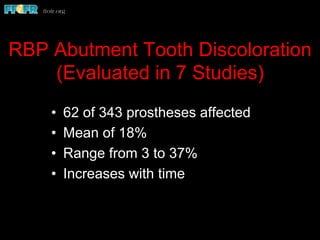
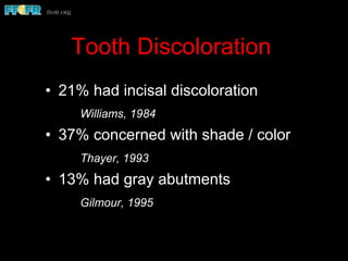
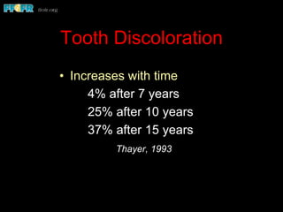
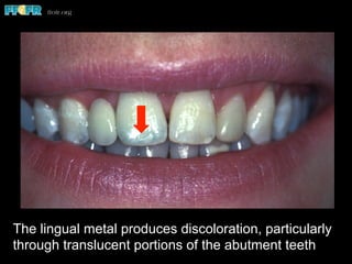
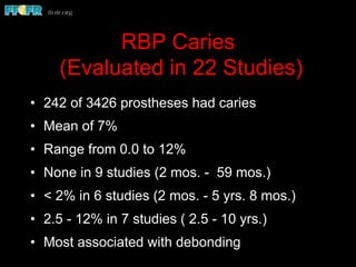
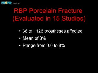
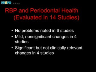
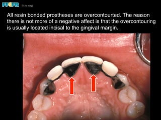
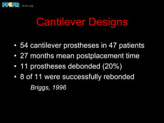
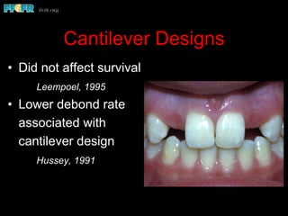
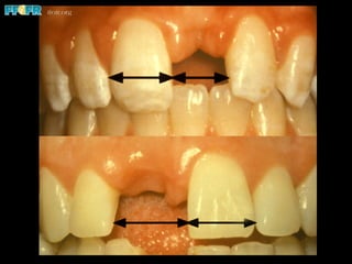
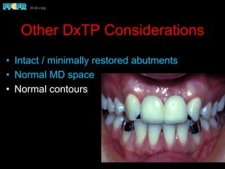
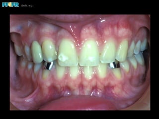
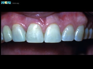
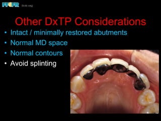
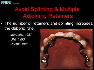
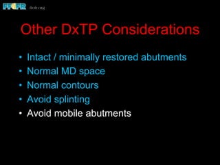
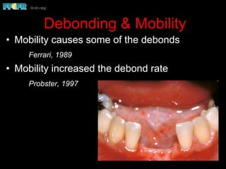
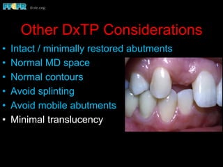
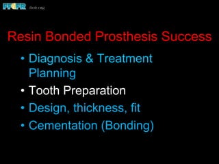
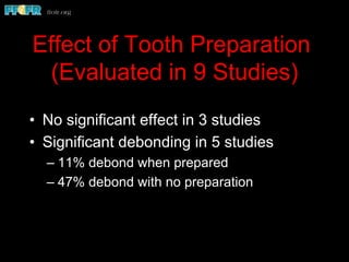
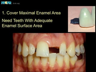
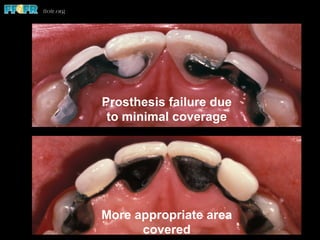
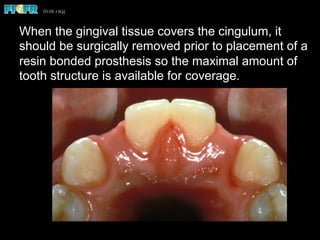
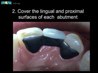
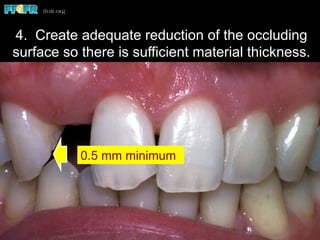
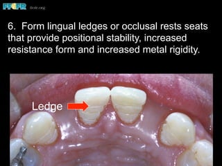
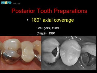
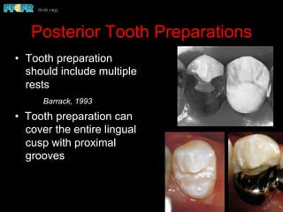
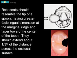
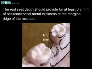
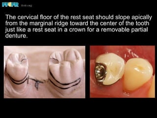
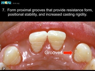
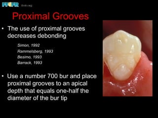
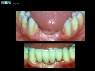
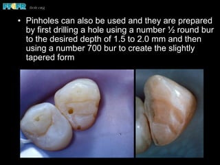
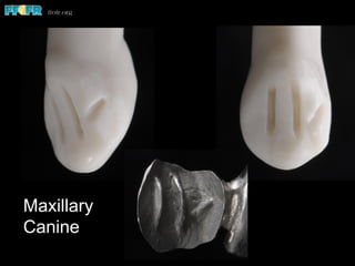
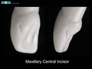
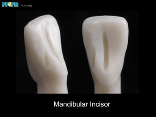
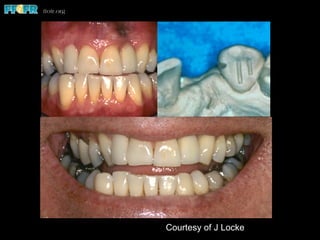
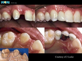
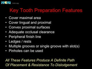
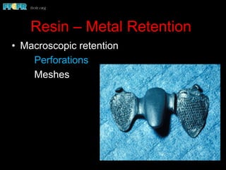
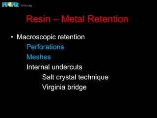
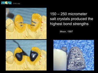
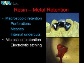
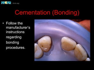
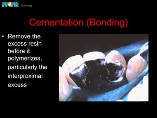
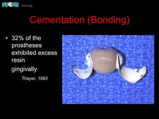
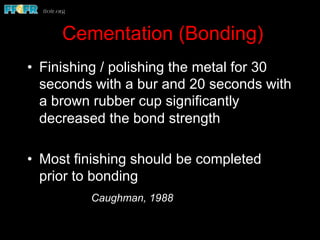
Charles J. Goodacre, DDS, MSD
Professor of Restorative Dentistry
Loma Linda University School of Dentistry
This program of instruction is protected by copyright ©. No portion of this
program of instruction may be reproduced, recorded or transferred by any
means electronic, digital, photographic, mechanical etc., or by any information
storage or retrieval system, without prior permission.
Optimizing success
with resin bonded prostheses
•  Rochette
•  Acid Etched
•  Resin Retained
Resin bonded prostheses names
•  Etched Cast
•  Maryland
•  Virginia
Resin Bonded Prosthesis Success
•  Diagnosis & Treatment Planning
•  Tooth Preparation
•  Design, thickness, fit
•  Cementation (Bonding)
RBP Complications
Incidence
•  48 studies
•  1823 of 7029
prostheses had
complications
•  Mean of 26%
•  1-4 years (25%)
•  5+ years (28%)
Most Common RBP
Complications
•  Debonding (21%)
•  Tooth discoloration (18%)
•  Caries (7%)
•  Porcelain fracture (3%)
•  Periodontal health (N.S.)
RBP Debonding
(Evaluated in 49 Studies)
•  1481 of 7029 prostheses debonded
•  Mean of 21%
•  Range from 0.0 to 52%
Systematic Review
of RBP
•  Review included 17 publications
•  19% debond rate after 5 years
•  Annual debond rate of 5% on posterior
teeth versus 3% on anterior teeth
Pjetursson, Clin Oral Imp Res 2008;19:141-141
Debond Rates Over Time
•  Initially a higher debond rate
•  Then a decrease up to 5 years
•  Increased rate after 5 years
•  “Wearin” for the first 5 years
•  “Wearout” after 5 years
Boyer, 1993
Debonding should not be considered a
failure since rebonding does not
constitute significant investment or
inconvenience to patients or dentists
Priest, 1988
Do you agree?
RBP Span Length
(Evaluated in 8 Studies)
•  Mean debond rate of 25% with short
spans
•  Mean debond rate of 52% when more
than 3 units, more than 1 pontic or
prostheses with more than 2 retainers
Span Length & Debonding
•  Increased debonding with multiple
pontics
Dunne, 1993
Mudassir, 1995
•  Increased debonding with 4 or more
units
Probster, 1997
Toooo Wide
Occlusal Forces
(Evaluted in 7 Studies)
•  In 2 studies, 70% and 45% of
debonding caused by heavy occlusal
forces
•  In 5 studies, 22% (31 of 143 debonds)
of debonds caused by heavy occlusal
forces
Maxillary/Mandibular Debond
Rates (Evaluated in 27 Studies)
•  á in maxilla (6 studies)
•  á in mandible (8 studies)
•  No significant difference in 13 studies
•  No conclusive trend
Anterior / Posterior Debond
Rates (Evaluated in 23 Studies)
•  á Anteriorly in 4 studies
•  á Posteriorly in 8 studies
•  No significant difference in 11 studies
•  No conclusive trend
Affect of Gender on Debonding
(Evaluated in 8 Studies)
•  No significant difference in 5 studies
•  Higher male debond rate in 3 studies
•  Possible male trend
Affect of Age on Debonding
(Evaluated in 6 Studies)
•  Higher debond rate in young patients in
4 studies
•  No significant difference in 2 studies
•  Possible age trend
Affect of Incisally
Positioned Gingiva
Produces Short
Clinical Crowns
RBP Abutment Tooth Discoloration
(Evaluated in 7 Studies)
•  62 of 343 prostheses affected
•  Mean of 18%
•  Range from 3 to 37%
•  Increases with time
Tooth Discoloration
•  21% had incisal discoloration
Williams, 1984
•  37% concerned with shade / color
Thayer, 1993
•  13% had gray abutments
Gilmour, 1995
Tooth Discoloration
•  Increases with time
4% after 7 years
25% after 10 years
37% after 15 years
Thayer, 1993
The lingual metal produces discoloration, particularly
through translucent portions of the abutment teeth
RBP Caries
(Evaluated in 22 Studies)
•  242 of 3426 prostheses had caries
•  Mean of 7%
•  Range from 0.0 to 12%
•  None in 9 studies (2 mos. - 59 mos.)
•  < 2% in 6 studies (2 mos. - 5 yrs. 8 mos.)
•  2.5 - 12% in 7 studies ( 2.5 - 10 yrs.)
•  Most associated with debonding
RBP Porcelain Fracture
(Evaluated in 15 Studies)
•  38 of 1126 prostheses affected
•  Mean of 3%
•  Range from 0.0 to 8%
RBP and Periodontal Health
(Evaluated in 14 Studies)
•  No problems noted in 6 studies
•  Mild, nonsignificant changes in 4
studies
•  Significant but not clinically relevant
changes in 4 studies
All resin bonded prostheses are overcontourted. The reason
there is not more of a negative affect is that the overcontouring
is usually located incisal to the gingival margin.
Cantilever Designs
•  54 cantilever prostheses in 47 patients
•  27 months mean postplacement time
•  11 prostheses debonded (20%)
•  8 of 11 were successfully rebonded
Briggs, 1996
Cantilever Designs
•  Did not affect survival
Leempoel, 1995
•  Lower debond rate
associated with
cantilever design
Hussey, 1991
Cantilever Success Data
•  32% debond rate
when the maxillary
central incisor was
cantilevered from the
central incisor or the
lateral incisor
(28 prostheses)
Hussey, 1996
Cantilever Success Data
•  3 year mean postplacement time
•  12% overall debond rate
•  24% debond rate when the maxillary
canine was cantilevered from the first
premolar (17 prostheses)
Hussey, 1996
Cantilever Success Data
•  6% debond rate when
the maxillary lateral
incisor was cantilevered
from the canine
(63 prostheses)
Hussey, 1996
•  6% debond rate when
the maxillary lateral
incisor was cantilevered
from the canine
(63 prostheses)
Hussey, 1996
Cantilever Designs Have Been Successfully
Used since 1988 by Dr. John Locke,
Prosthodontist in Melbourne, Australia
Cantilever Success Data
•  No debonds when
a maxillary
premolar was
cantilevered from
the other premolar
(8 prostheses)
Hussey, 1996
Cantilever Success Data
•  No debonds with
26 mandibular
prostheses
Hussey, 1996
Cantilever with Splinted Retainers due to
Orthodontic Rotation of Maxillary Canine
Other DxTP Considerations
•  Intact / minimally restored abutments
Other DxTP Considerations
•  Intact / minimally restored abutments
•  Normal MD space
Other DxTP Considerations
•  Intact / minimally restored abutments
•  Normal MD space
•  Normal contours
Other DxTP Considerations
•  Intact / minimally restored abutments
•  Normal MD space
•  Normal contours
•  Avoid splinting
Avoid Splinting & Multiple
Adjoining Retainers
•  The number of retainers and splinting increases
the debond rate
Marinello, 1987
Olin, 1990
Dunne, 1993
Other DxTP Considerations
•  Intact / minimally restored abutments
•  Normal MD space
•  Normal contours
•  Avoid splinting
•  Avoid mobile abutments
Debonding & Mobility
•  Mobility causes some of the debonds
Ferrari, 1989
•  Mobility increased the debond rate
Probster, 1997
Other DxTP Considerations
•  Intact / minimally restored abutments
•  Normal MD space
•  Normal contours
•  Avoid splinting
•  Avoid mobile abutments
•  Minimal translucency
Resin Bonded Prosthesis Success
•  Diagnosis & Treatment
Planning
•  Tooth Preparation
•  Design, thickness, fit
•  Cementation (Bonding)
Resin bonded prostheses were originally
introduced by Rochette as a prosthesis requiring
little or no tooth preparation. However, most of
the clinical studies that evaluated the effects of
tooth preparation indicate it decreases the
incidence of debonding and therefore increases
prosthesis longevity
Effect of Tooth Preparation
(Evaluated in 9 Studies)
•  No significant effect in 3 studies
•  Significant debonding in 5 studies
– 11% debond when prepared
– 47% debond with no preparation
1. Cover Maximal Enamel Area
Need Teeth With Adequate
Enamel Surface Area
Effect of Bonding Area
•  The area available for bonding affects success
Thayer, 1993
Priest, 1995
Ferrari, 1998
• The mean area of debonded retainers was 38
mm2 compared to retainers that did not debond
(45 mm2)
Thayer, 1993
• Age and gingival position affected the area
available for bonding
Prosthesis failure due
to minimal coverage
More appropriate area
covered
When the gingival tissue covers the cingulum, it
should be surgically removed prior to placement of a
resin bonded prosthesis so the maximal amount of
tooth structure is available for coverage.
2. Cover the lingual and proximal
surfaces of each abutment
3. Prepare the proximal surfaces so
they are convex faciolingually.
4. Create adequate occlusal clearance
so the metal casting can possess
adequate rigidity (0.5 millimeter
minimum). In situations where there is
tight interdigitation of opposing teeth, it
may be necessary to modify opposing
enamel surfaces to create sufficient
clearance.
0.5 mm minimum
4. Create adequate reduction of the occluding
surface so there is sufficient material thickness.
5. Form a perpheral finish line (0.2 - 0.3 mm) that
provides positional stabiliy, increases casting rigidity,
and decreases overcontouring.
•  Because the cervical enamel thickness on
mandibular incisors is minimal (0.1 – 0.3 mm),
placement of a chamfer finish line is not
recommended because it may remove all the
cervical enamel and adversely affect the bond.
Ledge
6. Form lingual ledges or occlusal rests seats
that provide positional stability, increased
resistance form and increased metal rigidity.
Posterior Tooth Preparations
• 180° axial coverage
Creugers, 1989
Crispin, 1991
Posterior Tooth Preparations
•  Tooth preparation
should include multiple
rests
Barrack, 1993
• Tooth preparation can
cover the entire lingual
cusp with proximal
grooves
Rest seats should
resemble the tip of a
spoon, having greater
faciolingual dimension at
the marginal ridge and
taper toward the center
of the tooth. They
should extend about
1/3rd of the distance
across the occlusal
surface.
0.5
mm
The rest seat depth should provide for at least 0.5 mm
of occlusocervical metal thickness at the marginal
ridge of the rest seat..
The cervical floor of the rest seat should slope apically
from the marginal ridge toward the center of the tooth
just like a rest seat in a crown for a removable partial
denture.
Grooves
7. Form proximal grooves that provide resistance form,
positional stability, and increased casting rigidity.
Proximal Grooves
•  The use of proximal grooves
decreases debonding
Simon, 1992
Rammelsberg, 1993
Besimo, 1993
Barrack, 1993
• Use a number 700 bur and place
proximal grooves to an apical
depth that equals one-half the
diameter of the bur tip
•  Pinholes can also be used and they are prepared
by first drilling a hole using a number ½ round bur
to the desired depth of 1.5 to 2.0 mm and then
using a number 700 bur to create the slightly
tapered form
Proximal Groove combined with
Lingual Groove(s)
•  One proximal groove
•  One or two lingual
grooves
(channels / furrows) that
are 1 mm deep
•  ½ round bur to penetrate
enamel and then 168 bur
to refine lingual furrows
• 
Technique developed by Dr. John Locke,
Prosthodontist, Melbourne, Australia
Courtesy of J Locke
Maxillary
Canine
Maxillary Central Incisor
Mandibular Incisor
Courtesy of J Locke
Courtesy of J Locke
Key Tooth Preparation Features
•  Cover maximal area
•  Cover lingual and proximal
•  Convex proximal surfaces
•  Adequate occlusal clearance
•  Peripheral finish line
•  Ledges / rests
•  Multiple grooves or single groove with slot(s)
•  Pinholes can be used
All These Features Produce A Definite Path
Of Placement & Resistance To Dislodgement
•  Chamfer finish line – 0.2 - 0.3 mm deep
•  Proximal grooves – apical depth of ½ the
tip diameter of a number 700 bur
•  Reduction for occlusal clearance –
0.5 mm minimal clearance
•  Lingual ledges (ant. teeth) –
0.2-0.5 mm deep
•  Occlusal rests (posterior teeth) – 0.5 mm
minimal thickness at the marginal ridge,
extend 1/3 of the MD dimension of the
occlusal surface, and slope apically toward
the tooth center
•  Pinholes (1.5 – 2.0 mm deep) can be used
Key Tooth Preparation Features
Resin Bonded Prosthesis
Success
•  Diagnosis & Treatment
Planning
•  Tooth Preparation
•  Design, thickness, fit
•  Cementation (Bonding)
Metal Thickness & Fit
•  The space created for occlusal
clearance should be at least 0.5 mm
•  Metal thickness over marginal ridges
should be 1.0 mm or more
•  The internal adaptation need to be
excellent so the retentive features can
engage tooth structure
Resin – Metal Retention
•  Macroscopic retention perforations
Resin – Metal Retention
•  Macroscopic retention
Perforations
Meshes
Resin – Metal Retention
•  Macroscopic retention
Perforations
Meshes
Internal undercuts
Salt crystal technique
Virginia bridge
150 – 250 micrometer
salt crystals produced the
highest bond strengths
Moon, 1987
Resin – Metal Retention
•  Macroscopic retention
Perforations
Meshes
Internal undercuts
• Microscopic retention
Electrolytic etching
Resin – Metal Bond
•  Electrochemical etch is best
LaBarre, 1984
Brantley, 1986
Creugers, 1986
Resin – Metal Retention
•  Macroscopic retention
Perforations
Meshes
Internal undercuts
• Microscopic retention
Electrolytic etching
Chemical etching
Electrolytically and
chemically etched bond
strengths were the
same. Three chemical
etchants were equally
effective
Aquilino, 1990
Resin – Metal Retention
•  Macroscopic retention
Perforations
Meshes
Internal undercuts
• Microscopic retention
Electrolytic etching
Chemical etching
Airborne particle
abrasion
Airborne particle abrasion
and electrochemically
etched surfaces were the
same
Atta, 1986
Covington, 1987
Resin – Metal Bond Consensus
•  Method of metal treatment is not critical
•  The enamel – resin bond is the weak link
Shaw, 1982
LaBarre, 1984
Aksu, 1982
Holland, 1984
Resin Bonded Prosthesis
Success
•  Diagnosis & Treatment
Planning
•  Tooth Preparation
•  Design, thickness, fit
•  Cementation (Bonding)
Cementation (Bonding)
•  Meticulous isolation and attention to
detail is required
Crispin, 1986
Cementation (Bonding)
•  The usual enamel etching is performed
and visually verified.
Cementation (Bonding)
•  If the etched enamel
becomes
contaminated
by saliva, the
surface should
be re-etched
since only drying
caused decreased
bond strengths
Hormati, 1980
Cementation (Bonding)
•  If the etched metal surface
becomes contaminated by
saliva, it can be dried
since research
has shown that
drying has no
harmful effect
on bond strength
Ballesteros, 1986
Cassidy, 1987
Select A Chemically Polymerized
Resin Cement
Cementation (Bonding)
•  Follow the
manufacturer’s
instructions
regarding
bonding
procedures.
Cementation (Bonding)
•  Remove the
excess resin
before it
polymerizes,
particularly the
interproximal
excess
Cementation (Bonding)
•  32% of the
prostheses
exhibited excess
resin
gingivally
Thayer, 1993
Cementation (Bonding)
•  Finishing / polishing the metal for 30
seconds with a bur and 20 seconds with
a brown rubber cup significantly
decreased the bond strength
•  Most finishing should be completed
prior to bonding
Caughman, 1988
Rebonding Prostheses
•  Remove existing resin using airborne
particle abrasion with 50 micrometer
aluminous oxide particles
•  Rebonded prostheses have a higher
debond rate
Kerschbaum, 1996
Believe in “good luck”, but also believe it happens
most often to those who work hard and keep their
eyes open.
Thank You
Charles J. Goodacre, DDS, MSD
Professor of Restorative Dentistry
Loma Linda University School of Dentistry
v Visit ffofr.org for hundreds of
additional lectures on Complete
Dentures, Implant Dentistry,
Removable Partial Dentures,
Esthetic Dentistry and
Maxillofacial Prosthetics.
v The lectures are free.
v Our objective is to create the
best and most comprehensive
online programs of instruction in
Prosthodontics

12.resin bonded prostheses

  • 1.
    Charles J. Goodacre,DDS, MSD Professor of Restorative Dentistry Loma Linda University School of Dentistry This program of instruction is protected by copyright ©. No portion of this program of instruction may be reproduced, recorded or transferred by any means electronic, digital, photographic, mechanical etc., or by any information storage or retrieval system, without prior permission. Optimizing success with resin bonded prostheses
  • 2.
    •  Rochette •  AcidEtched •  Resin Retained Resin bonded prostheses names •  Etched Cast •  Maryland •  Virginia
  • 3.
    Resin Bonded ProsthesisSuccess •  Diagnosis & Treatment Planning •  Tooth Preparation •  Design, thickness, fit •  Cementation (Bonding)
  • 4.
    RBP Complications Incidence •  48studies •  1823 of 7029 prostheses had complications •  Mean of 26% •  1-4 years (25%) •  5+ years (28%)
  • 5.
    Most Common RBP Complications • Debonding (21%) •  Tooth discoloration (18%) •  Caries (7%) •  Porcelain fracture (3%) •  Periodontal health (N.S.)
  • 6.
    RBP Debonding (Evaluated in49 Studies) •  1481 of 7029 prostheses debonded •  Mean of 21% •  Range from 0.0 to 52%
  • 7.
    Systematic Review of RBP • Review included 17 publications •  19% debond rate after 5 years •  Annual debond rate of 5% on posterior teeth versus 3% on anterior teeth Pjetursson, Clin Oral Imp Res 2008;19:141-141
  • 8.
    Debond Rates OverTime •  Initially a higher debond rate •  Then a decrease up to 5 years •  Increased rate after 5 years •  “Wearin” for the first 5 years •  “Wearout” after 5 years Boyer, 1993
  • 9.
    Debonding should notbe considered a failure since rebonding does not constitute significant investment or inconvenience to patients or dentists Priest, 1988 Do you agree?
  • 10.
    RBP Span Length (Evaluatedin 8 Studies) •  Mean debond rate of 25% with short spans •  Mean debond rate of 52% when more than 3 units, more than 1 pontic or prostheses with more than 2 retainers
  • 11.
    Span Length &Debonding •  Increased debonding with multiple pontics Dunne, 1993 Mudassir, 1995 •  Increased debonding with 4 or more units Probster, 1997
  • 13.
  • 14.
    Occlusal Forces (Evaluted in7 Studies) •  In 2 studies, 70% and 45% of debonding caused by heavy occlusal forces •  In 5 studies, 22% (31 of 143 debonds) of debonds caused by heavy occlusal forces
  • 17.
    Maxillary/Mandibular Debond Rates (Evaluatedin 27 Studies) •  á in maxilla (6 studies) •  á in mandible (8 studies) •  No significant difference in 13 studies •  No conclusive trend
  • 18.
    Anterior / PosteriorDebond Rates (Evaluated in 23 Studies) •  á Anteriorly in 4 studies •  á Posteriorly in 8 studies •  No significant difference in 11 studies •  No conclusive trend
  • 19.
    Affect of Genderon Debonding (Evaluated in 8 Studies) •  No significant difference in 5 studies •  Higher male debond rate in 3 studies •  Possible male trend
  • 20.
    Affect of Ageon Debonding (Evaluated in 6 Studies) •  Higher debond rate in young patients in 4 studies •  No significant difference in 2 studies •  Possible age trend
  • 21.
    Affect of Incisally PositionedGingiva Produces Short Clinical Crowns
  • 22.
    RBP Abutment ToothDiscoloration (Evaluated in 7 Studies) •  62 of 343 prostheses affected •  Mean of 18% •  Range from 3 to 37% •  Increases with time
  • 23.
    Tooth Discoloration •  21%had incisal discoloration Williams, 1984 •  37% concerned with shade / color Thayer, 1993 •  13% had gray abutments Gilmour, 1995
  • 24.
    Tooth Discoloration •  Increaseswith time 4% after 7 years 25% after 10 years 37% after 15 years Thayer, 1993
  • 25.
    The lingual metalproduces discoloration, particularly through translucent portions of the abutment teeth
  • 27.
    RBP Caries (Evaluated in22 Studies) •  242 of 3426 prostheses had caries •  Mean of 7% •  Range from 0.0 to 12% •  None in 9 studies (2 mos. - 59 mos.) •  < 2% in 6 studies (2 mos. - 5 yrs. 8 mos.) •  2.5 - 12% in 7 studies ( 2.5 - 10 yrs.) •  Most associated with debonding
  • 29.
    RBP Porcelain Fracture (Evaluatedin 15 Studies) •  38 of 1126 prostheses affected •  Mean of 3% •  Range from 0.0 to 8%
  • 30.
    RBP and PeriodontalHealth (Evaluated in 14 Studies) •  No problems noted in 6 studies •  Mild, nonsignificant changes in 4 studies •  Significant but not clinically relevant changes in 4 studies
  • 31.
    All resin bondedprostheses are overcontourted. The reason there is not more of a negative affect is that the overcontouring is usually located incisal to the gingival margin.
  • 32.
    Cantilever Designs •  54cantilever prostheses in 47 patients •  27 months mean postplacement time •  11 prostheses debonded (20%) •  8 of 11 were successfully rebonded Briggs, 1996
  • 33.
    Cantilever Designs •  Didnot affect survival Leempoel, 1995 •  Lower debond rate associated with cantilever design Hussey, 1991
  • 34.
    Cantilever Success Data • 32% debond rate when the maxillary central incisor was cantilevered from the central incisor or the lateral incisor (28 prostheses) Hussey, 1996
  • 35.
    Cantilever Success Data • 3 year mean postplacement time •  12% overall debond rate •  24% debond rate when the maxillary canine was cantilevered from the first premolar (17 prostheses) Hussey, 1996
  • 36.
    Cantilever Success Data • 6% debond rate when the maxillary lateral incisor was cantilevered from the canine (63 prostheses) Hussey, 1996
  • 37.
    •  6% debondrate when the maxillary lateral incisor was cantilevered from the canine (63 prostheses) Hussey, 1996
  • 38.
    Cantilever Designs HaveBeen Successfully Used since 1988 by Dr. John Locke, Prosthodontist in Melbourne, Australia
  • 39.
    Cantilever Success Data • No debonds when a maxillary premolar was cantilevered from the other premolar (8 prostheses) Hussey, 1996
  • 40.
    Cantilever Success Data • No debonds with 26 mandibular prostheses Hussey, 1996
  • 41.
    Cantilever with SplintedRetainers due to Orthodontic Rotation of Maxillary Canine
  • 42.
    Other DxTP Considerations • Intact / minimally restored abutments
  • 43.
    Other DxTP Considerations • Intact / minimally restored abutments •  Normal MD space
  • 45.
    Other DxTP Considerations • Intact / minimally restored abutments •  Normal MD space •  Normal contours
  • 48.
    Other DxTP Considerations • Intact / minimally restored abutments •  Normal MD space •  Normal contours •  Avoid splinting
  • 49.
    Avoid Splinting &Multiple Adjoining Retainers •  The number of retainers and splinting increases the debond rate Marinello, 1987 Olin, 1990 Dunne, 1993
  • 50.
    Other DxTP Considerations • Intact / minimally restored abutments •  Normal MD space •  Normal contours •  Avoid splinting •  Avoid mobile abutments
  • 51.
    Debonding & Mobility • Mobility causes some of the debonds Ferrari, 1989 •  Mobility increased the debond rate Probster, 1997
  • 52.
    Other DxTP Considerations • Intact / minimally restored abutments •  Normal MD space •  Normal contours •  Avoid splinting •  Avoid mobile abutments •  Minimal translucency
  • 53.
    Resin Bonded ProsthesisSuccess •  Diagnosis & Treatment Planning •  Tooth Preparation •  Design, thickness, fit •  Cementation (Bonding)
  • 54.
    Resin bonded prostheseswere originally introduced by Rochette as a prosthesis requiring little or no tooth preparation. However, most of the clinical studies that evaluated the effects of tooth preparation indicate it decreases the incidence of debonding and therefore increases prosthesis longevity
  • 55.
    Effect of ToothPreparation (Evaluated in 9 Studies) •  No significant effect in 3 studies •  Significant debonding in 5 studies – 11% debond when prepared – 47% debond with no preparation
  • 56.
    1. Cover MaximalEnamel Area Need Teeth With Adequate Enamel Surface Area
  • 57.
    Effect of BondingArea •  The area available for bonding affects success Thayer, 1993 Priest, 1995 Ferrari, 1998 • The mean area of debonded retainers was 38 mm2 compared to retainers that did not debond (45 mm2) Thayer, 1993 • Age and gingival position affected the area available for bonding
  • 58.
    Prosthesis failure due tominimal coverage More appropriate area covered
  • 59.
    When the gingivaltissue covers the cingulum, it should be surgically removed prior to placement of a resin bonded prosthesis so the maximal amount of tooth structure is available for coverage.
  • 60.
    2. Cover thelingual and proximal surfaces of each abutment
  • 61.
    3. Prepare theproximal surfaces so they are convex faciolingually.
  • 62.
    4. Create adequateocclusal clearance so the metal casting can possess adequate rigidity (0.5 millimeter minimum). In situations where there is tight interdigitation of opposing teeth, it may be necessary to modify opposing enamel surfaces to create sufficient clearance.
  • 63.
    0.5 mm minimum 4.Create adequate reduction of the occluding surface so there is sufficient material thickness.
  • 64.
    5. Form aperpheral finish line (0.2 - 0.3 mm) that provides positional stabiliy, increases casting rigidity, and decreases overcontouring.
  • 65.
    •  Because thecervical enamel thickness on mandibular incisors is minimal (0.1 – 0.3 mm), placement of a chamfer finish line is not recommended because it may remove all the cervical enamel and adversely affect the bond.
  • 66.
    Ledge 6. Form lingualledges or occlusal rests seats that provide positional stability, increased resistance form and increased metal rigidity.
  • 67.
    Posterior Tooth Preparations •180° axial coverage Creugers, 1989 Crispin, 1991
  • 68.
    Posterior Tooth Preparations • Tooth preparation should include multiple rests Barrack, 1993 • Tooth preparation can cover the entire lingual cusp with proximal grooves
  • 69.
    Rest seats should resemblethe tip of a spoon, having greater faciolingual dimension at the marginal ridge and taper toward the center of the tooth. They should extend about 1/3rd of the distance across the occlusal surface.
  • 70.
    0.5 mm The rest seatdepth should provide for at least 0.5 mm of occlusocervical metal thickness at the marginal ridge of the rest seat..
  • 71.
    The cervical floorof the rest seat should slope apically from the marginal ridge toward the center of the tooth just like a rest seat in a crown for a removable partial denture.
  • 72.
    Grooves 7. Form proximalgrooves that provide resistance form, positional stability, and increased casting rigidity.
  • 73.
    Proximal Grooves •  Theuse of proximal grooves decreases debonding Simon, 1992 Rammelsberg, 1993 Besimo, 1993 Barrack, 1993 • Use a number 700 bur and place proximal grooves to an apical depth that equals one-half the diameter of the bur tip
  • 75.
    •  Pinholes canalso be used and they are prepared by first drilling a hole using a number ½ round bur to the desired depth of 1.5 to 2.0 mm and then using a number 700 bur to create the slightly tapered form
  • 76.
    Proximal Groove combinedwith Lingual Groove(s) •  One proximal groove •  One or two lingual grooves (channels / furrows) that are 1 mm deep •  ½ round bur to penetrate enamel and then 168 bur to refine lingual furrows •  Technique developed by Dr. John Locke, Prosthodontist, Melbourne, Australia Courtesy of J Locke
  • 77.
  • 78.
  • 79.
  • 80.
  • 81.
  • 82.
    Key Tooth PreparationFeatures •  Cover maximal area •  Cover lingual and proximal •  Convex proximal surfaces •  Adequate occlusal clearance •  Peripheral finish line •  Ledges / rests •  Multiple grooves or single groove with slot(s) •  Pinholes can be used All These Features Produce A Definite Path Of Placement & Resistance To Dislodgement
  • 83.
    •  Chamfer finishline – 0.2 - 0.3 mm deep •  Proximal grooves – apical depth of ½ the tip diameter of a number 700 bur •  Reduction for occlusal clearance – 0.5 mm minimal clearance •  Lingual ledges (ant. teeth) – 0.2-0.5 mm deep •  Occlusal rests (posterior teeth) – 0.5 mm minimal thickness at the marginal ridge, extend 1/3 of the MD dimension of the occlusal surface, and slope apically toward the tooth center •  Pinholes (1.5 – 2.0 mm deep) can be used Key Tooth Preparation Features
  • 84.
    Resin Bonded Prosthesis Success • Diagnosis & Treatment Planning •  Tooth Preparation •  Design, thickness, fit •  Cementation (Bonding)
  • 85.
    Metal Thickness &Fit •  The space created for occlusal clearance should be at least 0.5 mm •  Metal thickness over marginal ridges should be 1.0 mm or more •  The internal adaptation need to be excellent so the retentive features can engage tooth structure
  • 87.
    Resin – MetalRetention •  Macroscopic retention perforations
  • 88.
    Resin – MetalRetention •  Macroscopic retention Perforations Meshes
  • 89.
    Resin – MetalRetention •  Macroscopic retention Perforations Meshes Internal undercuts Salt crystal technique Virginia bridge
  • 90.
    150 – 250micrometer salt crystals produced the highest bond strengths Moon, 1987
  • 91.
    Resin – MetalRetention •  Macroscopic retention Perforations Meshes Internal undercuts • Microscopic retention Electrolytic etching
  • 92.
    Resin – MetalBond •  Electrochemical etch is best LaBarre, 1984 Brantley, 1986 Creugers, 1986
  • 93.
    Resin – MetalRetention •  Macroscopic retention Perforations Meshes Internal undercuts • Microscopic retention Electrolytic etching Chemical etching Electrolytically and chemically etched bond strengths were the same. Three chemical etchants were equally effective Aquilino, 1990
  • 94.
    Resin – MetalRetention •  Macroscopic retention Perforations Meshes Internal undercuts • Microscopic retention Electrolytic etching Chemical etching Airborne particle abrasion Airborne particle abrasion and electrochemically etched surfaces were the same Atta, 1986 Covington, 1987
  • 95.
    Resin – MetalBond Consensus •  Method of metal treatment is not critical •  The enamel – resin bond is the weak link Shaw, 1982 LaBarre, 1984 Aksu, 1982 Holland, 1984
  • 96.
    Resin Bonded Prosthesis Success • Diagnosis & Treatment Planning •  Tooth Preparation •  Design, thickness, fit •  Cementation (Bonding)
  • 97.
    Cementation (Bonding) •  Meticulousisolation and attention to detail is required Crispin, 1986
  • 98.
    Cementation (Bonding) •  Theusual enamel etching is performed and visually verified.
  • 99.
    Cementation (Bonding) •  Ifthe etched enamel becomes contaminated by saliva, the surface should be re-etched since only drying caused decreased bond strengths Hormati, 1980
  • 100.
    Cementation (Bonding) •  Ifthe etched metal surface becomes contaminated by saliva, it can be dried since research has shown that drying has no harmful effect on bond strength Ballesteros, 1986 Cassidy, 1987
  • 101.
    Select A ChemicallyPolymerized Resin Cement
  • 102.
    Cementation (Bonding) •  Followthe manufacturer’s instructions regarding bonding procedures.
  • 103.
    Cementation (Bonding) •  Removethe excess resin before it polymerizes, particularly the interproximal excess
  • 104.
    Cementation (Bonding) •  32%of the prostheses exhibited excess resin gingivally Thayer, 1993
  • 105.
    Cementation (Bonding) •  Finishing/ polishing the metal for 30 seconds with a bur and 20 seconds with a brown rubber cup significantly decreased the bond strength •  Most finishing should be completed prior to bonding Caughman, 1988
  • 106.
    Rebonding Prostheses •  Removeexisting resin using airborne particle abrasion with 50 micrometer aluminous oxide particles •  Rebonded prostheses have a higher debond rate Kerschbaum, 1996
  • 107.
    Believe in “goodluck”, but also believe it happens most often to those who work hard and keep their eyes open. Thank You Charles J. Goodacre, DDS, MSD Professor of Restorative Dentistry Loma Linda University School of Dentistry
  • 108.
    v Visit ffofr.org forhundreds of additional lectures on Complete Dentures, Implant Dentistry, Removable Partial Dentures, Esthetic Dentistry and Maxillofacial Prosthetics. v The lectures are free. v Our objective is to create the best and most comprehensive online programs of instruction in Prosthodontics