Downloaded 1,590 times
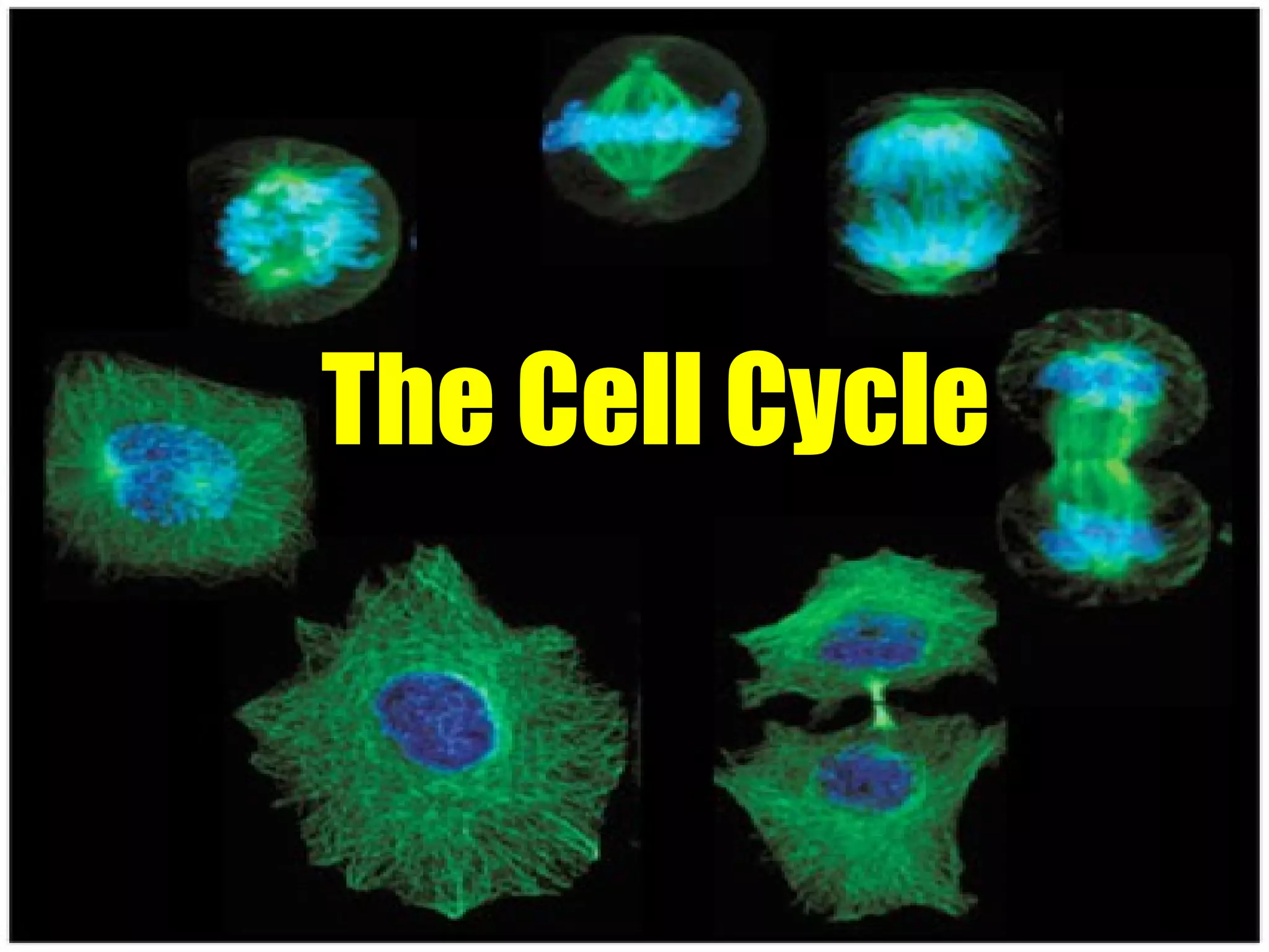
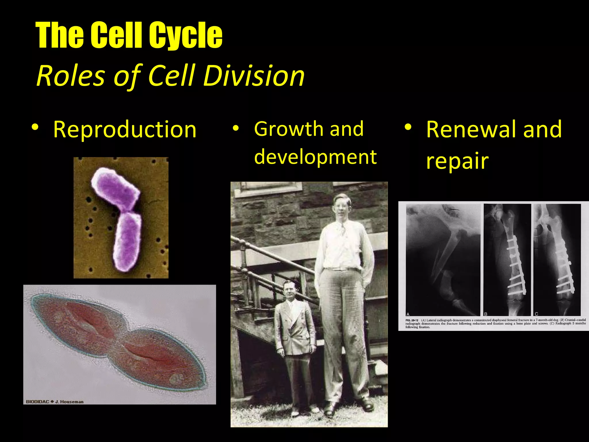
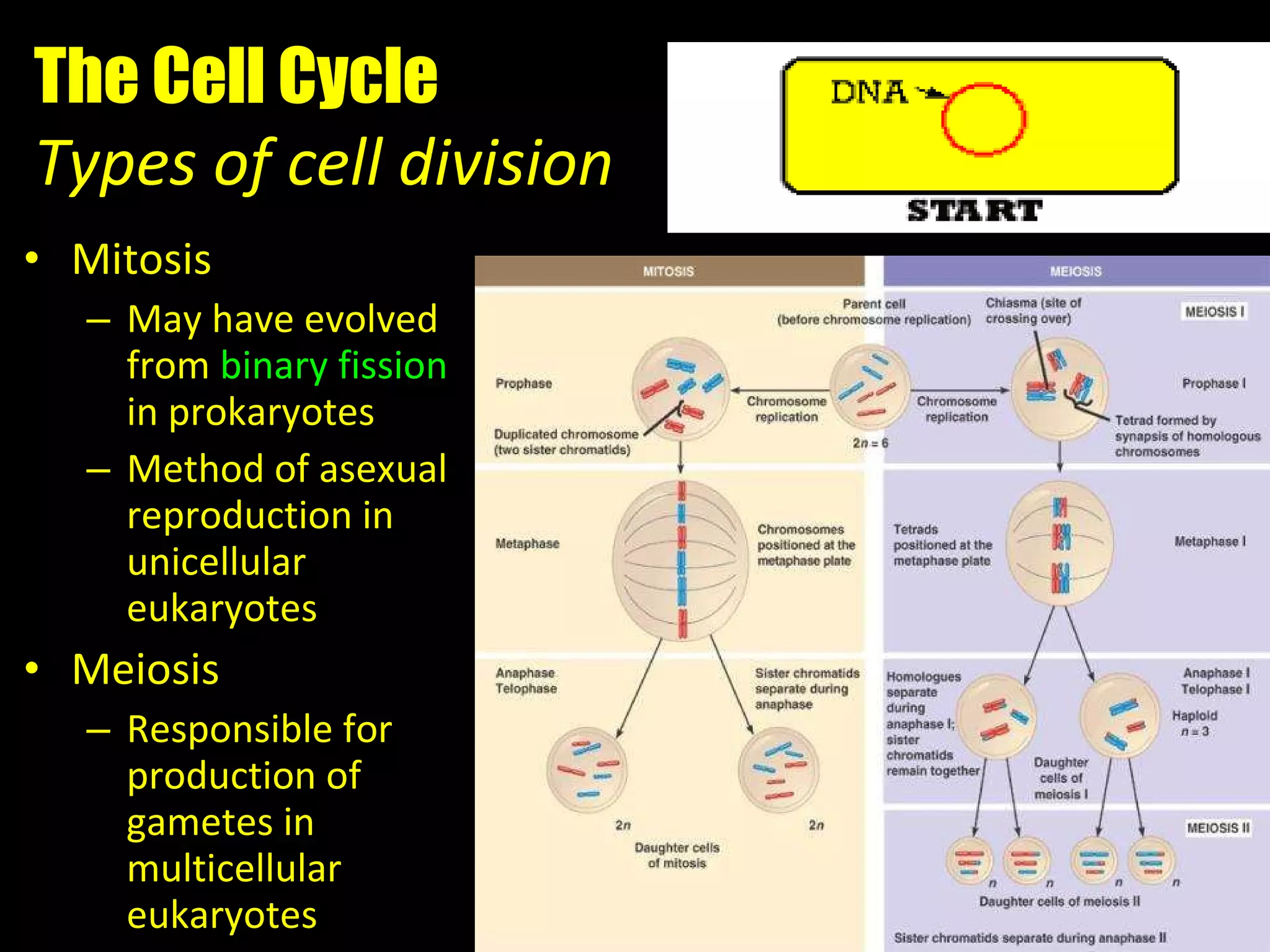
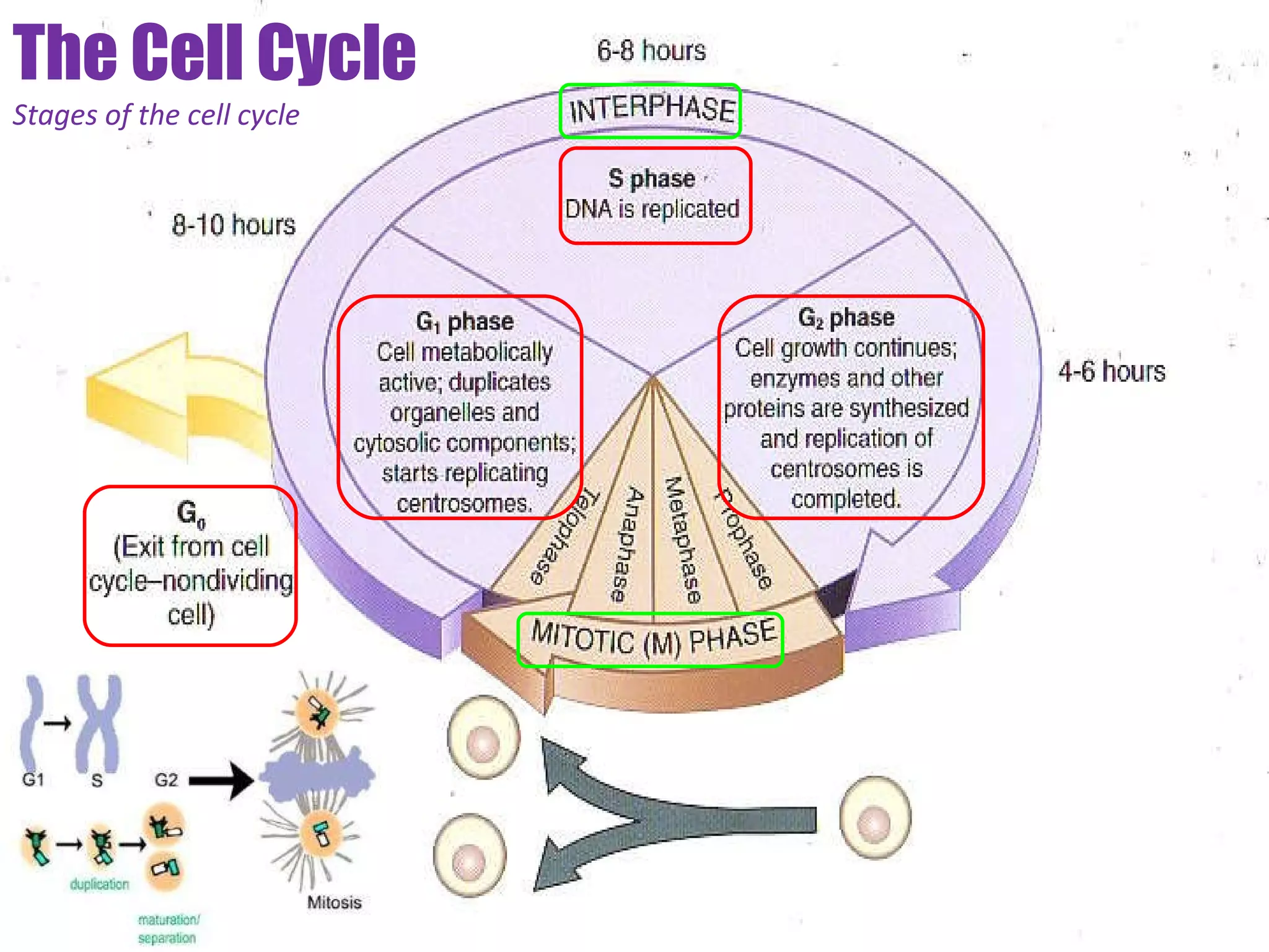
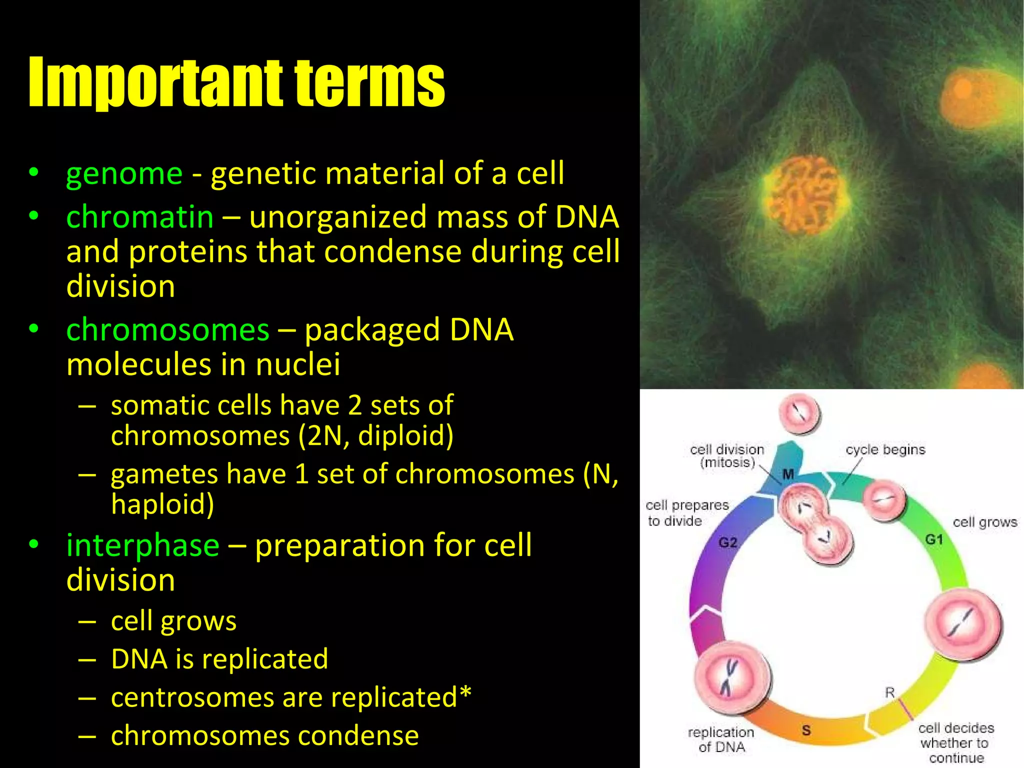
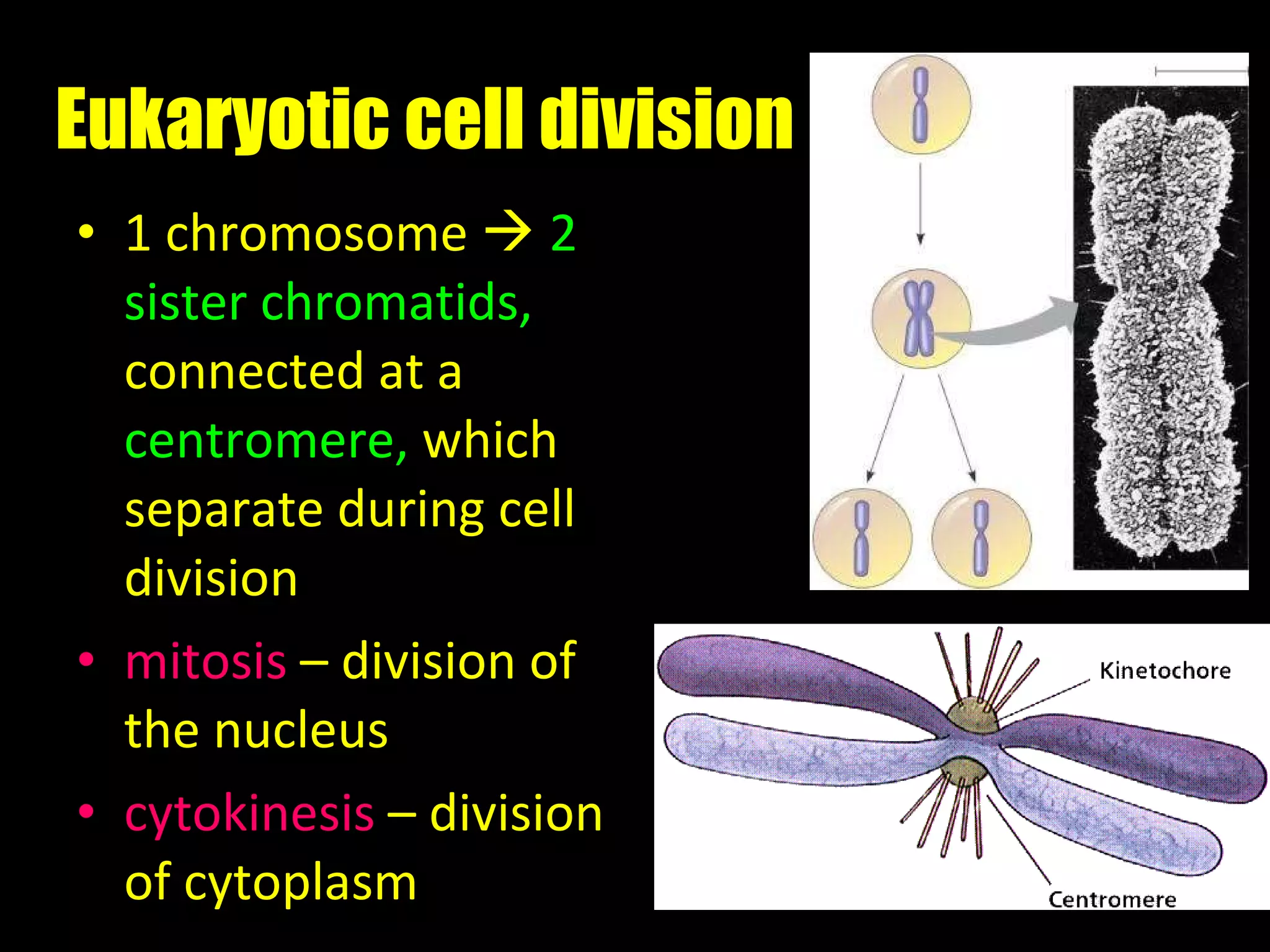
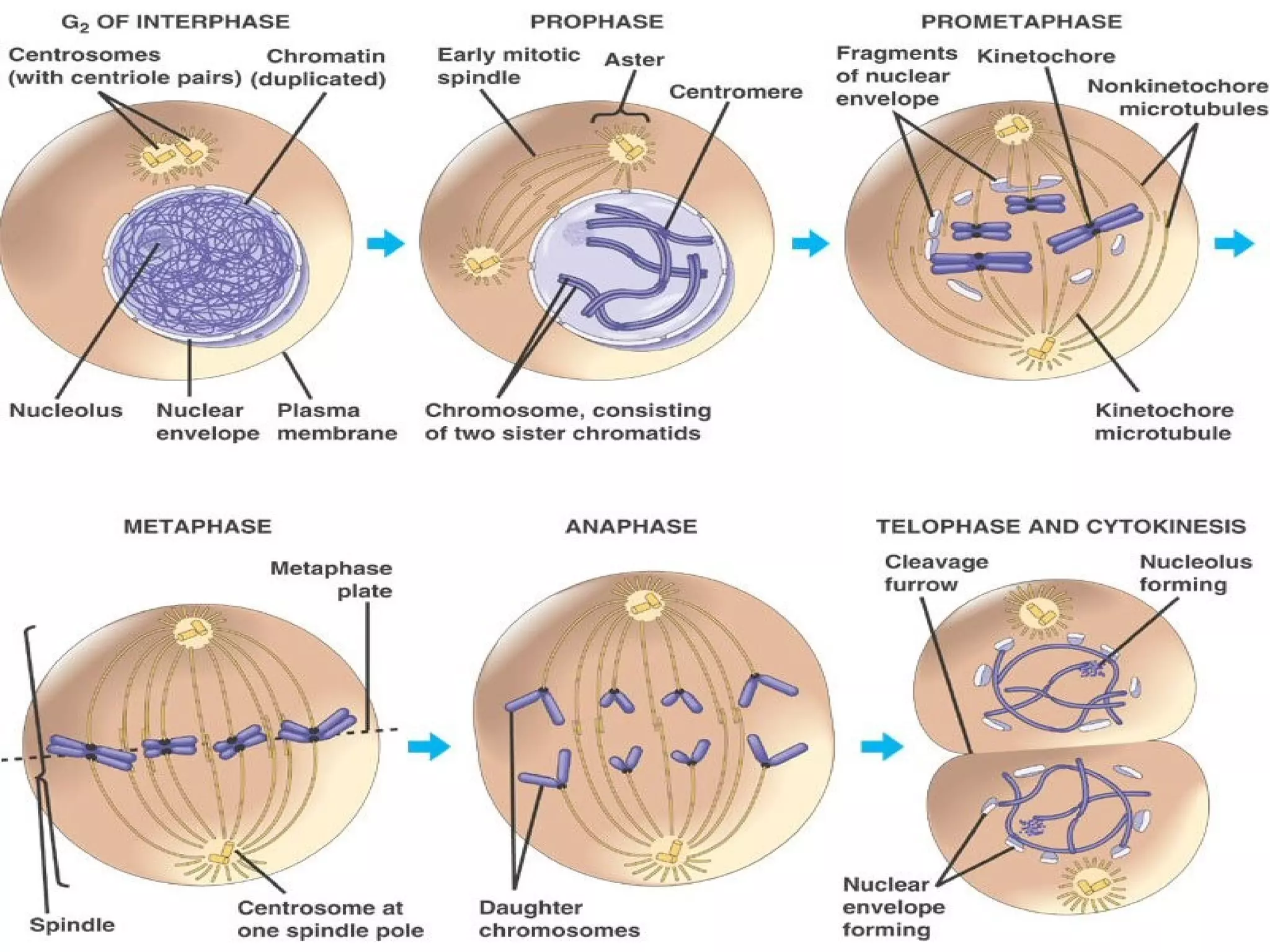
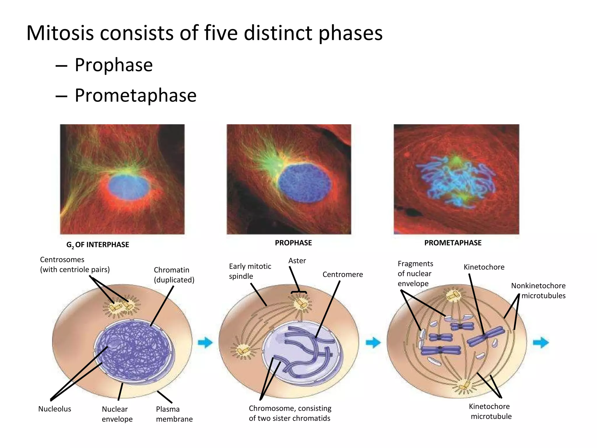
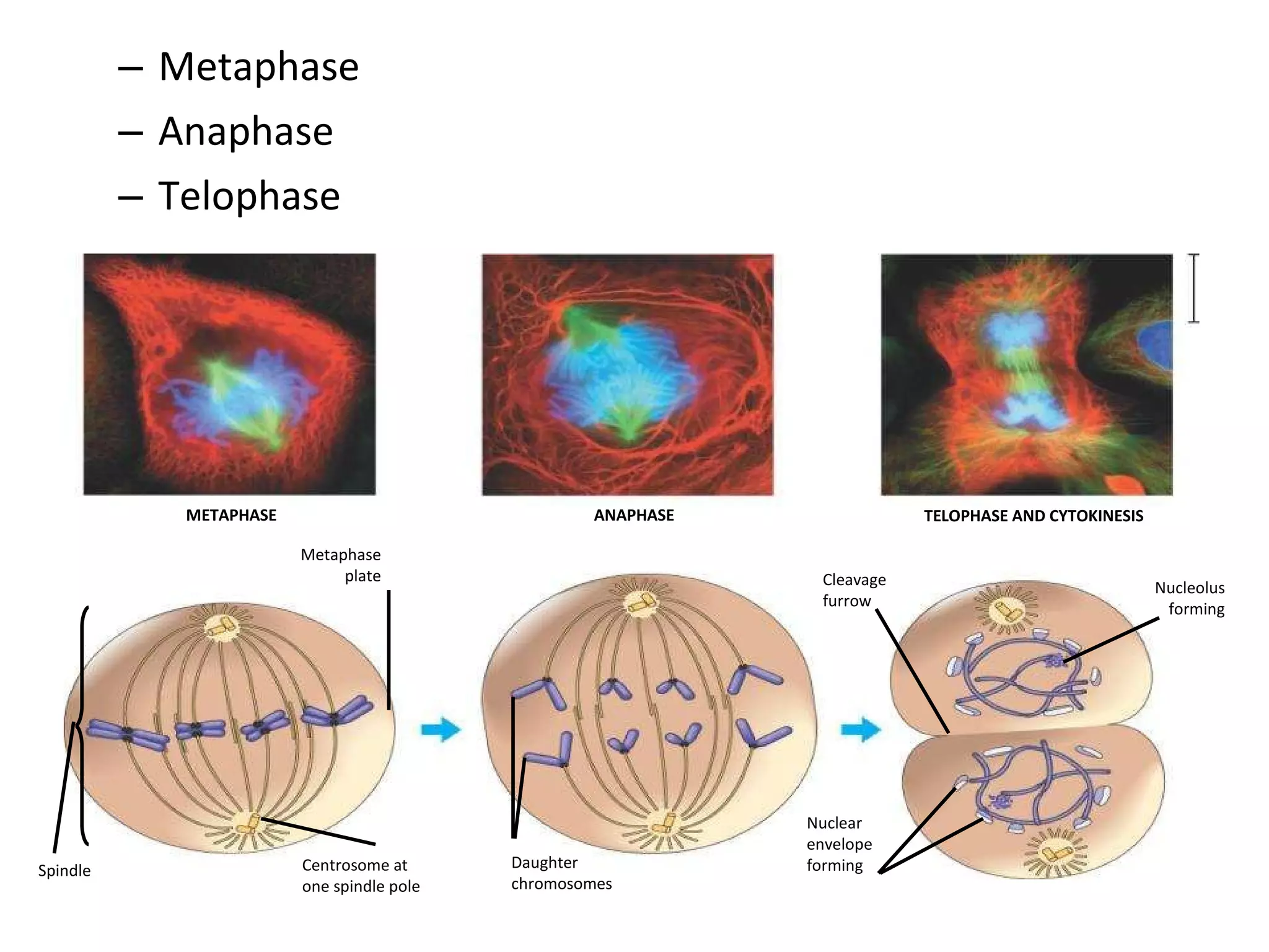
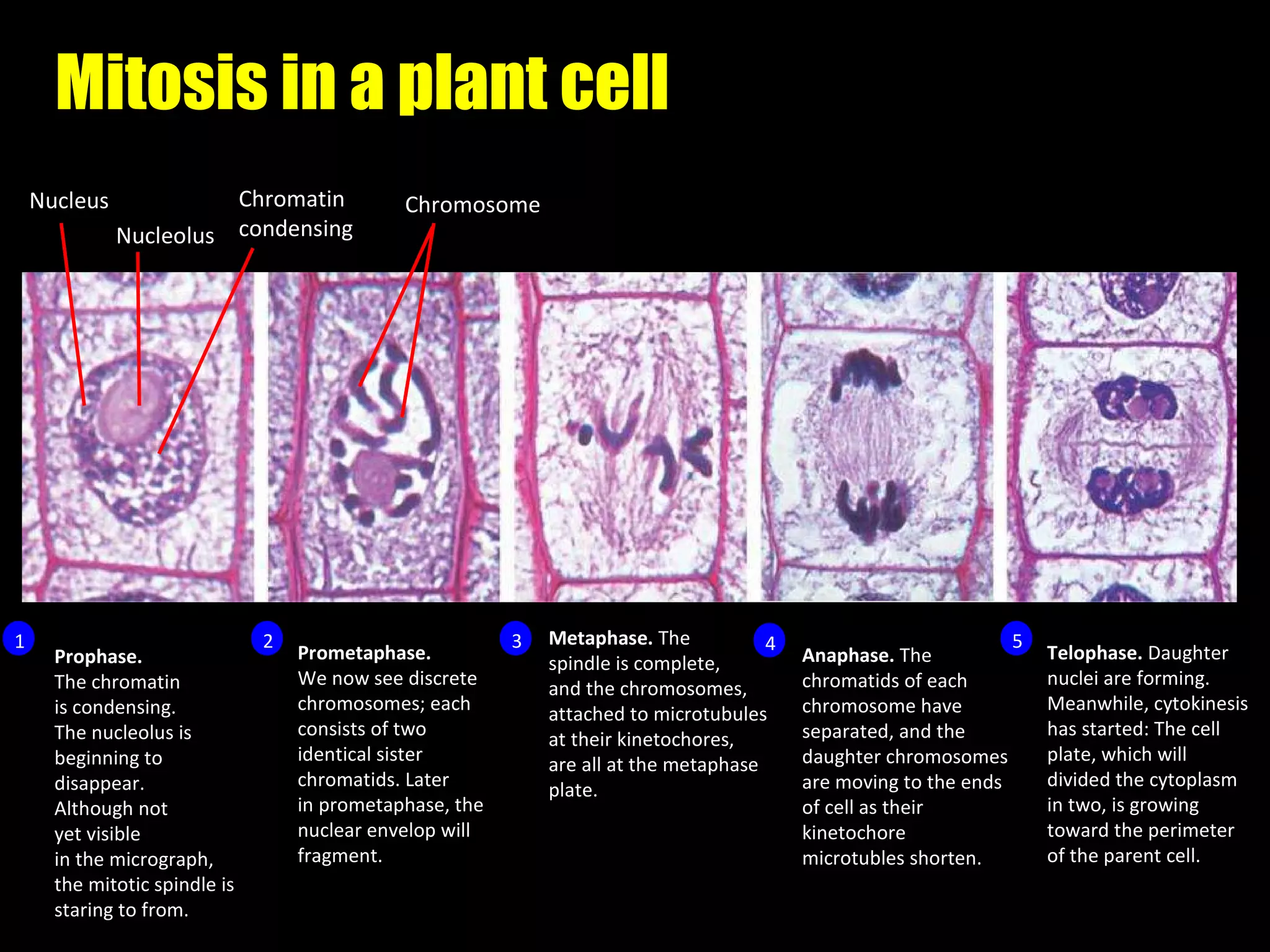
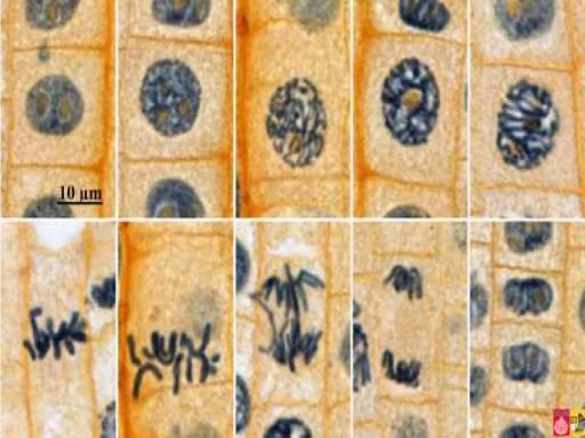
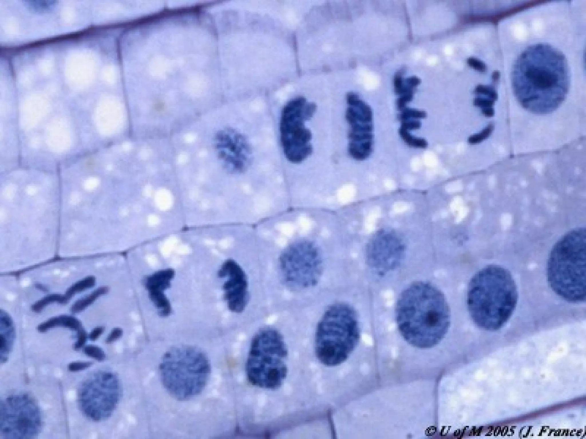
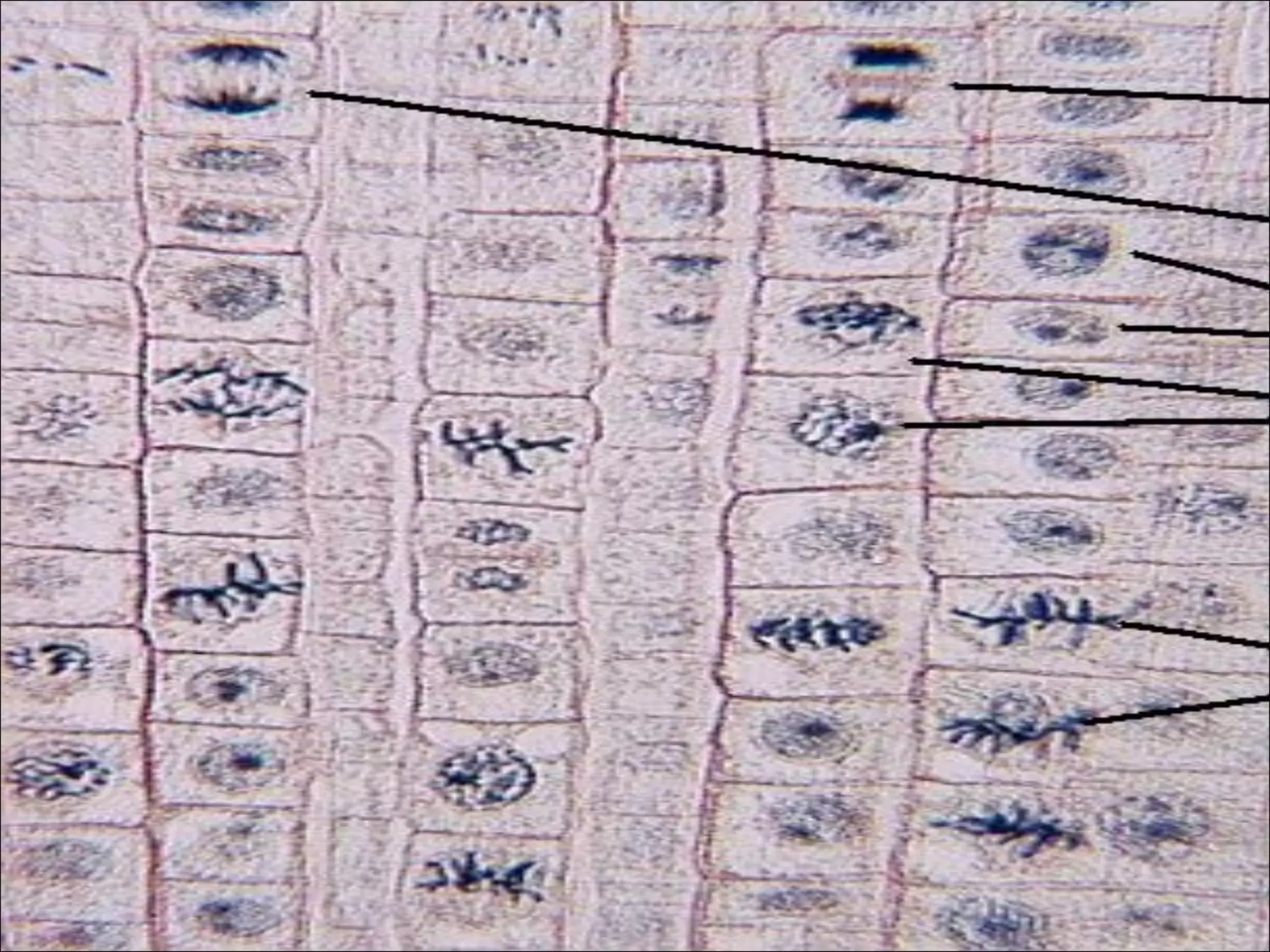
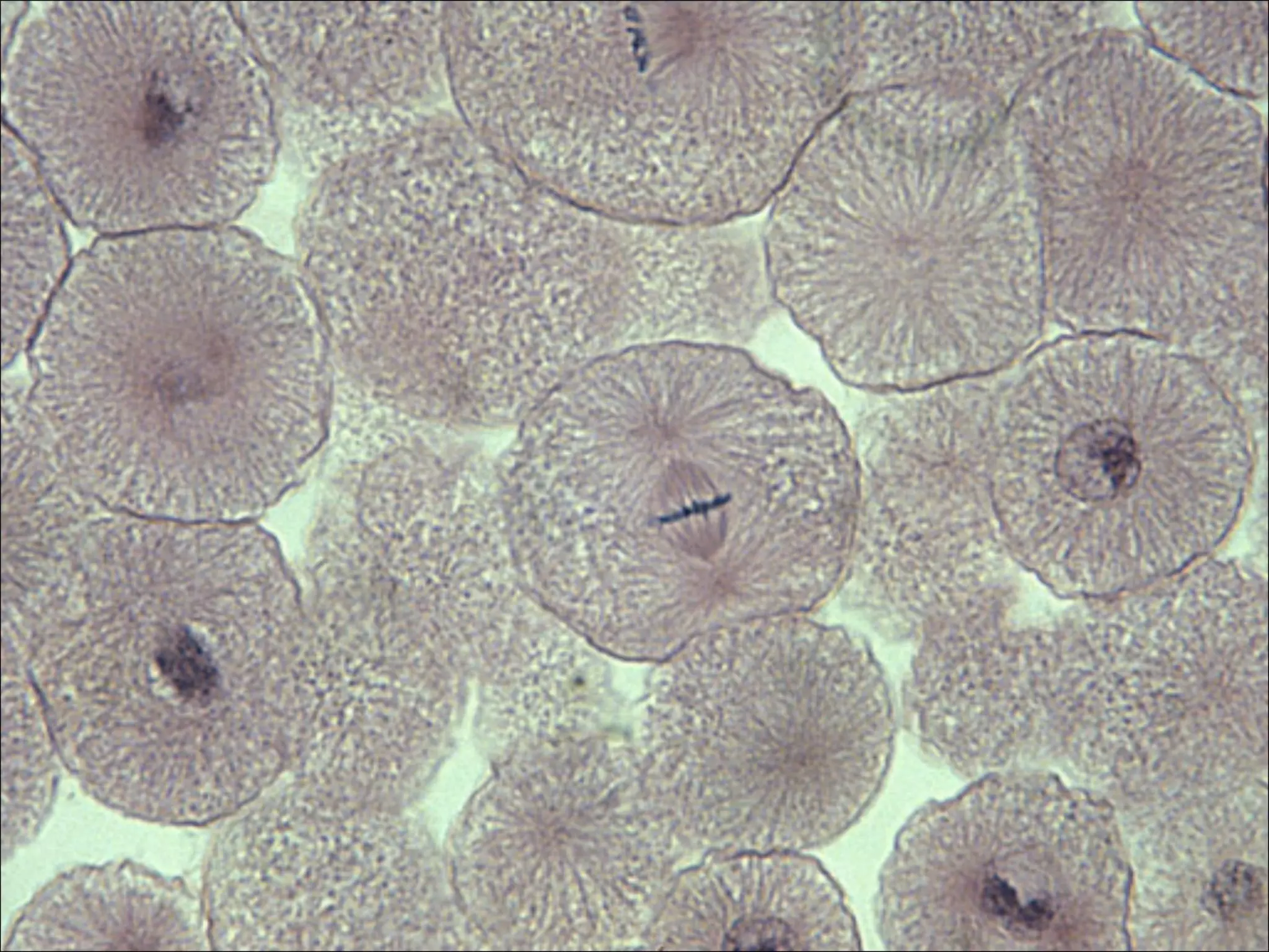
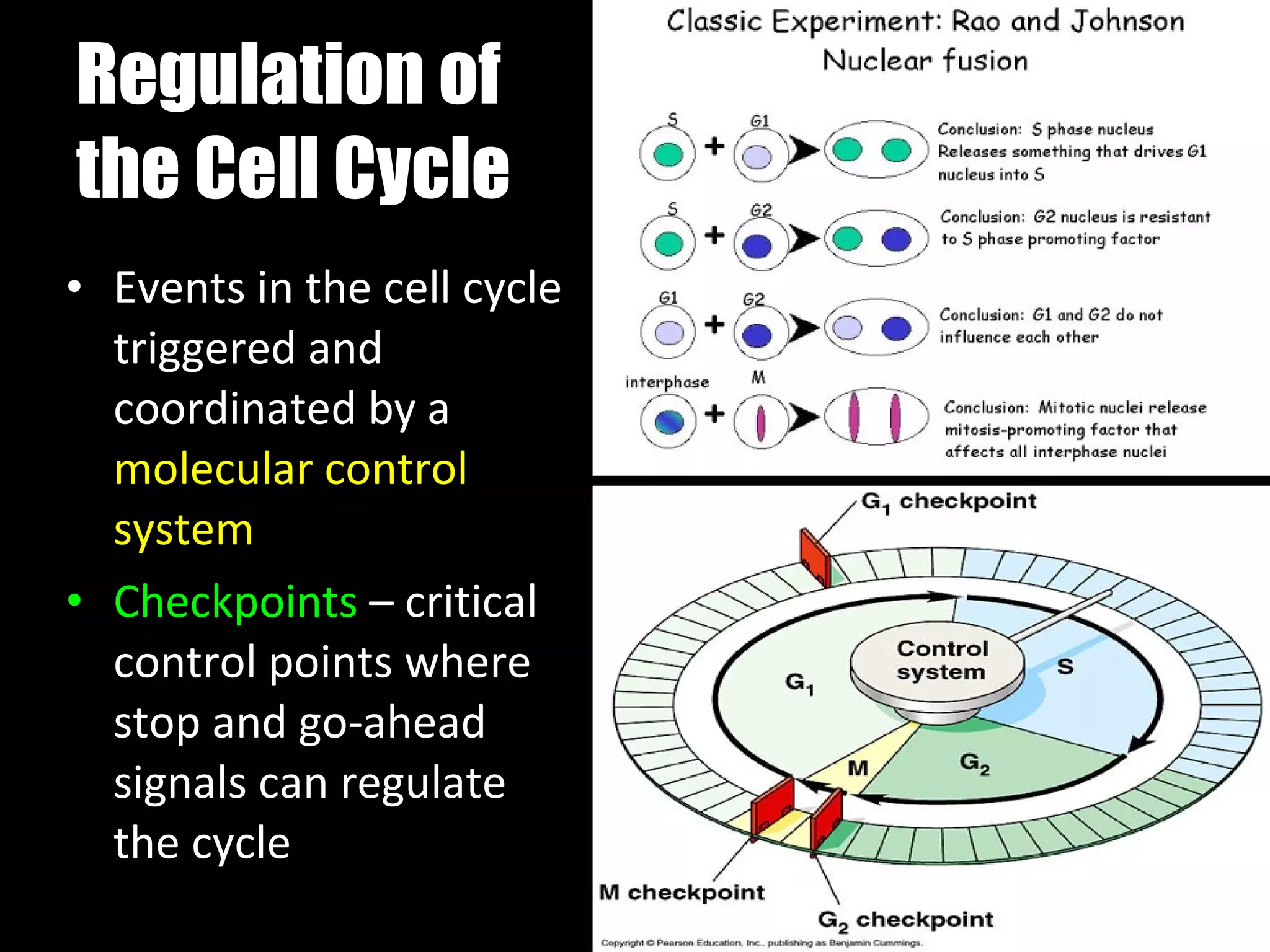
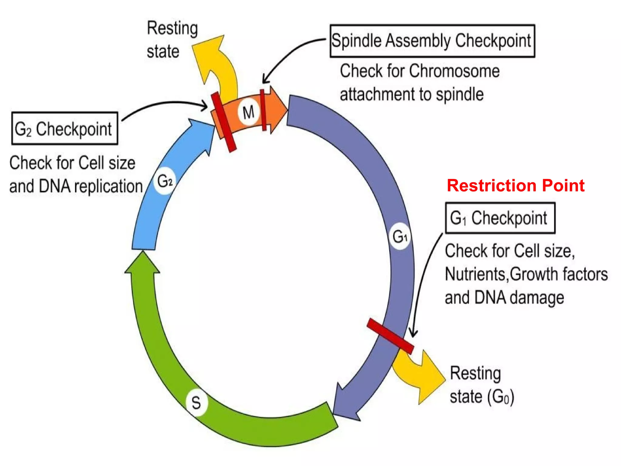
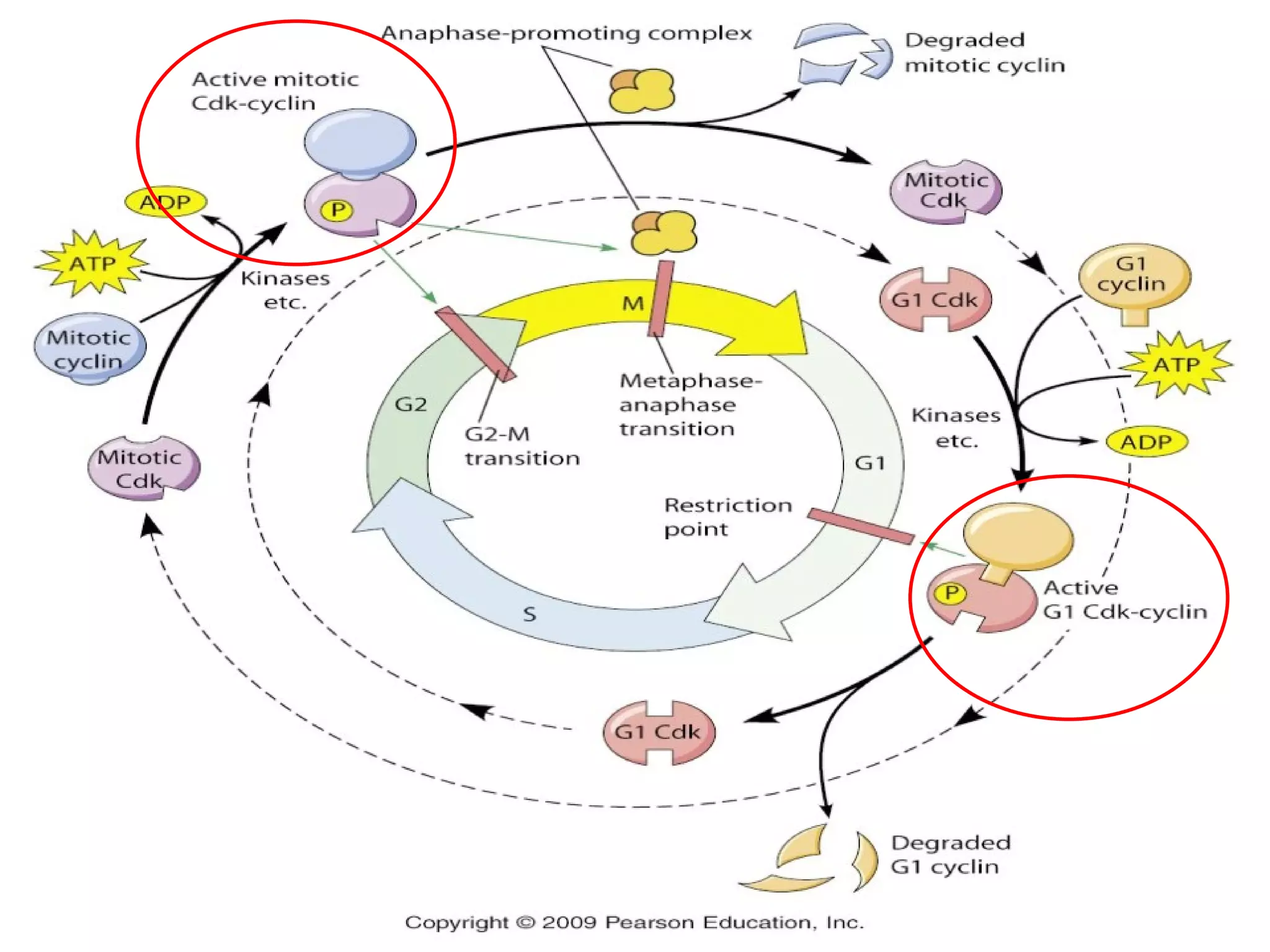
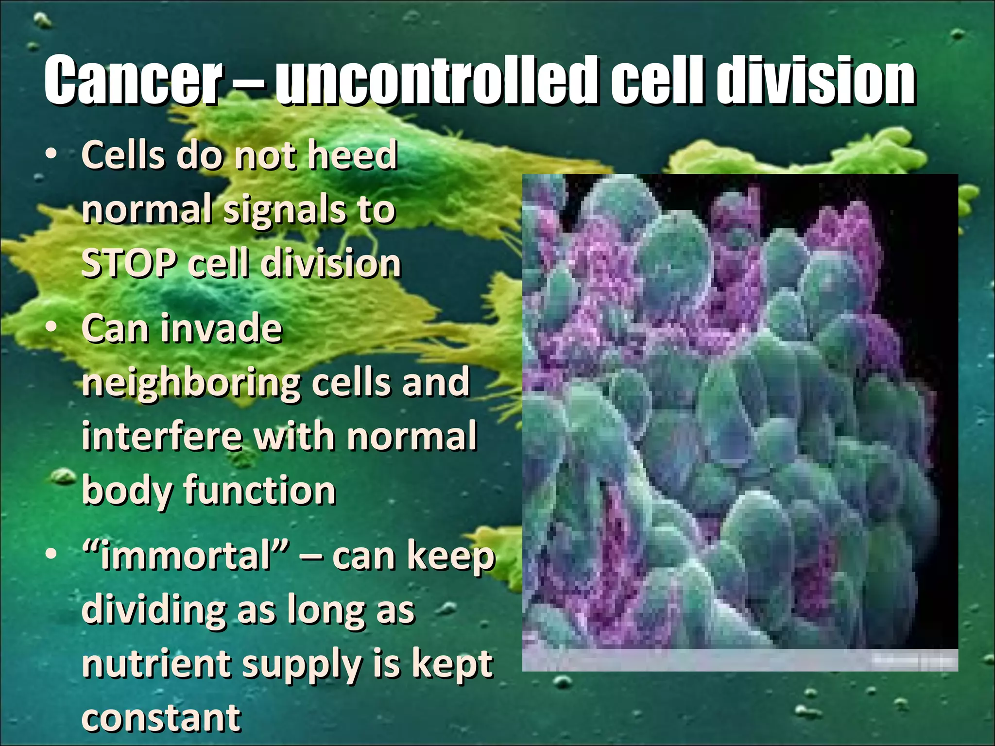
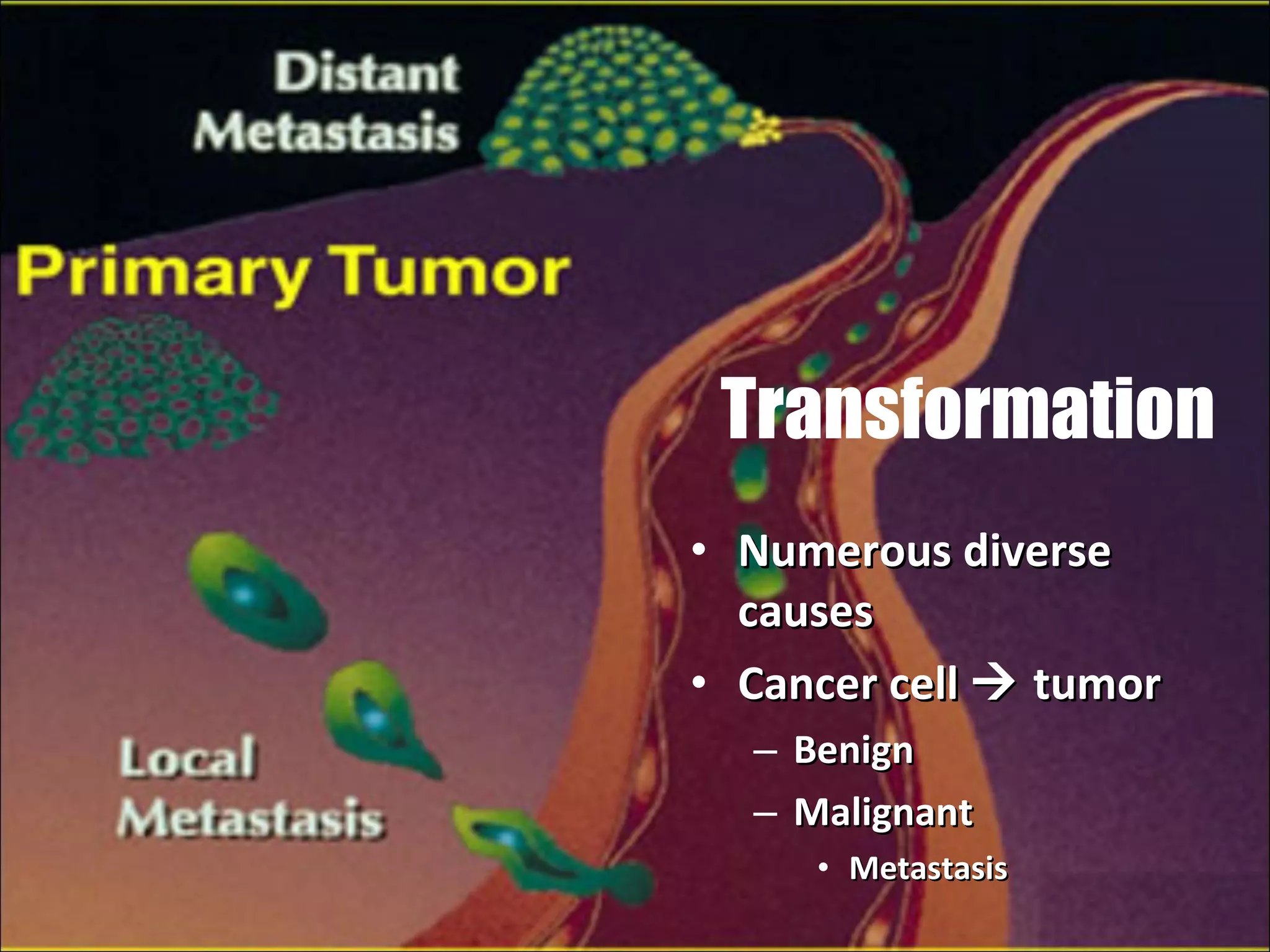
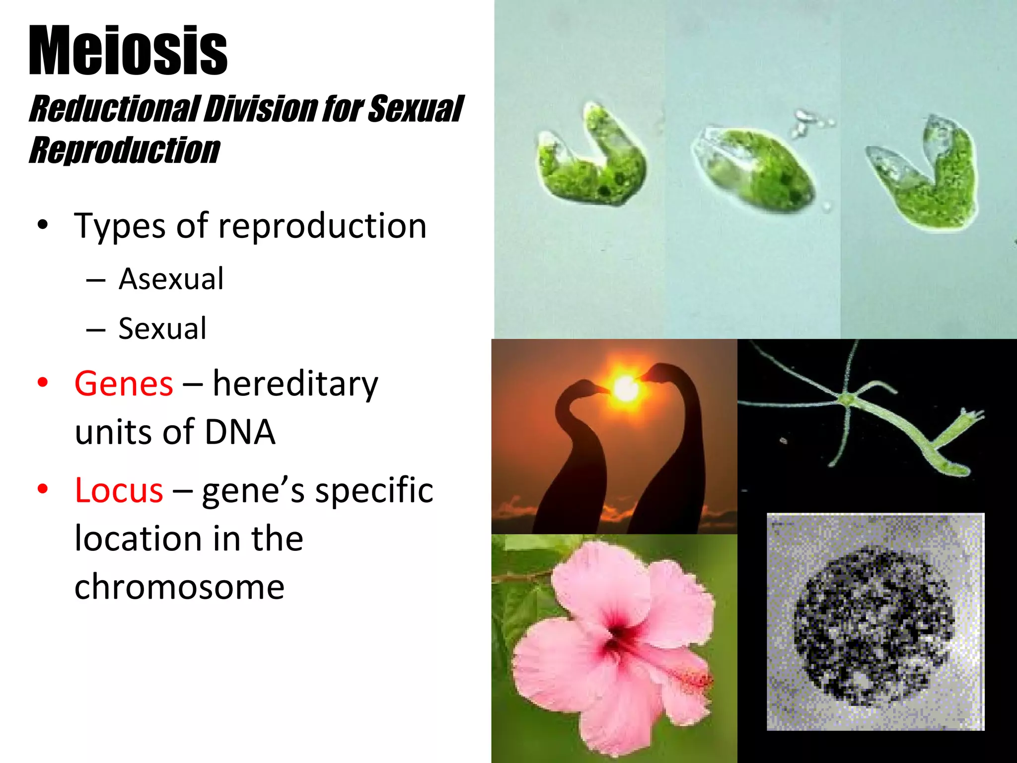
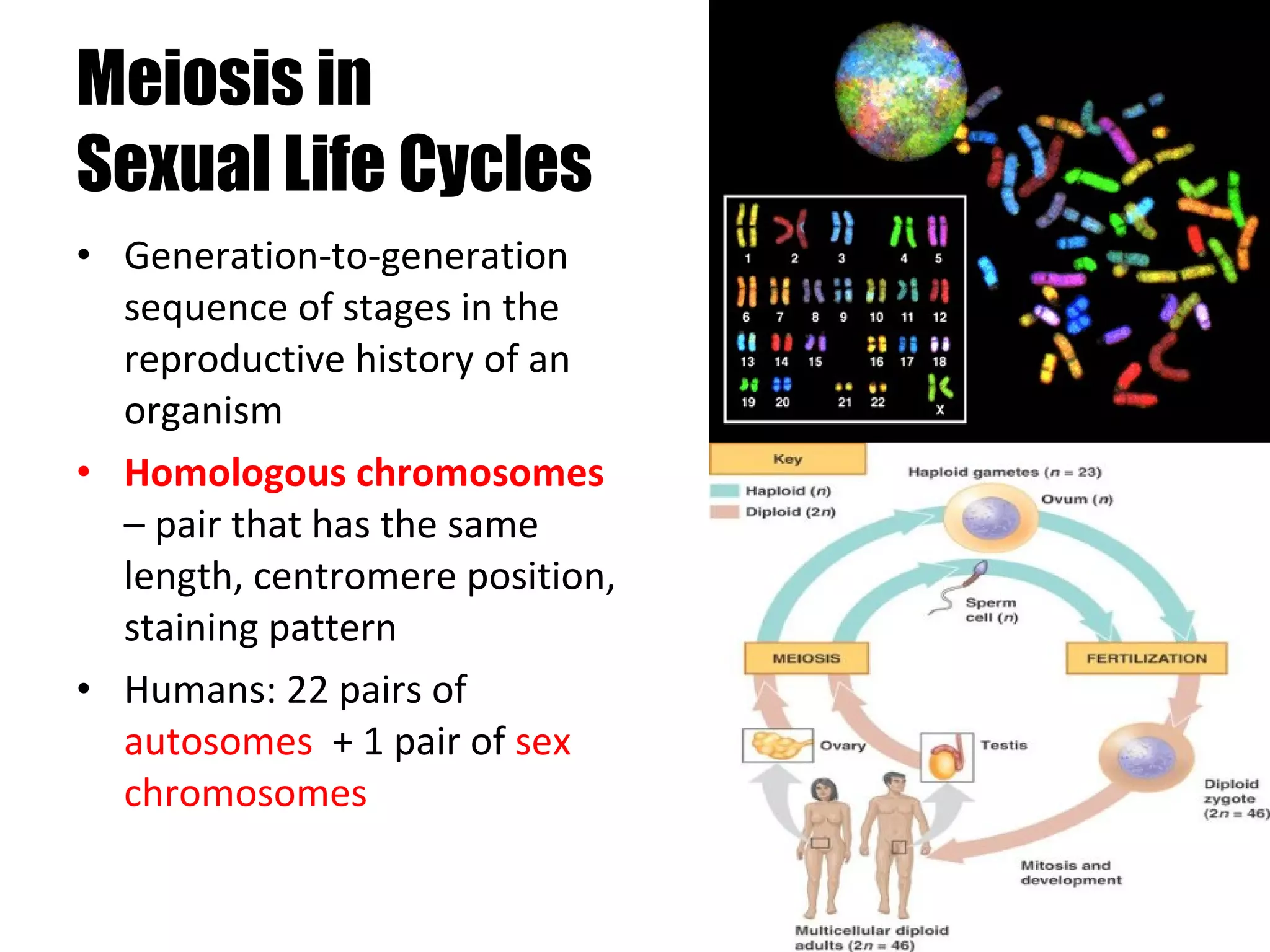
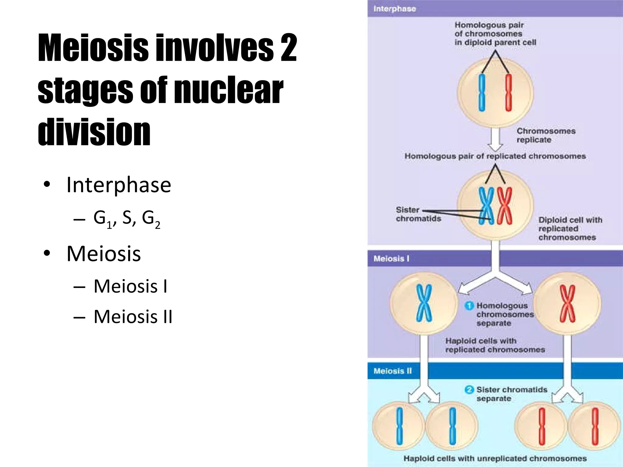
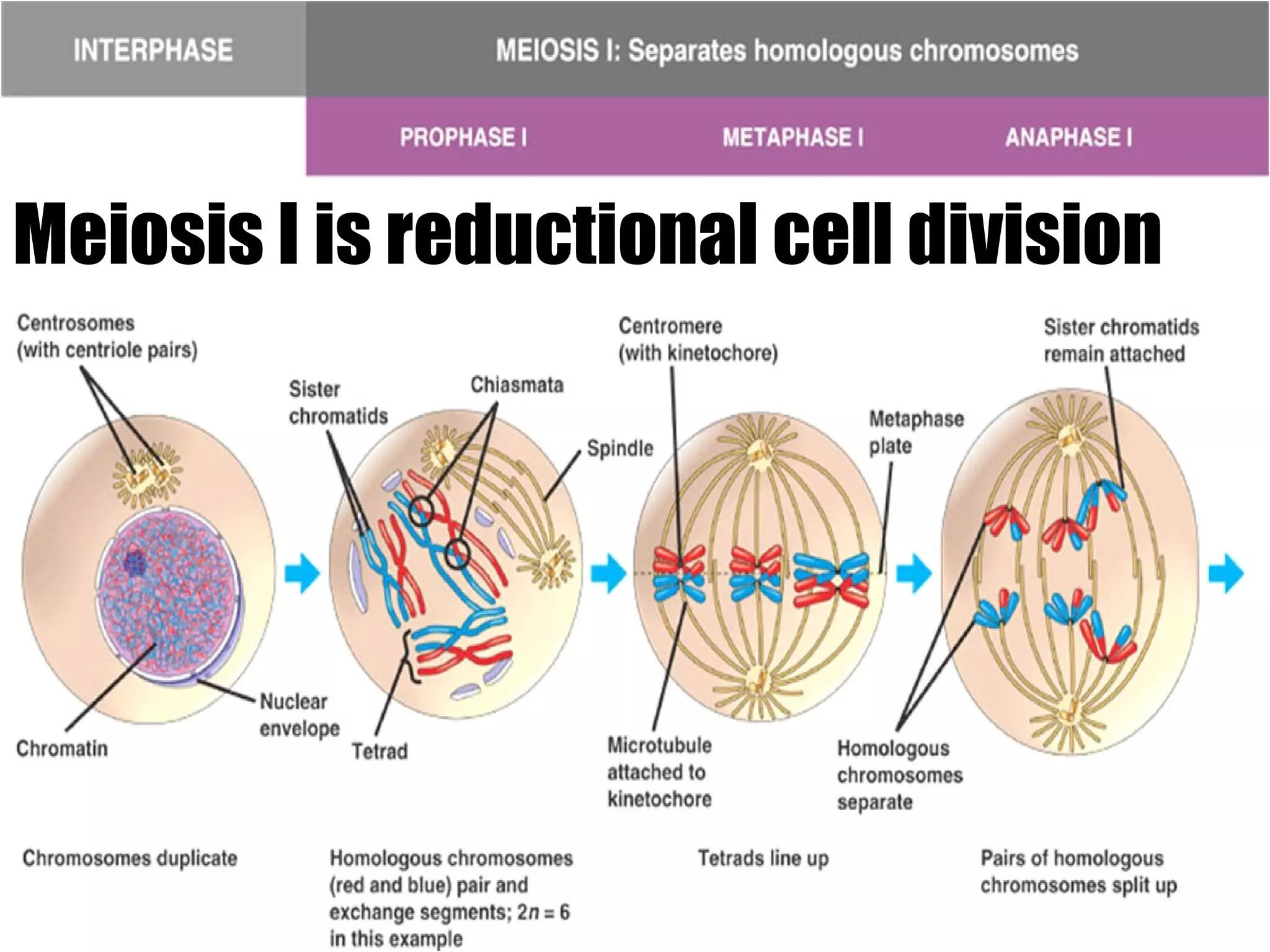
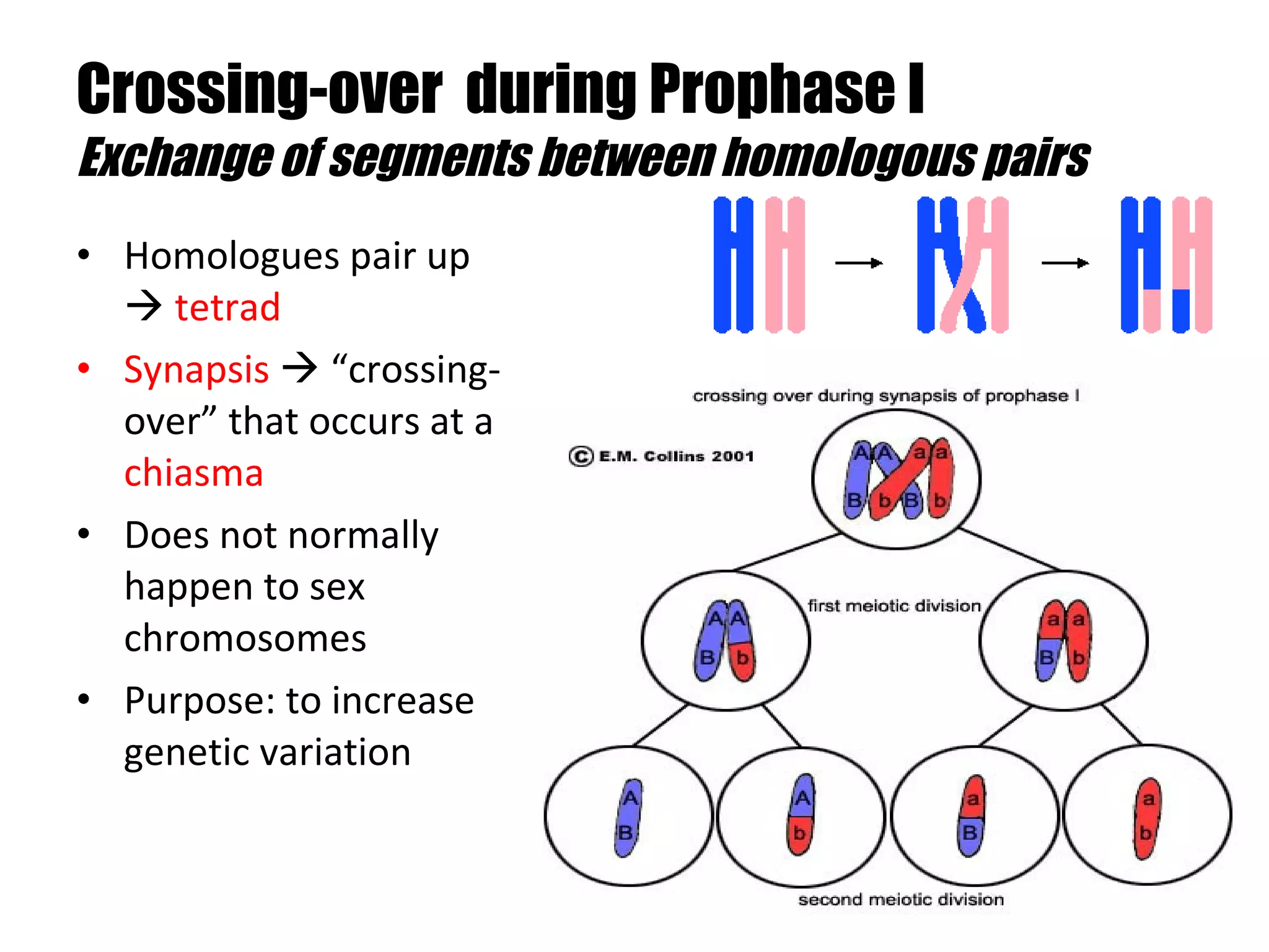
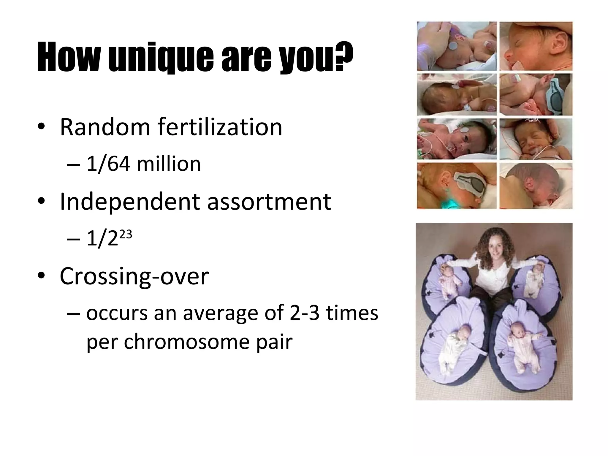
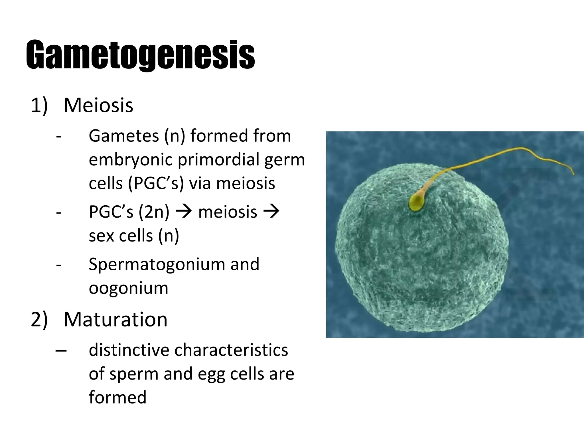
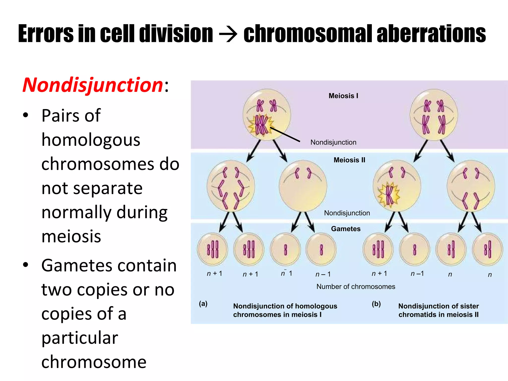
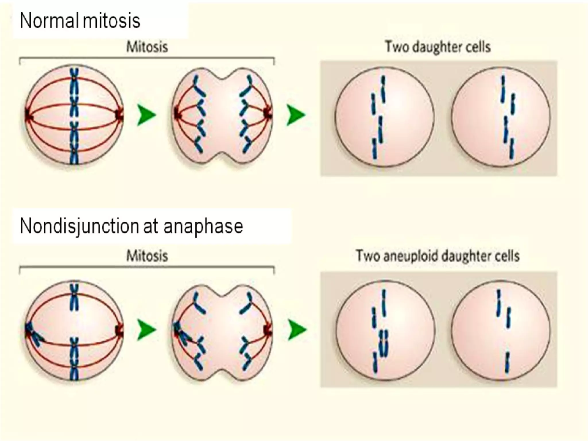
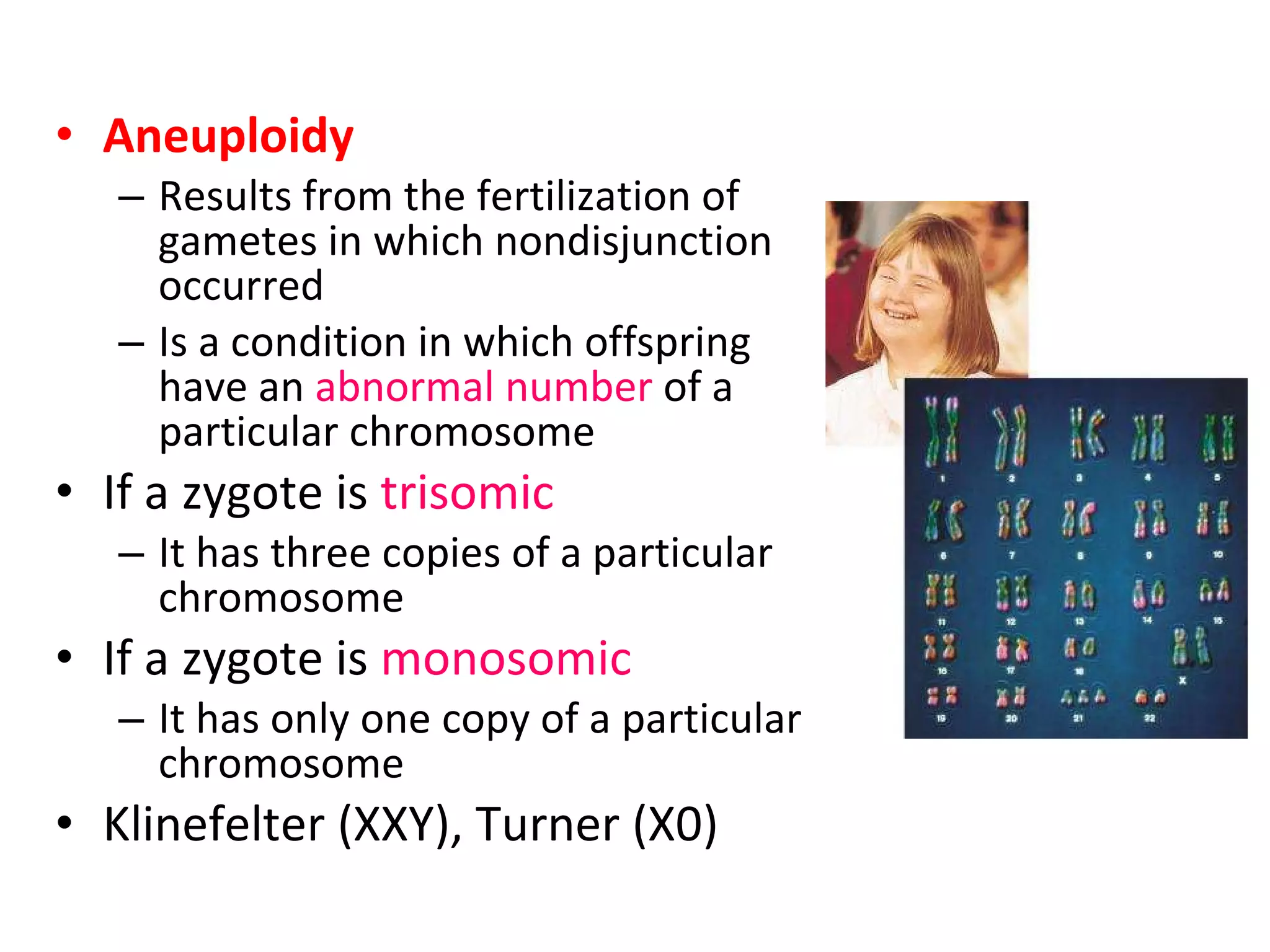
The document discusses the cell cycle, which involves growth, functioning, and division of cells. It has two main types of cell division - mitosis and meiosis. Mitosis produces two identical cells and is involved in growth and repair. Meiosis produces gametes through two divisions and involves genetic mixing through crossing over. Precise control mechanisms regulate the cell cycle, and errors can lead to genetic conditions.


