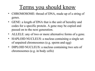
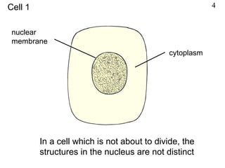
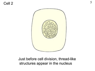
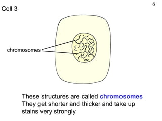
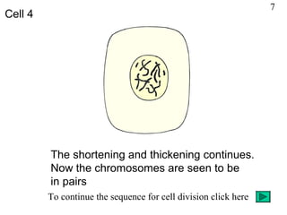
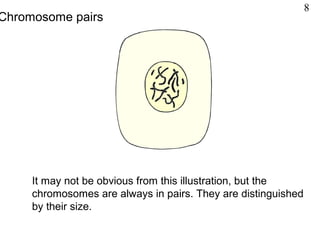
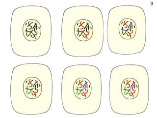
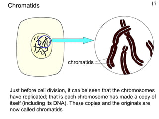
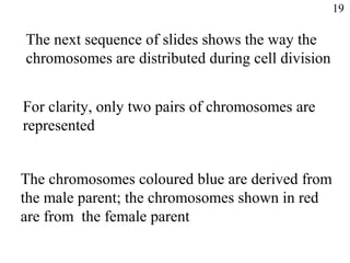
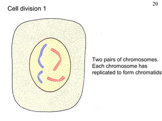
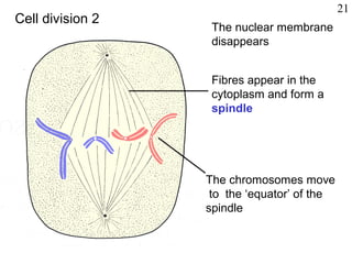
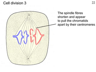
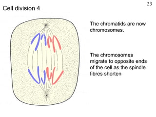
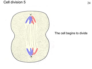
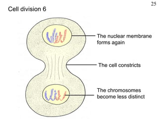
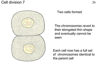
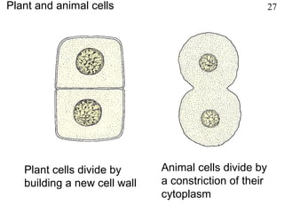
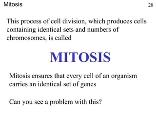
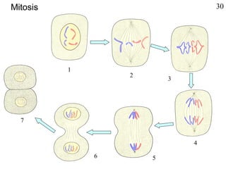
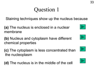
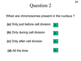
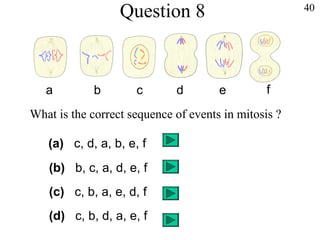
2. In a living cell seen under
the microscope, it is often
difficult to see the nucleus
When certain stains are applied, the
nucleus takes up the stain more
strongly than the cytoplasm does,
making the nucleus easily visible
Staining 2
3. By means of some special techniques of
illumination, the nucleus and other structures
may be seen in the living cell.
But in this presentation and in most textbooks, the
nucleus is shown dark, as if it had been stained
It is the differences in chemical composition
between nucleus and cytoplasm that make one take
up a particular stain more strongly than the other
3
4. INHERITANCE
• Is the transmission of genetic information
from generation to generation.
5. Terms you should know
• CHROMOSOME: thread of DNA, made up of a string of
genes.
• GENE: a length of DNA that is the unit of heredity and
codes for a specific protein. A gene may be copied and
passed on to the next generation.
• ALLELE: any of two or more alternative forms of a gene.
• HAPLOID NUCLEUS: a nucleus containing a single set
of unpaired chromosomes (e.g. sperm and egg)
• DIPLOID NUCLEUS: a nucleus containing two sets of
chromosomes (e.g. in body cells)
6. In a cell which is not about to divide, the
structures in the nucleus are not distinct
nuclear
membrane
cytoplasm
Cell 1 4
7. Just before cell division, thread-like
structures appear in the nucleus
Cell 2 5
8. These structures are called chromosomes
They get shorter and thicker and take up
stains very strongly
chromosomes
Cell 3
6
9. The shortening and thickening continues.
Now the chromosomes are seen to be
in pairs
To continue the sequence for cell division click here
Cell 4
7
10. It may not be obvious from this illustration, but the
chromosomes are always in pairs. They are distinguished
by their size.
Chromosome pairs
8
12. The chromosomes are always in pairs because one
of them is derived from the male parent and the
other from the female parent
Different species have different numbers and
shapes of chromosomes
Members of the same species have identical
sets of chromosomes
10
13. kangaroo (6 pairs) a sedge (21 pairs) hawkweed (4 pairs*)
chicken (18 pairs) fruit fly (4 pairs) human (23 pairs)
Chromosome numbers 11
14. Although chromosomes can be seen distinctly
only at the time of cell division, they are present
and active all the time
The chromosomes carry the DNA
DNA controls all the chemical reactions in the cell
DNA also determines the species of organism
and its individual characteristics (See the
presentation on ‘DNA’ for more detail)
12
15. Chromosomes consist of DNA molecules supported
by a ‘scaffold’ of proteins. The diagram illustrates such
an arrangement but it is really more complex than this
Chromosomes and DNA
chromosome
DNA
protein
double helix
13
16. The DNA in the chromosomes carries the genes
Consequently, the genes are spaced out along
the chromosome
The genes consist of distinct stretches of the DNA
By means of their DNA content, the genes control
the activities of the cell, the type of cell it becomes,
the species of the organism and the individual
characteristics of that organism
14
17. genes for eye colour
genes for hair colour
single gene
chromosome
Chromosomes and genes
genes for tallness
The diagram illustrates the relationship between chromosomes
and genes but it does not represent an actual chromosome.
The genes for these characteristics are not necessarily on the
same chromosome and the number of genes shown is arbitrary
15
18. A
B
C
D
E
F
G
H
I
a
b
c
d
e
f
g
h
I
Because the chromosomes are in
pairs, the genes they carry are also
in pairs
The individual genes of a pair, control
the same characteristic, e.g. B and b
could control eye colour; G and g could
control hair colour
Each member of a pair of genes comes
from either the male or the female
parent just as the chromosomes do
Chromosomes and genes 16
19. Just before cell division, it can be seen that the chromosomes
have replicated; that is each chromosome has made a copy of
itself (including its DNA). These copies and the originals are
now called chromatids
chromatids
Chromatids 17
20. The chromatids
tend to separate but
are held together by a
special region called
the centromere
chromatids
Centromere 18
21. CELL DIVISION:MITOSIS
• Is a nuclear division giving rise to
genetically identical cells in which the
chromosome number is maintained by the
exact duplication of chromosomes.
22. The next sequence of slides shows the way the
chromosomes are distributed during cell division
For clarity, only two pairs of chromosomes are
represented
The chromosomes coloured blue are derived from
the male parent; the chromosomes shown in red
are from the female parent
19
23. Two pairs of chromosomes.
Each chromosome has
replicated to form chromatids
Cell division 1
20
24. The nuclear membrane
disappears
Fibres appear in the
cytoplasm and form a
spindle
The chromosomes move
to the ‘equator’ of the
spindle
Cell division 2
21
25. The spindle fibres
shorten and appear
to pull the chromatids
apart by their centromeres
Cell division 3 22
26. The chromatids are now
chromosomes.
The chromosomes
migrate to opposite ends
of the cell as the spindle
fibres shorten
Cell division 4
23
28. The nuclear membrane
forms again
The cell constricts
The chromosomes
become less distinct
Cell division 6
25
29. Two cells formed
Each cell now has a full set
of chromosomes identical to
the parent cell
The chromosomes revert to
their elongated thin shape
and eventually cannot be
seen
Cell division 7 26
30. Plant cells divide by
building a new cell wall
Animal cells divide by
a constriction of their
cytoplasm
Plant and animal cells 27
31. This process of cell division, which produces cells
containing identical sets and numbers of
chromosomes, is called
MITOSIS
Mitosis 28
Mitosis ensures that every cell of an organism
carries an identical set of genes
Can you see a problem with this?
32. For example, what can a gene for brown eyes do
in a stomach cell?
The problem is that if every cell carries the same
set of genes, how do cells become specialised in
shape and function to do different jobs?
The answer is that the genes which are not relevant
to the cell’s function are not activated.
We say the gene for brown eyes is not expressed
in a stomach cell
29
34. The next slide shows a photomicrograph of onion root cells.
In a root tip, a great many cells are dividing by
mitosis, leading to rapid growth.
The preparation is made by softening the root tip tissue,
squashing it on a microscope slide and staining the
chromosomes and nuclei.
The ‘squash’ technique spreads the cells out.
See if you can associate the various stages of cell division
with the stages 1-6 in the previous slide.
Bear in mind that the previous slide is a purely
diagrammatic representation.
31
39. Question 1
Staining techniques show up the nucleus because
(a) The nucleus is enclosed in a nuclear
membrane
(b) Nucleus and cytoplasm have different
chemical properties
(c) The cytoplasm is less concentrated than
the nucleoplasm
(d) The nucleus is in the middle of the cell
33
40. Question 2
When are chromosomes present in the nucleus ?
(a) Only just before cell division
(b) Only during cell division
(c) Only after cell division
(d) All the time
34
41. Question 3
Chromosomes are in pairs because
(a) They have replicated
(b) Each is derived from either the male
or female parent
(c) They are joined at the centromere
(d) They have to be shared at cell division
35
42. Question 4
How many chromosomes are there in a human cell?
(a) 100+
(b) 92
(c) 46
(d) 23
36
43. Question 5
Which of these statements are correct ?
DNA controls
(a) The species of the organism
(b) The function of the cell
(c) Features of the organism such as size
(d) Chemical reactions in the cell
37
44. Question 6
Which of these statements are correct?
A chromosome contains
(a) Protein
(b) Cellulose
(c) DNA
(d) Genes
38
45. Question 7
The process by which a chromosome makes a
copy of itself is called
(a) Reproduction
(b) Recombination
(c) Relocation
(d) Replication
39
46. 40
What is the correct sequence of events in mitosis ?
a b c d e f
(c) c, b, a, e, d, f
(a) c, d, a, b, e, f
(b) b, c, a, d, e, f
(d) c, b, d, a, e, f
Question 8
The illustration represents a cell in the process of dividing. The coloured structures are chromosomes. Full details are given in slides 20-27
Stains can be chosen for particular parts of the cell; the cytoplasm, the nucleus, the cell wall or structures within the cytoplasm. Staining usually kills the cell.
The living content of the nucleus is called nucleoplasm to distinguish it from cytoplasm.
* The 8 chromosomes have already replicated so some of them appear ‘double’.
The centromere appears as a constriction in the chromosome but in subsequent drawings it is represented by a circle to make its position clear.
Although the shortening of the spindle fibres and the corresponding movement of the chromosomes are clearly associated, it should not be assumed that the fibres are physically tugging the chromosomes apart.
Although No.1 doesn’t look much like the drawing, it represents the stage when the chromosomes are clearly visible but not yet organised on the spindle. Possible web sites www.microscopy-uk.org.uk/mag/artaug99/mitosis.html Photomicrographs of cell division in broad bean root tip www.johnkyrk.com/mitosis.html Simple animation of mitosis www.biologyinmotion.com/cell_division Interactive ‘drag and drop’ test on mitosis and meiosis www.tcb.cl/1535/article-56384.html Time-lapse video of mitosis in newt lung cell Possible web sites www.microscopy-uk.or.uk/mag/artaug99/mitosis.html