SlideShare a Scribd company logo
Cell Membrane & ion transport
By Ranim Mira
Plasma membrane/cell membrane
The Plasma membrane or the cell membrane is a thin,
biological membrane present in all eukaryotic and prokaryotic
cells that forms a boundary between the cell and its
environment and regulating the flow of materials in to and out
of cell.
The plasma membrane exhibits selective permeability,
allowing some substances to cross it more easily than others
Objectives
I. Membrane Models
II. Membrane Structure
III. Membrane Function
IV. Factors affecting membrane fluidity
V. Permeability of the membrane
VI. Traffic (Transport) Across Membranes
VII. Disorders of cell membrane
II. Membrane Models
• In 1935, Hugh Davson and James Danielli proposed a
sandwich model in which the phospholipid bilayer lies
between two layers of globular proteins
• Later studies found problems with this model, particularly the
placement of membrane proteins, which have hydrophilic and
hydrophobic regions
• In 1972, S. J. Singer and G. Nicolson proposed that the
membrane is a mosaic of proteins dispersed within the
bilayer, with only the hydrophilic regions exposed to water
II. Membrane Models
fluid mosaic model
• Phospholipids are the most abundant lipid in the plasma
membrane
• Phospholipids are amphipathic molecules, containing
hydrophobic and hydrophilic regions
• The fluid mosaic model states that a membrane is a fluid
structure with a “mosaic” of various proteins embedded in it.
• The fluid mosaic model theory thereby states that plasma
membrane structure is a lipid bilayer with mosaic of proteins
embedded in it and moves freely Laterally parallel to the surface
of the membrane.
fluid mosaic model
Phospholipid
bilayer
Hydrophobic regions
of protein
Hydrophilic
regions of protein
Copyright @ BioNinja
fluid mosaic model
Membranes are mosaics of floating proteins in a lipid
bilayer. 2 ways:
•Integral Proteins: transmembrane, have both
hydrophilic and hydrophobic parts
•Peripheral Proteins: Attached to membrane’s
surface by:
- Attachment to integral proteins or ECM fibers (outside).
- Attachment to filaments of cytoskeleton (inside).
fluid mosaic model
copyright@ Biology wise
fluid mosaic model
Freeze-fracture studies of the
plasma membrane supported
the fluid mosaic model
Freeze-fracture is a
specialized preparation
technique that splits a
membrane along the middle of
the phospholipid bilayer.
Figure 7.4
Knife
Plasma membrane Cytoplasmic layer
Proteins
Extracellular
layer
Inside of extracellular layer Inside of cytoplasmic layer
TECHNIQUE
RESULTS
Membrane Structure
Membranes are complex structures composed of:
• Membrane Lipids
• Membrane Proteins
• Carbohydrates
Membrane Structure
*Membrane Lipids
• Phospholipids:
can form micelle
- Phosphoglycerides: consist of two fatty acids joined to glycerol (ester
linkage).
- Sphingophospholipids: consist of single fatty acid joined to
sphingosine(amide linkage).
• Cholesterol
• Glycolipids e.g; glycosphingolipids
Membrane Structure
*Membrane Protein
• Integral Proteins: transmembrane, have both hydrophilic and
hydrophobic parts.
• Peripheral Proteins: loosely attached to membrane’s surface.
RBC cytoskeleton contains peripheral proteins spectrin as well
as ankyrin, both are responsible on the biconcave shape of RBC.
Genetic defective or loss of spectrin protein leads to hereditary
spherocytosis.
• Glycoprotein: e.g; glycophorin which is erythrocyte
transmembrane glycoprotein that is important in blood group
identification (ABO)
I. Membrane Function
Generally, the cell membrane is responsible on:
• Cell shape
• Barrier keeping the constituents of the cell in and unwanted
substances out.
• Biological activities such as flexibility, break and reseal,
Fission & Selective permeability
Membrane function (protein)
Major functions of membrane proteins
• Transport
• Enzymatic activity
• Signal transduction
• Cell-cell recognition
• Intercellular joining
• Attachment to the cytoskeleton and extracellular matrix (ECM)
• Involvement of proteins in the membrane repair pathway.
Membran function (protein)
Enzymes
Signaling molecule
Receptor
Signal transduction
ATP
(a) Transport (b) Enzymatic activity (c) Signal transduction
Membran function (protein)
Glyco-
protein
(d) Cell-cell recognition (e) Intercellular joining (f) Attachment to
the cytoskeleton
and extracellular
matrix (ECM)
Membrane function (Lipids)
Lipids function as essential structural components of membranes,
as :
• Structural role in shaping the physical properties of the plasma
membrane.
• Maintaining plasma membrane integrity.
• Role of facilitating plasma membrane repair.
• signaling molecules.
• chemical identifiers of specific membranes.
• energy storage molecules.
Membrane function (Carbohydrates)
Cell-Cell Recognition: cells recognize each other by binding
to surface molecules, often containing carbohydrates, on the
extracellular surface of the plasma membrane. This cell-cell
recognition is the basis for:
- sorting an embryo’s cells into tissues/organs.
- rejection of foreign cells by immune system.
*Carbohydrates on the external side of the plasma membrane
vary among species, individuals, and even cell types in an
individual.
Membrane function (Carbohydrates)
This cell-cell recognition is the basis for:
- sorting an embryo’s cells into tissues/organs.
- rejection of foreign cells by immune system.
- RBC blood grouping system
The Role of Membrane Carbohydrates in Cell-Cell Recognition
Receptor
(CD4)
Co-receptor
(CCR5)
HIV
Receptor (CD4)
but no CCR5 Plasma
membrane
HIV can infect a cell that
has CCR5 on its surface,
as in most people.
HIV cannot infect a cell lacking
CCR5 on its surface, as in
resistant individuals.
Factors affecting membrane fluidity
• The fluidity of lipid bilayer was shown by the technique
of fluorescence recovery.
• The fluorescent dye is used to tag the lipids and a high-
density laser beam is used to bleach the dye in a tiny spot
on the cell surface.
• When observed under fluorescent microscope, it is seen
that within seconds the bleached spot became
fluorescent again.
• This explained the lateral diffusion of phospholipids.
Membrane Fluidity
Other factors that increase the fluidity:
• Increased temperature.
• Double bonds in the cis configuration increase it than trans
configuration.
• Short saturated FA tail rather than the long ones.
Membrane Potential
• Voltage across membranes happens when anions/cations are
unequally distributed across cell membranes
• Potential ranges from -50 to -200 mv
• Negative sign indicates the inside of the cell is – charged.
• Affects traffic of charged subs. across membrane, favors
diffusion of anions out, cations in.
Membrane Potential
Factors affecting Membrane Potential
• Neg. charged proteins in the cell interior
• Plasma membrane’s selective permeability to various ions
• The Sodium-Potassium Pump is an ELECTROGENIC
PUMP: a transport protein which generates voltage across a
membrane. Na+/K+ ATPase is the major one in animals, a
Proton pump is the major one in Plants, bacteria, fungi
(also Mitochondria, Chloroplasts use it to make ATP)
Permeability of the cell membrane
• Hydrophobic (nonpolar) molecules, such as hydrocarbons, can dissolve
in the lipid bilayer and pass through the membrane rapidly.
• Polar molecules, such as sugars, do not cross the membrane easily.
*Chemicals that can pass through the membrane are:-
- Small non-polar molecules such as carbon dioxide, Oxygen, nitrogen,..
- Small polar molecules such as water, ammonia, glycerol,...
- Lipids such as cholesterol.
*Chemicals that cannot pass through the membrane are:-
- All ions including hydrogen ions
- Large polar molecules like glucose
- Amino acids
- Macromolecules such as proteins, polysacharides
Traffic (Transport) Across Membranes
• To maintain cell functions, many
biological molecules enter and
leave the cell.
• All materials that the cell gets from
its environment or sends to the
environment, Passes through this
semipermeable plasma membrane.
• Membrane transport is essetial
for cellular life.
Traffic (Transport) Across Membranes
The membrane transport depends on:
• Permeability if cell membrane.
• Transmembrane solute concentration.
• Size of solute.
• Charge of solute
Transport Across Membranes
Passive transport: Concentration Gradient, Net directional movement,
diffusion
Smalluncharged polar molecules like
water, urea, ethanol, have an exceptions as
they can diffuse through the lipid bilayer.
There are certain factors that affect the
diffusion across the cell membrane:
Sizeof solute
Solute polarity
Temperature
Lipid solubility
Thesubstances to be moved binds to these
proteins and this complex will bind to a
receptor site and then be transported across
the membrane.
Thisprocess does not require energy as
molecules are moving down the concentration
gradient.
Polarand charged solutes such as glucose,
fructose, galactose and some vitamins are
transported by facilitated diffusion.
EXTRACELLULAR
FLUID
CYTOPLASM
Channel protein
Solute
Solute
Carrier protein
(a) A channel
protein
(b) A carrier protein
A
solution with lower solute concentration
than inside of cell is called hypotonic
solution.
Itcauses the cell to swell and burst as it
causes movement of water to inside of cell.
A
solution with higher solute concentration
than inside of cell is called hypertonic
solution.
Thiscauses osmosis of water from inside of
cell to outside leading to shrinkage of cell.
Osmosis
• Tonicity is the ability of a surrounding solution to cause a
cell to gain or lose water
• Isotonic solution: Solute concentration is the same as that
inside the cell; no net water movement across the plasma
membrane
• Hypertonic solution: Solute concentration is greater than that
inside the cell; cell loses water
• Hypotonic solution: Solute concentration is less than that
inside the cell; cell gains water
Osmosis
Hypotonic
solution
Isotonic
solution
Hypertonic
solution
(a) Animal cell
(b) Plant cell
H2O H2O H2O H2O
H2O H2O H2O H2O
Cell wall
Lysed Normal Shriveled
Turgid (normal) Flaccid Plasmolyzed
There are two forms of active transports:-
Active transport
When the process uses chemical energy in
the form of ATP, redox energy or photon
energy to transport substances across the
membrane, it is called primary active
transport.
The energy is derived directly from the
breaskdown of ATP or some other high
energy phosphate compounds.
The proteins act as pumps to transport
ions.
Most of the enzymes that perform this
transport are transmembrane ATP-ase.
A primary ATP-ase which is universal to all
animal cells is sodium- potassium pump
which maintains the cell potential.
CYTOPLASM
ATP EXTRACELLULAR
FLUID
Proton pump
H
H
H
H
H
H








When the process uses electrochemical
gradient to transport substances, it is called
secondary active transport.
Here the energy is derived secondarily from
energy that has been stored in the form of
ionic concentration differences between the
two sides of a membrane, created in the first
place by primary active transport.
The pore forming proteins act as channels
across the cell membrane for transporting
substances.
The energy stored in Na+, H+ concentration
gradient is used to transport other solutes
or ions.
This pump is called a P-type ion pump because the ATP interactions
phosphorylate the transport protein and causes a change in its
confirmation.
It is an antiporter enzyme located in the
plasma membrane of the cells, which
transport potassium ions from the extra
cellular fluid to the cytoplasm and sodium
ions from the cytoplasm to outside of the
cell.
The pump is present in all the cells of the
body, and it is responsible for maintaining
the sodium and potassium concentration
difference across the cell membrane as well
as establishing a negative electrolyte
potential inside the cells.
It was discovered by Danish scientist Jens
Christian Skou in 1950.
It was investigated by the passage of
radioactively labelled ions across the plasma
membrane.
It showed that the sodium and potassium
ions on both sides were interdependent
which suggested that the same carrier
protein transported both the ions.
This carrier protein is a complex of two
globular proteins namely αsubunit
andβsubunit which has receptor sites for
transport of three sodium ions out of cell for
every two potassium ions pumped in.
4. Now, two potassium ions binds at the
receptor sites present on the portion of
protein that is near to outside of the
carrier protein.
5. The ATP is then activated and the energy
released causes confirmational change in
the protein causing potassium ions to be
released into the cell.
6. The returns to its first stage-steady to
receive new sodium ions, so that the
cycle can begin all over again.
• It is the movement of substances out of the
cell in the form of the secondary vesicles,
which fuses with the plasma membrane and
then releases its contents into the
extracellular fluid.
• It is important in the expulsion of waste
materials out of the cell, and the secretion of
enzymes and hormones.
• Neurotransmitters, digestive enzymes,
hormones are released from cell by
exocytosis.
• It is the movement of substances from extra
cellular fluid into cell in the form of vesicles.
• The large polar molecules that cannot pass
through the plasma membrane enters the cell
by endocytosis.
• This process requires energy in the form of
ATP.
Pinocytosis
It attracts the substance tobe absorbed
by forming a membrane depression or a
coated pit on the membrane.
When sufficient molecules have been
attracted, the pocket will pinch off
forming a coated vesicle in the
cytoplasm.
Inside the cytoplams the vesicle shed off
their coats and then fuse with other
membrane bound structures releasing
their contents.
E.g, Uptake of iron, cholesterol by the
cell occurs by receptor mediated
endocytosis.
• Cell junction is a type of structure that
exists in the tissues and organs.
• It is a multi-protein complex that
occurs between the neighbouring cells
which helps in communication between
them.
• There occurs a specialized
modification of the plasma membrane
at the point of contact, forming a
function or a bridge.
• Also known as occluding junction, is
the closest contact between adjacent
cells providing a tight seal, preventing
the leakage of mlecules cross the cells.
• It is found just beneath the apical
region (portion of cell exposed to
lumen is apical surface) of cell around
the cell circumference.
• Since they are tight seals limiting the
passage of molecules and ions, most
materials actually enter the cells by diffusion
or active transport.
• The tight junction is formed by proteins
called claudins and occludins which are
arranged in strands along the line of junction
creating a tight seal.
• It is usually seen in epithelial cells, ducts of
liver, pancreas and urinary bladder.
• They are specialized intracellular
channels which are brought into
intimate contact with a gap of about 2-3
nm between the adjacent cells.
• They directly form a connection
between the cytoplasm of adjacent cell
so that molecules, ions, electrical
impulse pass directly from cell to cell.
• The intracellular channels are like hollow
cylinders and they are called as connexons.
• These connexons are madeup of proteins
called connexin.
• The two adjcent connexons form a
hydrophilic channel of 3 nm diameter and it
is through this channel that the ions and
molecule pass.
• Gap junction is seen in muscles and nerves.
In heart tissue helps in regular heart beat, in
brain it is seen in cerebellum and it helps in
muscular activity.
• These are intracellular junctions which form
a strong adhesion between adjacent cells.
• It enables the cell to resist any stress.
• The intermediate filaments (presents
intracellularly) of adjacents cells join with
eachother to form the strong adhesions so
that they can function as a single unit.
• They are usually seen in orgns subjected to
mechanical stress like skin, heart and neck
of uterus
Endomembrane system
*Cells have extensive sets of
intracellular membranes, which
together compose
the endomembrane system.
*It is a group of membranes and
organelles in eukaryotic cells that
works together to modify, package,
and transport lipids and proteins. It
includes a variety of organelles,
such as ER, the nuclear envelope,
the Golgi apparatus, and lysosomes.
* the endomembrane system does
not include mitochondria,
chloroplasts, or peroxisomes.
Disorders of the cell membrane
-Peripheral Proteins: loosely attached to membrane’s surface. RBC cytoskeleton
contains peripheral proteins spectrin as well as ankyrin, both are responsiple on the
biconcave shape of RBC. Genetic defective or loss of spectrin protein leads to
hereditary spherocytosis.
-Transmembrane protein complexes within the lipid membrane (channels). They
are divided into distinct protein units called subunits. Each subunit has a specific
function and is encoded by a different gene.
-Channels can be classified into:
• Non-gated: K+ leak channels.
• Directly gated: voltage gated (Na(+), K(+), Ca(2+), Cl(-))
• Ligand gated (ACh, Glutamate, GABA, Glycine) channels
+/-Second messenger gated channels: Ca, cGMP…
Disorders of the cell membrane
The following inherited channelopathies are described.
(1) Sodium channelopathies: familial generalized epilepsy with febrile seizures plus, hyperkalemic
periodic paralysis, para-myotonia, hypokalemic periodic paralysis, long QT syndrome.
(2) potassium channelopathies: benign infantile epilepsy, episodic ataxia type 1, dominant deafness.
(3) calcium channelopathies: episodic ataxia type 2, spinocerebellar ataxia type 6, familial
hemiplegic migraine, hypokalemic periodic paralysis, central core disease, malignant hyperthermia
syndrome, congenital stationary night blindness, polycystic kidney diseases,
(4) chloride channelopathies: myotonia congenital, cystic fibrosis.
(5) cGMP gated : retinitis pigmentosa
(6) ACh receptor channelopathies: autosomal dominant frontal lobe nocturnal epilepsy, congenital
myasthenic syndromes.
(7) glycine receptor channelopathies: hyperekplexia.
(8) Gap junction channels: autosomal dominant hearing loss
Disorders of the cell membrane
Thank you
Cell Membrane & ion transport.pptx

More Related Content

Similar to Cell Membrane & ion transport.pptx

membrane_structure_and_function.ppt
membrane_structure_and_function.pptmembrane_structure_and_function.ppt
membrane_structure_and_function.ppt
AliCemGer1
 
membrane_structure_and_function.ppt
membrane_structure_and_function.pptmembrane_structure_and_function.ppt
membrane_structure_and_function.ppt
PINCForBurquasAbayas
 
Cell the unit of life ncert cell envelop to cell membrane
Cell the unit of life ncert  cell envelop to cell membraneCell the unit of life ncert  cell envelop to cell membrane
Cell the unit of life ncert cell envelop to cell membrane
HARINATHA REDDY ASWARTHA
 
New
NewNew
The Cell Membrane
The Cell MembraneThe Cell Membrane
The Cell Membrane
BrunoFreire61
 
Lecture 2-PM-Cellular Membranes_structure.ppt
Lecture 2-PM-Cellular Membranes_structure.pptLecture 2-PM-Cellular Membranes_structure.ppt
Lecture 2-PM-Cellular Membranes_structure.ppt
Sachin Teotia
 
membrana celular y sus componentes estructurarles
membrana celular y sus componentes estructurarlesmembrana celular y sus componentes estructurarles
membrana celular y sus componentes estructurarles
brayancriollo6
 
chapter 7.ppt
chapter 7.pptchapter 7.ppt
chapter 7.ppt
Walled1
 
The cell and Cell's Organells
The cell and Cell's OrganellsThe cell and Cell's Organells
The cell and Cell's Organells
Alkesh Patel
 
chapter7.ppt for mechanical engineering gtu
chapter7.ppt for mechanical engineering gtuchapter7.ppt for mechanical engineering gtu
chapter7.ppt for mechanical engineering gtu
abhishagi22
 
Membrane structure and transport for medical school
Membrane structure and transport  for medical schoolMembrane structure and transport  for medical school
Membrane structure and transport for medical school
Ravi Kiran
 
cell membrane.pptx
cell membrane.pptxcell membrane.pptx
cell membrane.pptx
JonasCabusbusan1
 
Cell membbrane
Cell membbraneCell membbrane
Cell membbrane
Hafeezarana
 
Ap bio ch 5 ppt Cell Membranes
Ap bio ch 5 ppt Cell MembranesAp bio ch 5 ppt Cell Membranes
Ap bio ch 5 ppt Cell Membraneszernwoman
 
cell organelles new.pptx
cell organelles new.pptxcell organelles new.pptx
cell organelles new.pptx
VishalAmheda1
 
cell structure.pptx
cell structure.pptxcell structure.pptx
cell structure.pptx
KowsikaS8
 
Biological membrane and transport BY Mohammadali
Biological membrane and transport BY Mohammadali Biological membrane and transport BY Mohammadali
Biological membrane and transport BY Mohammadali
mohammadali783
 
Cell Membrane.pptx
Cell Membrane.pptxCell Membrane.pptx
Cell Membrane.pptx
alizain9604
 
Biological Basis of Oral Sciences .pptx
Biological Basis of Oral Sciences .pptxBiological Basis of Oral Sciences .pptx
Biological Basis of Oral Sciences .pptx
KENWONGSIONGHOU
 
cell structure and cell membrain // cell
cell structure and cell membrain // cellcell structure and cell membrain // cell
cell structure and cell membrain // cell
StarletMG
 

Similar to Cell Membrane & ion transport.pptx (20)

membrane_structure_and_function.ppt
membrane_structure_and_function.pptmembrane_structure_and_function.ppt
membrane_structure_and_function.ppt
 
membrane_structure_and_function.ppt
membrane_structure_and_function.pptmembrane_structure_and_function.ppt
membrane_structure_and_function.ppt
 
Cell the unit of life ncert cell envelop to cell membrane
Cell the unit of life ncert  cell envelop to cell membraneCell the unit of life ncert  cell envelop to cell membrane
Cell the unit of life ncert cell envelop to cell membrane
 
New
NewNew
New
 
The Cell Membrane
The Cell MembraneThe Cell Membrane
The Cell Membrane
 
Lecture 2-PM-Cellular Membranes_structure.ppt
Lecture 2-PM-Cellular Membranes_structure.pptLecture 2-PM-Cellular Membranes_structure.ppt
Lecture 2-PM-Cellular Membranes_structure.ppt
 
membrana celular y sus componentes estructurarles
membrana celular y sus componentes estructurarlesmembrana celular y sus componentes estructurarles
membrana celular y sus componentes estructurarles
 
chapter 7.ppt
chapter 7.pptchapter 7.ppt
chapter 7.ppt
 
The cell and Cell's Organells
The cell and Cell's OrganellsThe cell and Cell's Organells
The cell and Cell's Organells
 
chapter7.ppt for mechanical engineering gtu
chapter7.ppt for mechanical engineering gtuchapter7.ppt for mechanical engineering gtu
chapter7.ppt for mechanical engineering gtu
 
Membrane structure and transport for medical school
Membrane structure and transport  for medical schoolMembrane structure and transport  for medical school
Membrane structure and transport for medical school
 
cell membrane.pptx
cell membrane.pptxcell membrane.pptx
cell membrane.pptx
 
Cell membbrane
Cell membbraneCell membbrane
Cell membbrane
 
Ap bio ch 5 ppt Cell Membranes
Ap bio ch 5 ppt Cell MembranesAp bio ch 5 ppt Cell Membranes
Ap bio ch 5 ppt Cell Membranes
 
cell organelles new.pptx
cell organelles new.pptxcell organelles new.pptx
cell organelles new.pptx
 
cell structure.pptx
cell structure.pptxcell structure.pptx
cell structure.pptx
 
Biological membrane and transport BY Mohammadali
Biological membrane and transport BY Mohammadali Biological membrane and transport BY Mohammadali
Biological membrane and transport BY Mohammadali
 
Cell Membrane.pptx
Cell Membrane.pptxCell Membrane.pptx
Cell Membrane.pptx
 
Biological Basis of Oral Sciences .pptx
Biological Basis of Oral Sciences .pptxBiological Basis of Oral Sciences .pptx
Biological Basis of Oral Sciences .pptx
 
cell structure and cell membrain // cell
cell structure and cell membrain // cellcell structure and cell membrain // cell
cell structure and cell membrain // cell
 

Recently uploaded

erythropoiesis-I_mechanism& clinical significance.pptx
erythropoiesis-I_mechanism& clinical significance.pptxerythropoiesis-I_mechanism& clinical significance.pptx
erythropoiesis-I_mechanism& clinical significance.pptx
muralinath2
 
Hemostasis_importance& clinical significance.pptx
Hemostasis_importance& clinical significance.pptxHemostasis_importance& clinical significance.pptx
Hemostasis_importance& clinical significance.pptx
muralinath2
 
Comparing Evolved Extractive Text Summary Scores of Bidirectional Encoder Rep...
Comparing Evolved Extractive Text Summary Scores of Bidirectional Encoder Rep...Comparing Evolved Extractive Text Summary Scores of Bidirectional Encoder Rep...
Comparing Evolved Extractive Text Summary Scores of Bidirectional Encoder Rep...
University of Maribor
 
GBSN - Biochemistry (Unit 5) Chemistry of Lipids
GBSN - Biochemistry (Unit 5) Chemistry of LipidsGBSN - Biochemistry (Unit 5) Chemistry of Lipids
GBSN - Biochemistry (Unit 5) Chemistry of Lipids
Areesha Ahmad
 
platelets- lifespan -Clot retraction-disorders.pptx
platelets- lifespan -Clot retraction-disorders.pptxplatelets- lifespan -Clot retraction-disorders.pptx
platelets- lifespan -Clot retraction-disorders.pptx
muralinath2
 
Earliest Galaxies in the JADES Origins Field: Luminosity Function and Cosmic ...
Earliest Galaxies in the JADES Origins Field: Luminosity Function and Cosmic ...Earliest Galaxies in the JADES Origins Field: Luminosity Function and Cosmic ...
Earliest Galaxies in the JADES Origins Field: Luminosity Function and Cosmic ...
Sérgio Sacani
 
Body fluids_tonicity_dehydration_hypovolemia_hypervolemia.pptx
Body fluids_tonicity_dehydration_hypovolemia_hypervolemia.pptxBody fluids_tonicity_dehydration_hypovolemia_hypervolemia.pptx
Body fluids_tonicity_dehydration_hypovolemia_hypervolemia.pptx
muralinath2
 
(May 29th, 2024) Advancements in Intravital Microscopy- Insights for Preclini...
(May 29th, 2024) Advancements in Intravital Microscopy- Insights for Preclini...(May 29th, 2024) Advancements in Intravital Microscopy- Insights for Preclini...
(May 29th, 2024) Advancements in Intravital Microscopy- Insights for Preclini...
Scintica Instrumentation
 
Orion Air Quality Monitoring Systems - CWS
Orion Air Quality Monitoring Systems - CWSOrion Air Quality Monitoring Systems - CWS
Orion Air Quality Monitoring Systems - CWS
Columbia Weather Systems
 
RNA INTERFERENCE: UNRAVELING GENETIC SILENCING
RNA INTERFERENCE: UNRAVELING GENETIC SILENCINGRNA INTERFERENCE: UNRAVELING GENETIC SILENCING
RNA INTERFERENCE: UNRAVELING GENETIC SILENCING
AADYARAJPANDEY1
 
extra-chromosomal-inheritance[1].pptx.pdfpdf
extra-chromosomal-inheritance[1].pptx.pdfpdfextra-chromosomal-inheritance[1].pptx.pdfpdf
extra-chromosomal-inheritance[1].pptx.pdfpdf
DiyaBiswas10
 
Hemoglobin metabolism_pathophysiology.pptx
Hemoglobin metabolism_pathophysiology.pptxHemoglobin metabolism_pathophysiology.pptx
Hemoglobin metabolism_pathophysiology.pptx
muralinath2
 
EY - Supply Chain Services 2018_template.pptx
EY - Supply Chain Services 2018_template.pptxEY - Supply Chain Services 2018_template.pptx
EY - Supply Chain Services 2018_template.pptx
AlguinaldoKong
 
Leaf Initiation, Growth and Differentiation.pdf
Leaf Initiation, Growth and Differentiation.pdfLeaf Initiation, Growth and Differentiation.pdf
Leaf Initiation, Growth and Differentiation.pdf
RenuJangid3
 
insect taxonomy importance systematics and classification
insect taxonomy importance systematics and classificationinsect taxonomy importance systematics and classification
insect taxonomy importance systematics and classification
anitaento25
 
Structures and textures of metamorphic rocks
Structures and textures of metamorphic rocksStructures and textures of metamorphic rocks
Structures and textures of metamorphic rocks
kumarmathi863
 
Richard's entangled aventures in wonderland
Richard's entangled aventures in wonderlandRichard's entangled aventures in wonderland
Richard's entangled aventures in wonderland
Richard Gill
 
in vitro propagation of plants lecture note.pptx
in vitro propagation of plants lecture note.pptxin vitro propagation of plants lecture note.pptx
in vitro propagation of plants lecture note.pptx
yusufzako14
 
Lab report on liquid viscosity of glycerin
Lab report on liquid viscosity of glycerinLab report on liquid viscosity of glycerin
Lab report on liquid viscosity of glycerin
ossaicprecious19
 
ESR_factors_affect-clinic significance-Pathysiology.pptx
ESR_factors_affect-clinic significance-Pathysiology.pptxESR_factors_affect-clinic significance-Pathysiology.pptx
ESR_factors_affect-clinic significance-Pathysiology.pptx
muralinath2
 

Recently uploaded (20)

erythropoiesis-I_mechanism& clinical significance.pptx
erythropoiesis-I_mechanism& clinical significance.pptxerythropoiesis-I_mechanism& clinical significance.pptx
erythropoiesis-I_mechanism& clinical significance.pptx
 
Hemostasis_importance& clinical significance.pptx
Hemostasis_importance& clinical significance.pptxHemostasis_importance& clinical significance.pptx
Hemostasis_importance& clinical significance.pptx
 
Comparing Evolved Extractive Text Summary Scores of Bidirectional Encoder Rep...
Comparing Evolved Extractive Text Summary Scores of Bidirectional Encoder Rep...Comparing Evolved Extractive Text Summary Scores of Bidirectional Encoder Rep...
Comparing Evolved Extractive Text Summary Scores of Bidirectional Encoder Rep...
 
GBSN - Biochemistry (Unit 5) Chemistry of Lipids
GBSN - Biochemistry (Unit 5) Chemistry of LipidsGBSN - Biochemistry (Unit 5) Chemistry of Lipids
GBSN - Biochemistry (Unit 5) Chemistry of Lipids
 
platelets- lifespan -Clot retraction-disorders.pptx
platelets- lifespan -Clot retraction-disorders.pptxplatelets- lifespan -Clot retraction-disorders.pptx
platelets- lifespan -Clot retraction-disorders.pptx
 
Earliest Galaxies in the JADES Origins Field: Luminosity Function and Cosmic ...
Earliest Galaxies in the JADES Origins Field: Luminosity Function and Cosmic ...Earliest Galaxies in the JADES Origins Field: Luminosity Function and Cosmic ...
Earliest Galaxies in the JADES Origins Field: Luminosity Function and Cosmic ...
 
Body fluids_tonicity_dehydration_hypovolemia_hypervolemia.pptx
Body fluids_tonicity_dehydration_hypovolemia_hypervolemia.pptxBody fluids_tonicity_dehydration_hypovolemia_hypervolemia.pptx
Body fluids_tonicity_dehydration_hypovolemia_hypervolemia.pptx
 
(May 29th, 2024) Advancements in Intravital Microscopy- Insights for Preclini...
(May 29th, 2024) Advancements in Intravital Microscopy- Insights for Preclini...(May 29th, 2024) Advancements in Intravital Microscopy- Insights for Preclini...
(May 29th, 2024) Advancements in Intravital Microscopy- Insights for Preclini...
 
Orion Air Quality Monitoring Systems - CWS
Orion Air Quality Monitoring Systems - CWSOrion Air Quality Monitoring Systems - CWS
Orion Air Quality Monitoring Systems - CWS
 
RNA INTERFERENCE: UNRAVELING GENETIC SILENCING
RNA INTERFERENCE: UNRAVELING GENETIC SILENCINGRNA INTERFERENCE: UNRAVELING GENETIC SILENCING
RNA INTERFERENCE: UNRAVELING GENETIC SILENCING
 
extra-chromosomal-inheritance[1].pptx.pdfpdf
extra-chromosomal-inheritance[1].pptx.pdfpdfextra-chromosomal-inheritance[1].pptx.pdfpdf
extra-chromosomal-inheritance[1].pptx.pdfpdf
 
Hemoglobin metabolism_pathophysiology.pptx
Hemoglobin metabolism_pathophysiology.pptxHemoglobin metabolism_pathophysiology.pptx
Hemoglobin metabolism_pathophysiology.pptx
 
EY - Supply Chain Services 2018_template.pptx
EY - Supply Chain Services 2018_template.pptxEY - Supply Chain Services 2018_template.pptx
EY - Supply Chain Services 2018_template.pptx
 
Leaf Initiation, Growth and Differentiation.pdf
Leaf Initiation, Growth and Differentiation.pdfLeaf Initiation, Growth and Differentiation.pdf
Leaf Initiation, Growth and Differentiation.pdf
 
insect taxonomy importance systematics and classification
insect taxonomy importance systematics and classificationinsect taxonomy importance systematics and classification
insect taxonomy importance systematics and classification
 
Structures and textures of metamorphic rocks
Structures and textures of metamorphic rocksStructures and textures of metamorphic rocks
Structures and textures of metamorphic rocks
 
Richard's entangled aventures in wonderland
Richard's entangled aventures in wonderlandRichard's entangled aventures in wonderland
Richard's entangled aventures in wonderland
 
in vitro propagation of plants lecture note.pptx
in vitro propagation of plants lecture note.pptxin vitro propagation of plants lecture note.pptx
in vitro propagation of plants lecture note.pptx
 
Lab report on liquid viscosity of glycerin
Lab report on liquid viscosity of glycerinLab report on liquid viscosity of glycerin
Lab report on liquid viscosity of glycerin
 
ESR_factors_affect-clinic significance-Pathysiology.pptx
ESR_factors_affect-clinic significance-Pathysiology.pptxESR_factors_affect-clinic significance-Pathysiology.pptx
ESR_factors_affect-clinic significance-Pathysiology.pptx
 

Cell Membrane & ion transport.pptx

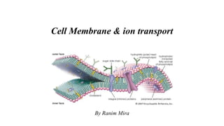
  • 1. Cell Membrane & ion transport By Ranim Mira
  • 2. Plasma membrane/cell membrane The Plasma membrane or the cell membrane is a thin, biological membrane present in all eukaryotic and prokaryotic cells that forms a boundary between the cell and its environment and regulating the flow of materials in to and out of cell. The plasma membrane exhibits selective permeability, allowing some substances to cross it more easily than others
  • 3. Objectives I. Membrane Models II. Membrane Structure III. Membrane Function IV. Factors affecting membrane fluidity V. Permeability of the membrane VI. Traffic (Transport) Across Membranes VII. Disorders of cell membrane
  • 4. II. Membrane Models • In 1935, Hugh Davson and James Danielli proposed a sandwich model in which the phospholipid bilayer lies between two layers of globular proteins • Later studies found problems with this model, particularly the placement of membrane proteins, which have hydrophilic and hydrophobic regions • In 1972, S. J. Singer and G. Nicolson proposed that the membrane is a mosaic of proteins dispersed within the bilayer, with only the hydrophilic regions exposed to water
  • 6. fluid mosaic model • Phospholipids are the most abundant lipid in the plasma membrane • Phospholipids are amphipathic molecules, containing hydrophobic and hydrophilic regions • The fluid mosaic model states that a membrane is a fluid structure with a “mosaic” of various proteins embedded in it. • The fluid mosaic model theory thereby states that plasma membrane structure is a lipid bilayer with mosaic of proteins embedded in it and moves freely Laterally parallel to the surface of the membrane.
  • 7. fluid mosaic model Phospholipid bilayer Hydrophobic regions of protein Hydrophilic regions of protein Copyright @ BioNinja
  • 8. fluid mosaic model Membranes are mosaics of floating proteins in a lipid bilayer. 2 ways: •Integral Proteins: transmembrane, have both hydrophilic and hydrophobic parts •Peripheral Proteins: Attached to membrane’s surface by: - Attachment to integral proteins or ECM fibers (outside). - Attachment to filaments of cytoskeleton (inside).
  • 10. fluid mosaic model Freeze-fracture studies of the plasma membrane supported the fluid mosaic model Freeze-fracture is a specialized preparation technique that splits a membrane along the middle of the phospholipid bilayer. Figure 7.4 Knife Plasma membrane Cytoplasmic layer Proteins Extracellular layer Inside of extracellular layer Inside of cytoplasmic layer TECHNIQUE RESULTS
  • 11. Membrane Structure Membranes are complex structures composed of: • Membrane Lipids • Membrane Proteins • Carbohydrates
  • 12. Membrane Structure *Membrane Lipids • Phospholipids: can form micelle - Phosphoglycerides: consist of two fatty acids joined to glycerol (ester linkage). - Sphingophospholipids: consist of single fatty acid joined to sphingosine(amide linkage). • Cholesterol • Glycolipids e.g; glycosphingolipids
  • 13. Membrane Structure *Membrane Protein • Integral Proteins: transmembrane, have both hydrophilic and hydrophobic parts. • Peripheral Proteins: loosely attached to membrane’s surface. RBC cytoskeleton contains peripheral proteins spectrin as well as ankyrin, both are responsible on the biconcave shape of RBC. Genetic defective or loss of spectrin protein leads to hereditary spherocytosis. • Glycoprotein: e.g; glycophorin which is erythrocyte transmembrane glycoprotein that is important in blood group identification (ABO)
  • 14. I. Membrane Function Generally, the cell membrane is responsible on: • Cell shape • Barrier keeping the constituents of the cell in and unwanted substances out. • Biological activities such as flexibility, break and reseal, Fission & Selective permeability
  • 15. Membrane function (protein) Major functions of membrane proteins • Transport • Enzymatic activity • Signal transduction • Cell-cell recognition • Intercellular joining • Attachment to the cytoskeleton and extracellular matrix (ECM) • Involvement of proteins in the membrane repair pathway.
  • 16. Membran function (protein) Enzymes Signaling molecule Receptor Signal transduction ATP (a) Transport (b) Enzymatic activity (c) Signal transduction
  • 17. Membran function (protein) Glyco- protein (d) Cell-cell recognition (e) Intercellular joining (f) Attachment to the cytoskeleton and extracellular matrix (ECM)
  • 18. Membrane function (Lipids) Lipids function as essential structural components of membranes, as : • Structural role in shaping the physical properties of the plasma membrane. • Maintaining plasma membrane integrity. • Role of facilitating plasma membrane repair. • signaling molecules. • chemical identifiers of specific membranes. • energy storage molecules.
  • 19. Membrane function (Carbohydrates) Cell-Cell Recognition: cells recognize each other by binding to surface molecules, often containing carbohydrates, on the extracellular surface of the plasma membrane. This cell-cell recognition is the basis for: - sorting an embryo’s cells into tissues/organs. - rejection of foreign cells by immune system. *Carbohydrates on the external side of the plasma membrane vary among species, individuals, and even cell types in an individual.
  • 20. Membrane function (Carbohydrates) This cell-cell recognition is the basis for: - sorting an embryo’s cells into tissues/organs. - rejection of foreign cells by immune system. - RBC blood grouping system
  • 21. The Role of Membrane Carbohydrates in Cell-Cell Recognition Receptor (CD4) Co-receptor (CCR5) HIV Receptor (CD4) but no CCR5 Plasma membrane HIV can infect a cell that has CCR5 on its surface, as in most people. HIV cannot infect a cell lacking CCR5 on its surface, as in resistant individuals.
  • 22. Factors affecting membrane fluidity • The fluidity of lipid bilayer was shown by the technique of fluorescence recovery. • The fluorescent dye is used to tag the lipids and a high- density laser beam is used to bleach the dye in a tiny spot on the cell surface. • When observed under fluorescent microscope, it is seen that within seconds the bleached spot became fluorescent again. • This explained the lateral diffusion of phospholipids.
  • 23.
  • 24. Membrane Fluidity Other factors that increase the fluidity: • Increased temperature. • Double bonds in the cis configuration increase it than trans configuration. • Short saturated FA tail rather than the long ones.
  • 25. Membrane Potential • Voltage across membranes happens when anions/cations are unequally distributed across cell membranes • Potential ranges from -50 to -200 mv • Negative sign indicates the inside of the cell is – charged. • Affects traffic of charged subs. across membrane, favors diffusion of anions out, cations in.
  • 26. Membrane Potential Factors affecting Membrane Potential • Neg. charged proteins in the cell interior • Plasma membrane’s selective permeability to various ions • The Sodium-Potassium Pump is an ELECTROGENIC PUMP: a transport protein which generates voltage across a membrane. Na+/K+ ATPase is the major one in animals, a Proton pump is the major one in Plants, bacteria, fungi (also Mitochondria, Chloroplasts use it to make ATP)
  • 27. Permeability of the cell membrane • Hydrophobic (nonpolar) molecules, such as hydrocarbons, can dissolve in the lipid bilayer and pass through the membrane rapidly. • Polar molecules, such as sugars, do not cross the membrane easily. *Chemicals that can pass through the membrane are:- - Small non-polar molecules such as carbon dioxide, Oxygen, nitrogen,.. - Small polar molecules such as water, ammonia, glycerol,... - Lipids such as cholesterol. *Chemicals that cannot pass through the membrane are:- - All ions including hydrogen ions - Large polar molecules like glucose - Amino acids - Macromolecules such as proteins, polysacharides
  • 28.
  • 29. Traffic (Transport) Across Membranes • To maintain cell functions, many biological molecules enter and leave the cell. • All materials that the cell gets from its environment or sends to the environment, Passes through this semipermeable plasma membrane. • Membrane transport is essetial for cellular life.
  • 30. Traffic (Transport) Across Membranes The membrane transport depends on: • Permeability if cell membrane. • Transmembrane solute concentration. • Size of solute. • Charge of solute
  • 33. Transport Across Membranes Passive transport: Concentration Gradient, Net directional movement, diffusion
  • 34.
  • 35. Smalluncharged polar molecules like water, urea, ethanol, have an exceptions as they can diffuse through the lipid bilayer. There are certain factors that affect the diffusion across the cell membrane: Sizeof solute Solute polarity Temperature Lipid solubility
  • 36.
  • 37.
  • 38.
  • 39. Thesubstances to be moved binds to these proteins and this complex will bind to a receptor site and then be transported across the membrane. Thisprocess does not require energy as molecules are moving down the concentration gradient. Polarand charged solutes such as glucose, fructose, galactose and some vitamins are transported by facilitated diffusion.
  • 41.
  • 42. A solution with lower solute concentration than inside of cell is called hypotonic solution. Itcauses the cell to swell and burst as it causes movement of water to inside of cell. A solution with higher solute concentration than inside of cell is called hypertonic solution. Thiscauses osmosis of water from inside of cell to outside leading to shrinkage of cell.
  • 43. Osmosis • Tonicity is the ability of a surrounding solution to cause a cell to gain or lose water • Isotonic solution: Solute concentration is the same as that inside the cell; no net water movement across the plasma membrane • Hypertonic solution: Solute concentration is greater than that inside the cell; cell loses water • Hypotonic solution: Solute concentration is less than that inside the cell; cell gains water
  • 44. Osmosis Hypotonic solution Isotonic solution Hypertonic solution (a) Animal cell (b) Plant cell H2O H2O H2O H2O H2O H2O H2O H2O Cell wall Lysed Normal Shriveled Turgid (normal) Flaccid Plasmolyzed
  • 45.
  • 46. There are two forms of active transports:- Active transport
  • 47. When the process uses chemical energy in the form of ATP, redox energy or photon energy to transport substances across the membrane, it is called primary active transport. The energy is derived directly from the breaskdown of ATP or some other high energy phosphate compounds. The proteins act as pumps to transport ions.
  • 48. Most of the enzymes that perform this transport are transmembrane ATP-ase. A primary ATP-ase which is universal to all animal cells is sodium- potassium pump which maintains the cell potential. CYTOPLASM ATP EXTRACELLULAR FLUID Proton pump H H H H H H        
  • 49. When the process uses electrochemical gradient to transport substances, it is called secondary active transport. Here the energy is derived secondarily from energy that has been stored in the form of ionic concentration differences between the two sides of a membrane, created in the first place by primary active transport.
  • 50. The pore forming proteins act as channels across the cell membrane for transporting substances. The energy stored in Na+, H+ concentration gradient is used to transport other solutes or ions.
  • 51.
  • 52.
  • 53.
  • 54.
  • 55. This pump is called a P-type ion pump because the ATP interactions phosphorylate the transport protein and causes a change in its confirmation.
  • 56. It is an antiporter enzyme located in the plasma membrane of the cells, which transport potassium ions from the extra cellular fluid to the cytoplasm and sodium ions from the cytoplasm to outside of the cell. The pump is present in all the cells of the body, and it is responsible for maintaining the sodium and potassium concentration difference across the cell membrane as well as establishing a negative electrolyte potential inside the cells.
  • 57. It was discovered by Danish scientist Jens Christian Skou in 1950. It was investigated by the passage of radioactively labelled ions across the plasma membrane. It showed that the sodium and potassium ions on both sides were interdependent which suggested that the same carrier protein transported both the ions. This carrier protein is a complex of two globular proteins namely αsubunit andβsubunit which has receptor sites for transport of three sodium ions out of cell for every two potassium ions pumped in.
  • 58.
  • 59. 4. Now, two potassium ions binds at the receptor sites present on the portion of protein that is near to outside of the carrier protein. 5. The ATP is then activated and the energy released causes confirmational change in the protein causing potassium ions to be released into the cell. 6. The returns to its first stage-steady to receive new sodium ions, so that the cycle can begin all over again.
  • 60.
  • 61.
  • 62.
  • 63. • It is the movement of substances out of the cell in the form of the secondary vesicles, which fuses with the plasma membrane and then releases its contents into the extracellular fluid. • It is important in the expulsion of waste materials out of the cell, and the secretion of enzymes and hormones. • Neurotransmitters, digestive enzymes, hormones are released from cell by exocytosis.
  • 64. • It is the movement of substances from extra cellular fluid into cell in the form of vesicles. • The large polar molecules that cannot pass through the plasma membrane enters the cell by endocytosis. • This process requires energy in the form of ATP.
  • 66.
  • 67.
  • 68.
  • 69.
  • 70.
  • 71. It attracts the substance tobe absorbed by forming a membrane depression or a coated pit on the membrane. When sufficient molecules have been attracted, the pocket will pinch off forming a coated vesicle in the cytoplasm. Inside the cytoplams the vesicle shed off their coats and then fuse with other membrane bound structures releasing their contents. E.g, Uptake of iron, cholesterol by the cell occurs by receptor mediated endocytosis.
  • 72.
  • 73. • Cell junction is a type of structure that exists in the tissues and organs. • It is a multi-protein complex that occurs between the neighbouring cells which helps in communication between them. • There occurs a specialized modification of the plasma membrane at the point of contact, forming a function or a bridge.
  • 74.
  • 75. • Also known as occluding junction, is the closest contact between adjacent cells providing a tight seal, preventing the leakage of mlecules cross the cells. • It is found just beneath the apical region (portion of cell exposed to lumen is apical surface) of cell around the cell circumference.
  • 76. • Since they are tight seals limiting the passage of molecules and ions, most materials actually enter the cells by diffusion or active transport. • The tight junction is formed by proteins called claudins and occludins which are arranged in strands along the line of junction creating a tight seal. • It is usually seen in epithelial cells, ducts of liver, pancreas and urinary bladder.
  • 77.
  • 78.
  • 79. • They are specialized intracellular channels which are brought into intimate contact with a gap of about 2-3 nm between the adjacent cells. • They directly form a connection between the cytoplasm of adjacent cell so that molecules, ions, electrical impulse pass directly from cell to cell.
  • 80. • The intracellular channels are like hollow cylinders and they are called as connexons. • These connexons are madeup of proteins called connexin. • The two adjcent connexons form a hydrophilic channel of 3 nm diameter and it is through this channel that the ions and molecule pass. • Gap junction is seen in muscles and nerves. In heart tissue helps in regular heart beat, in brain it is seen in cerebellum and it helps in muscular activity.
  • 81.
  • 82.
  • 83. • These are intracellular junctions which form a strong adhesion between adjacent cells. • It enables the cell to resist any stress. • The intermediate filaments (presents intracellularly) of adjacents cells join with eachother to form the strong adhesions so that they can function as a single unit. • They are usually seen in orgns subjected to mechanical stress like skin, heart and neck of uterus
  • 84.
  • 85. Endomembrane system *Cells have extensive sets of intracellular membranes, which together compose the endomembrane system. *It is a group of membranes and organelles in eukaryotic cells that works together to modify, package, and transport lipids and proteins. It includes a variety of organelles, such as ER, the nuclear envelope, the Golgi apparatus, and lysosomes. * the endomembrane system does not include mitochondria, chloroplasts, or peroxisomes.
  • 86. Disorders of the cell membrane -Peripheral Proteins: loosely attached to membrane’s surface. RBC cytoskeleton contains peripheral proteins spectrin as well as ankyrin, both are responsiple on the biconcave shape of RBC. Genetic defective or loss of spectrin protein leads to hereditary spherocytosis. -Transmembrane protein complexes within the lipid membrane (channels). They are divided into distinct protein units called subunits. Each subunit has a specific function and is encoded by a different gene. -Channels can be classified into: • Non-gated: K+ leak channels. • Directly gated: voltage gated (Na(+), K(+), Ca(2+), Cl(-)) • Ligand gated (ACh, Glutamate, GABA, Glycine) channels +/-Second messenger gated channels: Ca, cGMP…
  • 87. Disorders of the cell membrane The following inherited channelopathies are described. (1) Sodium channelopathies: familial generalized epilepsy with febrile seizures plus, hyperkalemic periodic paralysis, para-myotonia, hypokalemic periodic paralysis, long QT syndrome. (2) potassium channelopathies: benign infantile epilepsy, episodic ataxia type 1, dominant deafness. (3) calcium channelopathies: episodic ataxia type 2, spinocerebellar ataxia type 6, familial hemiplegic migraine, hypokalemic periodic paralysis, central core disease, malignant hyperthermia syndrome, congenital stationary night blindness, polycystic kidney diseases, (4) chloride channelopathies: myotonia congenital, cystic fibrosis. (5) cGMP gated : retinitis pigmentosa (6) ACh receptor channelopathies: autosomal dominant frontal lobe nocturnal epilepsy, congenital myasthenic syndromes. (7) glycine receptor channelopathies: hyperekplexia. (8) Gap junction channels: autosomal dominant hearing loss
  • 88. Disorders of the cell membrane