This document discusses evidence that membrane proteins have fluidity and freedom of movement within cell membranes. It provides three key points:
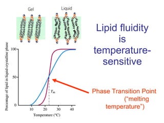
1) Classic indirect evidence from experiments with artificial lipid vesicles shows that membrane proteins require lipid fluidity to function properly.
2) Classic direct evidence from antibody labeling experiments demonstrates that some membrane proteins diffuse freely within the membrane.
3) However, membrane protein mobility can be restricted by interactions with other proteins, the cytoskeleton, surrounding lipids, and the lipid composition within membrane domains.