Downloaded 46 times


![Copyright © 2005 Pearson Education, Inc. publishing as Benjamin Cummings
• The sodium-potassium pump
– Is one type of active transport system
Figure 7.16
P
P i
EXTRACELLULAR
FLUID
Na+ binding stimulates
phosphorylation by ATP.
2
Na+
Cytoplasmic Na+
binds to
the sodium-potassium pump.
1
K+
is released and Na+
sites are receptive again;
the cycle repeats.
3 Phosphorylation causes the
protein to change its conformation,
expelling Na+
to the outside.
4
Extracellular K+
binds to the
protein, triggering release of the
Phosphate group.
6Loss of the phosphate
restores the protein’s
original conformation.
5
CYTOPLASM
[Na+
] low
[K+
] high
Na+
Na+
Na+
Na+
Na+
P ATP
Na+
Na+
Na+
P
ADP
K+
K+
K+
K+ K+
K+
[Na+
] high
[K+
] low](https://image.slidesharecdn.com/chapter7-membranestructureandfunction-140817092804-phpapp01/85/Membrane-Structure-and-Function-44-320.jpg)


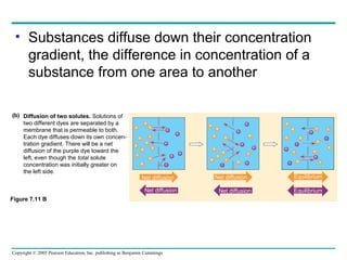
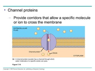
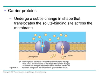
The document summarizes key concepts about membrane structure and function from Chapter 7 of Biology, Seventh Edition. It discusses the fluid mosaic model of membrane structure, which states that membranes are fluid structures composed of a phospholipid bilayer with various proteins embedded within. Membranes exhibit selective permeability, allowing some substances to pass through freely via diffusion or facilitated diffusion while actively transporting other substances against their gradients using transport proteins and cellular energy. Membrane proteins play important roles including transport, signaling, cell-cell recognition and attachment to the cytoskeleton. Membrane fluidity and composition impact these functions.