The document summarizes key concepts about cell membranes:
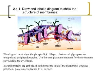
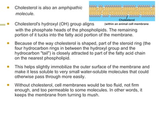
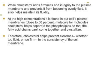
1. Cell membranes are made of a phospholipid bilayer with integral and peripheral proteins embedded. Cholesterol adds structure and prevents extremes of fluidity.
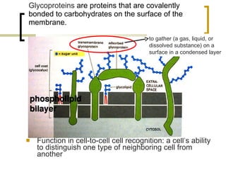
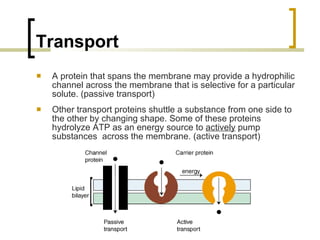
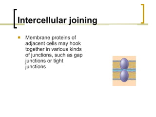
2. Membrane proteins perform important functions like transport, signaling, and attachment to the cytoskeleton.
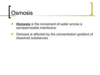
3. Passive transport like diffusion and facilitated diffusion moves molecules down concentration gradients without energy. Active transport uses protein pumps and ATP to move molecules against gradients.


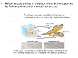


![The sodium-potassium pump is one type of active transport system (2) Na+ binding stimulates phosphorylation by ATP. Na + 1 (1) Cytoplasmic Na + binds to the sodium-potassium pump. 3 (3) K + is released and Na + sites are receptive again; the cycle repeats. (4) Phosphorylation causes the protein to change its conformation, expelling Na + to the outside. (6) Extracellular K + binds to the protein, triggering release of the Phosphate group. (5) Loss of the phosphate restores the protein’s original conformation. CYTOPLASM [Na + ] low [K + ] high Na + Na + Na + Na + Na + P ATP Na + Na + Na + P ADP K + K + K + K + K + K + [Na + ] high [K + ] low](https://image.slidesharecdn.com/membranes-110119001427-phpapp01/85/Membranes-31-320.jpg)