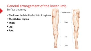
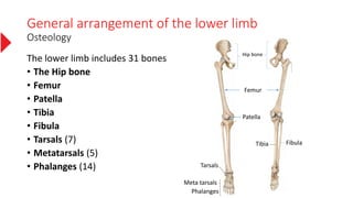
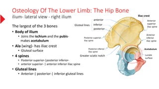
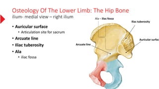
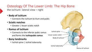
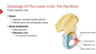
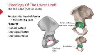
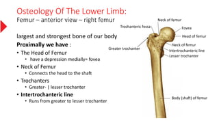
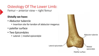
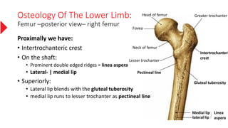
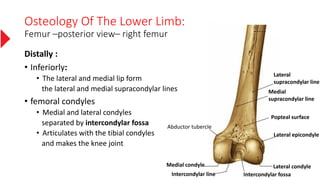
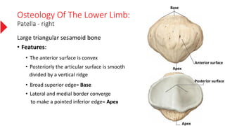
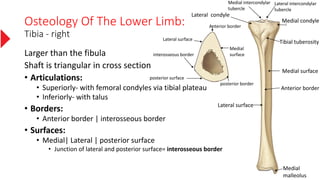
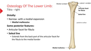
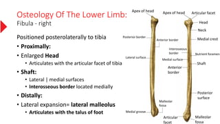
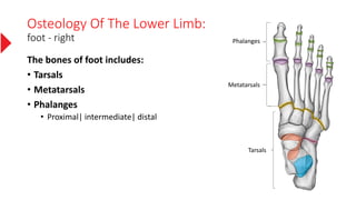
The Lower Limb
By Hasib SAMADİ
General arrangement of the lower limb
Surface anatomy
• The lower limb is divided into 4 regions
• The Gluteal region
• Thigh
• Leg
• Foot
Gluteal region
Thigh
Leg
Foot
General arrangement of the lower limb
Osteology
The lower limb includes 31 bones
• The Hip bone
• Femur
• Patella
• Tibia
• Fibula
• Tarsals (7)
• Metatarsals (5)
• Phalanges (14) Tarsals
Meta tarsals
Tibia Fibula
Femur
Hip bone
Patella
Phalanges
Osteology Of The Lower Limb: The Hip Bone
ilium- lateral view - right ilium
The largest of the 3 bones
• Body of ilium
• Joins the ischium and the pubis-
makes acetabulum
• Ala (wing)- has iliac crest
• Gluteal surface
• 4 spines
• Posterior superior-|posterior inferior-
• anterior superior- | anterior inferior iliac spine
• Gluteal lines
• Anterior-| posterior-| inferior gluteal lines
anterior
inferior
posterior
Gluteal lines
Posterior superior
iliac spine
Posterior inferior
iliac spine
Greater sciatic notch
Anterior
superior
iliac spine
Anterior
inferior
iliac spine
Acetabulum
Lunate
surface
Iliac crest
Osteology Of The Lower Limb: The Hip Bone
ilium- medial view – right ilium
• Auricular surface
• Articulation site for sacrum
• Arcuate line
• Iliac tuberosity
• Ala
• iliac fossa
Iliac tuberosity
Auricular surface
Arcuate line
Ala – iliac fossa
Osteology Of The Lower Limb: The Hip Bone
the ischium- lateral view – right
• Body of ischium
• Connects the ischium to ilium and pubis
• Sciatic notches
• Greater-| lesser sciatic notch
• Ramus of ischium
• Connects to the inferior pubic ramus
and forms the ischiopubic ramus
• Bony landmarks
• Ischial spine | ischial tuberosity
Ischial spine
Lesser sciatic notch
Ischial tuberosity
Ramus of ischium
Obturator
foramen
Body of ischium
Osteology Of The Lower Limb: The Hip Bone
Pubis-lateral view
• Rami:
• Superior- connects to the ischium
• Inferior forms the ischiopubic ramus
• Bony landmarks:
• Pubic tubercle
• Obturator crest
• For obturator membrane
Inferior pubic ramus
Pubic tubercle
Superior pubic ramus
Obturator crest
Osteology Of The Lower Limb: The Hip Bone
pubis- medial view
• Pectineal line
• proximal origin of the pectineus muscle
• Iliopubic eminence
• Marks the union of pubis and ilium
• Obturator groove
• Symphysial surface
• articulates with its contralateral
partner by means of the interpubic disc
Symphysial
surface
Iliopubic
eminence
Obturator groove
Pectineal
line
Osteology Of The Lower Limb:
The Hip Bone (Acetabulum)
Receives the head of Femur
• Makes the hip joint
Features
• Lunate surface
• Acetabular notch
• Acetabular fossa
Acetabulum
Lunate surface
Acetabular fossa
Acetabular
notch
Osteology Of The Lower Limb:
Femur – anterior view – right femur
largest and strongest bone of our body
Proximally we have :
• The Head of Femur
• have a depression medially= fovea
• Neck of Femur
• Connects the head to the shaft
• Trochanters
• Greater- | lesser trochanter
• Intertrochanteric line
• Runs from greater to lesser trochanter
Fovea
Head of femur
Intertrochanteric line
Neck of femur
Neck of femur
Lesser trochanter
Trochanteric fossa
Greater trochanter
Body (shaft) of femur
Osteology Of The Lower Limb:
Femur – anterior view – right femur
Distally we have:
• Abductor tubercle
• Insertion site for tendon of abductor meganus
• patellar surface
• Two Epicondyles
• Lateral- | medial epicondyle
Abductor tubercle
Medial
epicondyle
Lateral epicondyle
Patellar surface
Osteology Of The Lower Limb:
Femur –posterior view– right femur
Proximally we have:
• Intertrochanteric crest
• On the shaft:
• Prominent double edged ridges = linea aspera
• Lateral- | medial lip
• Superiorly:
• Lateral lip blends with the gluteal tuberosity
• medial lip runs to lesser trochanter as pectineal line
Gluteal tuberosity
Pectineal line
Medial lip
lateral lip
Linea
aspera
Intertrochanteri
crest
Greater trochanter
Lesser trochanter
Neck of femur
Fovea
Head of femur
Osteology Of The Lower Limb:
Femur –posterior view– right femur
Distally :
• Inferiorly:
• The lateral and medial lip form
the lateral and medial supracondylar lines
• femoral condyles
• Medial and lateral condyles
separated by intercondylar fossa
• Articulates with the tibial condyles
and makes the knee joint
Lateral
supracondylar line
Medial
supracondylar line
Popteal surface
Lateral condyleMedial condyle
Intercondylar line Intercondylar fossa
Abductor tubercle
Lateral epicondyle
Osteology Of The Lower Limb:
Patella - right
Large triangular sesamoid bone
• Features:
• The anterior surface is convex
• Posteriorly the articular surface is smooth
divided by a vertical ridge
• Broad superior edge= Base
• Lateral and medial border converge
to make a pointed inferior edge= Apex
Base
Apex
Anterior surface
Apex
Posterior surface
Osteology Of The Lower Limb:
Tibia - right
Larger than the fibula
Shaft is triangular in cross section
• Articulations:
• Superiorly- with femoral condyles via tibial plateau
• Inferiorly- with talus
• Borders:
• Anterior border | interosseous border
• Surfaces:
• Medial| Lateral | posterior surface
• Junction of lateral and posterior surface= interosseous border
Tibial tuberosity
Medial surface
Lateral surface
Medial condyle
Lateral condyle
Medial
malleolus
Anterior border
Medial
surface
posterior border
Lateral surface
interosseous border
posterior surface
Anterior border
Lateral intercondylar
tubercle
Medial intercondylar
tubercle
Osteology Of The Lower Limb:
Tibia - right
Distally:
• Narrow- with a medial expansion
• Medial malleoulus
Some posterior features:
• Articular facet for fibula
• Soleal line
• Extends from the back part of the articular facet for
the fibula to the medial border
Medial malleolus
Medial condyle Lateral condyle
posterior surface
Articular facet
for fibula
Soleal line
Osteology Of The Lower Limb:
Fibula - right
Positioned posterolaterally to tibia
• Proximally:
• Enlarged Head
• Articulates with the articular facet of tibia
• Shaft:
• Lateral | medial surfaces
• Interosseous border located medially
• Distally:
• Lateral expansion= lateral malleolus
• Articulates with the talus of foot
Articular facetApex of head
Head
Neck
Medial crest
Nutrient foramen
Interosseous
border
Medial surface
Anterior
border
Shaft
Posterior
surface
Malleolar
fossa
Articular
facet
Malleolar
fossa
Medial groove
Lateral surface
Anterior border
Posterior border
Apex of head
Osteology Of The Lower Limb:
foot - right
The bones of foot includes:
• Tarsals
• Metatarsals
• Phalanges
• Proximal| intermediate| distal
Phalanges
Metatarsals
Tarsals
Osteology Of The Lower Limb:
foot - right
Seven tarsal bones:
• Talus
• Calcaneus
• Navicular
• Cuboid
• Cuneiform
• medial | intermediate | lateral Talus
Navicular
Medial cuneiform
intermediate cuneiform
Lateral cuneiform
Calcaneus
Cuboid
Osteology Of The Lower Limb:
foot - right
Metatarsals:
• Connects the tarsals to the phalanges
• Toe1 has the shortest metatarsal bone
Phalanges:
• 3 phalanges on each toe
• Proximal| intermediate| Distal
• Except toe 1 which has only 2
I II
IV
III
V
Talus
Navicular
Medial cuneiform
intermediate cuneiform
Lateral cuneiform
Cuboid
Calcaneus
Credits
The illustrations used in this presentation are taken from:

The lower limb

  • 1.
    The Lower Limb ByHasib SAMADİ
  • 2.
    General arrangement ofthe lower limb Surface anatomy • The lower limb is divided into 4 regions • The Gluteal region • Thigh • Leg • Foot Gluteal region Thigh Leg Foot
  • 3.
    General arrangement ofthe lower limb Osteology The lower limb includes 31 bones • The Hip bone • Femur • Patella • Tibia • Fibula • Tarsals (7) • Metatarsals (5) • Phalanges (14) Tarsals Meta tarsals Tibia Fibula Femur Hip bone Patella Phalanges
  • 4.
    Osteology Of TheLower Limb: The Hip Bone ilium- lateral view - right ilium The largest of the 3 bones • Body of ilium • Joins the ischium and the pubis- makes acetabulum • Ala (wing)- has iliac crest • Gluteal surface • 4 spines • Posterior superior-|posterior inferior- • anterior superior- | anterior inferior iliac spine • Gluteal lines • Anterior-| posterior-| inferior gluteal lines anterior inferior posterior Gluteal lines Posterior superior iliac spine Posterior inferior iliac spine Greater sciatic notch Anterior superior iliac spine Anterior inferior iliac spine Acetabulum Lunate surface Iliac crest
  • 5.
    Osteology Of TheLower Limb: The Hip Bone ilium- medial view – right ilium • Auricular surface • Articulation site for sacrum • Arcuate line • Iliac tuberosity • Ala • iliac fossa Iliac tuberosity Auricular surface Arcuate line Ala – iliac fossa
  • 6.
    Osteology Of TheLower Limb: The Hip Bone the ischium- lateral view – right • Body of ischium • Connects the ischium to ilium and pubis • Sciatic notches • Greater-| lesser sciatic notch • Ramus of ischium • Connects to the inferior pubic ramus and forms the ischiopubic ramus • Bony landmarks • Ischial spine | ischial tuberosity Ischial spine Lesser sciatic notch Ischial tuberosity Ramus of ischium Obturator foramen Body of ischium
  • 7.
    Osteology Of TheLower Limb: The Hip Bone Pubis-lateral view • Rami: • Superior- connects to the ischium • Inferior forms the ischiopubic ramus • Bony landmarks: • Pubic tubercle • Obturator crest • For obturator membrane Inferior pubic ramus Pubic tubercle Superior pubic ramus Obturator crest
  • 8.
    Osteology Of TheLower Limb: The Hip Bone pubis- medial view • Pectineal line • proximal origin of the pectineus muscle • Iliopubic eminence • Marks the union of pubis and ilium • Obturator groove • Symphysial surface • articulates with its contralateral partner by means of the interpubic disc Symphysial surface Iliopubic eminence Obturator groove Pectineal line
  • 9.
    Osteology Of TheLower Limb: The Hip Bone (Acetabulum) Receives the head of Femur • Makes the hip joint Features • Lunate surface • Acetabular notch • Acetabular fossa Acetabulum Lunate surface Acetabular fossa Acetabular notch
  • 10.
    Osteology Of TheLower Limb: Femur – anterior view – right femur largest and strongest bone of our body Proximally we have : • The Head of Femur • have a depression medially= fovea • Neck of Femur • Connects the head to the shaft • Trochanters • Greater- | lesser trochanter • Intertrochanteric line • Runs from greater to lesser trochanter Fovea Head of femur Intertrochanteric line Neck of femur Neck of femur Lesser trochanter Trochanteric fossa Greater trochanter Body (shaft) of femur
  • 11.
    Osteology Of TheLower Limb: Femur – anterior view – right femur Distally we have: • Abductor tubercle • Insertion site for tendon of abductor meganus • patellar surface • Two Epicondyles • Lateral- | medial epicondyle Abductor tubercle Medial epicondyle Lateral epicondyle Patellar surface
  • 12.
    Osteology Of TheLower Limb: Femur –posterior view– right femur Proximally we have: • Intertrochanteric crest • On the shaft: • Prominent double edged ridges = linea aspera • Lateral- | medial lip • Superiorly: • Lateral lip blends with the gluteal tuberosity • medial lip runs to lesser trochanter as pectineal line Gluteal tuberosity Pectineal line Medial lip lateral lip Linea aspera Intertrochanteri crest Greater trochanter Lesser trochanter Neck of femur Fovea Head of femur
  • 13.
    Osteology Of TheLower Limb: Femur –posterior view– right femur Distally : • Inferiorly: • The lateral and medial lip form the lateral and medial supracondylar lines • femoral condyles • Medial and lateral condyles separated by intercondylar fossa • Articulates with the tibial condyles and makes the knee joint Lateral supracondylar line Medial supracondylar line Popteal surface Lateral condyleMedial condyle Intercondylar line Intercondylar fossa Abductor tubercle Lateral epicondyle
  • 14.
    Osteology Of TheLower Limb: Patella - right Large triangular sesamoid bone • Features: • The anterior surface is convex • Posteriorly the articular surface is smooth divided by a vertical ridge • Broad superior edge= Base • Lateral and medial border converge to make a pointed inferior edge= Apex Base Apex Anterior surface Apex Posterior surface
  • 15.
    Osteology Of TheLower Limb: Tibia - right Larger than the fibula Shaft is triangular in cross section • Articulations: • Superiorly- with femoral condyles via tibial plateau • Inferiorly- with talus • Borders: • Anterior border | interosseous border • Surfaces: • Medial| Lateral | posterior surface • Junction of lateral and posterior surface= interosseous border Tibial tuberosity Medial surface Lateral surface Medial condyle Lateral condyle Medial malleolus Anterior border Medial surface posterior border Lateral surface interosseous border posterior surface Anterior border Lateral intercondylar tubercle Medial intercondylar tubercle
  • 16.
    Osteology Of TheLower Limb: Tibia - right Distally: • Narrow- with a medial expansion • Medial malleoulus Some posterior features: • Articular facet for fibula • Soleal line • Extends from the back part of the articular facet for the fibula to the medial border Medial malleolus Medial condyle Lateral condyle posterior surface Articular facet for fibula Soleal line
  • 17.
    Osteology Of TheLower Limb: Fibula - right Positioned posterolaterally to tibia • Proximally: • Enlarged Head • Articulates with the articular facet of tibia • Shaft: • Lateral | medial surfaces • Interosseous border located medially • Distally: • Lateral expansion= lateral malleolus • Articulates with the talus of foot Articular facetApex of head Head Neck Medial crest Nutrient foramen Interosseous border Medial surface Anterior border Shaft Posterior surface Malleolar fossa Articular facet Malleolar fossa Medial groove Lateral surface Anterior border Posterior border Apex of head
  • 18.
    Osteology Of TheLower Limb: foot - right The bones of foot includes: • Tarsals • Metatarsals • Phalanges • Proximal| intermediate| distal Phalanges Metatarsals Tarsals
  • 19.
    Osteology Of TheLower Limb: foot - right Seven tarsal bones: • Talus • Calcaneus • Navicular • Cuboid • Cuneiform • medial | intermediate | lateral Talus Navicular Medial cuneiform intermediate cuneiform Lateral cuneiform Calcaneus Cuboid
  • 20.
    Osteology Of TheLower Limb: foot - right Metatarsals: • Connects the tarsals to the phalanges • Toe1 has the shortest metatarsal bone Phalanges: • 3 phalanges on each toe • Proximal| intermediate| Distal • Except toe 1 which has only 2 I II IV III V Talus Navicular Medial cuneiform intermediate cuneiform Lateral cuneiform Cuboid Calcaneus
  • 21.
    Credits The illustrations usedin this presentation are taken from: