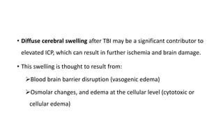
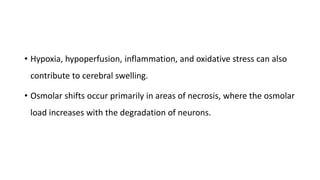
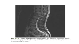
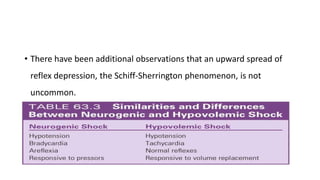
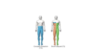
4. • Diffuse cerebral swelling after TBI may be a significant contributor to
elevated ICP, which can result in further ischemia and brain damage.
• This swelling is thought to result from:
Blood brain barrier disruption (vasogenic edema)
Osmolar changes, and edema at the cellular level (cytotoxic or
cellular edema)
5. • Hypoxia, hypoperfusion, inflammation, and oxidative stress can also
contribute to cerebral swelling.
• Osmolar shifts occur primarily in areas of necrosis, where the osmolar
load increases with the degradation of neurons.
6. • As reperfusion and recovery occur, water is drawn into the area
secondary to the high osmolar load, and the surrounding neurons
become edematous.
• Cellular swelling independent of osmolar load occurs primarily in
astrocyte foot processes and is thought to be brought on by
excitotoxicity and the uptake of glutamate.
7. • After a TBI, excitotoxicity occurs upon the release of excessive
amounts of excitatory amino acids such as glutamate, resulting in
neuronal injury.
• Excitotoxic damage occurs in two phases:
• (1) sodium-dependent neuronal swelling followed by
• (2) delayed calcium-dependent neuronal degeneration
8. • These effects are mediated through the activation of N-methyl-d-
aspartate (NMDA) and glutamate receptors, leading to a rise in the
intracellular calcium-mediated activation of proteases and lipases,
which facilitates neuronal degeneration and necrotic cell death.
9. • In contrast to necrotic cell death, apoptosis or programmed cell death
is marked not by swelling and dissolution of cell membranes but
rather by DNA fragmentation and the formation of apoptotic cell
bodies associated with neuronal shrinkage.
10. • Apoptosis is a cellular event triggered by intrinsic mechanisms
(initiated in the mitochondria) or extrinsic mechanisms (the tumor
necrosis factor superfamily of cell-surface death receptors), which
activate a cascade of enzymes called caspases and lead to cell
termination.
• Apoptosis is thought to contribute to secondary neuronal injury after
a TBI event.
11. • Animal studies have shown that developing neurons are more
susceptible than mature neurons to excitotoxic injury, probably
because more calcium is transmitted via the NMDA-mediated calcium
channel in the immature brain. 7
12. • However, although the administration of NMDA antagonists following
TBI in immature rats led to decreased excitotoxic-mediated neuronal
death, apoptotic cell death increased.
• The role of excitotoxicity and apoptosis following trauma to the
developing brain warrants further investigation.
13. TBI
• Patients with simple (“closed”) depressed cranial fractures may be
treated nonoperatively.
• Indication for operative treatment:
clinical or radiographic evidence of dural penetration
(pneumocephalus)
15. • Compound (open) depressed skull fractures are associated with
infections and the development of late epilepsy.
• They undergo surgical debridement and elevation to decrease the
possibility of infection.
16. • Also, any neurological deficit that can be associated with the skull
fracture, whether open or closed, is an indication for surgery.
• The Guidelines for Surgical Management of Depressed Cranial
Fractures recommend the routine administration of antibiotics.
17. LATE COMPLICATIONS
• Late complications from head injury include
seizures
hormonal disturbances
posttraumatic hydrocephalus
postconcussion syndrome,and psychosocial problems
18. • Posttraumatic seizures (PTSs) can be categorized as early (<7 days
after TBI) or late (>7 days after TBI).
• Early PTSs are seen in 30% of all patients with a severe TBI and in
about 1% of those with mild or moderate TBI.
19. • The estimated incidence of late-onset PTS is 13% within 2 years after
severe TBI.
• PTSs are more common in children and after penetrating head
injuries (50%).
• Anticonvulsants are used to prevent early seizures but should not be
given for more than 7 days because this practice does not decrease
the frequency of late PTSs.
20. • Posttraumatic hormonal disturbances can contribute to fatigue and
impede recovery after a TBI.
• Hypopituitarism occurs in approximately 25% of patients with a
severe TBI.
• It may be underrecognized due to its subtle clinical manifestations.
21. • Most common are deficiencies in growth hormone, gonadotropin,
and corticotropin levels.
• A routine neuroendocrine evaluation should be included in all follow-
up examinations of patients with a severe TBI.
• Posttraumatic communicating hydrocephalus has an incidence of
14% in severe head injuries.
22. • Postconcussion syndrome has an incidence of 4%–59%.
• The entire spectrum of TBI severity is associated with a risk for
psychiatric conditions and long-standing neurological deficits.
• Functional MRI has shown that working memory recruitment is
different even 1 year after mild TBI compared with healthy controls.
23. • Therefore thorough neurocognitive assessment after these patients
are released from an acute care hospital is imperative to offer them
the best chance for recovery.
• Approaches used to treat neurocognitve deficits have included
cognitive and behavioral therapy along with pharmacological
treatment.
24. • Psychosocial problems such as decreased social contact, anxiety,
depression, and loneliness create a major challenge for the majority
of TBI victims and can lead to aggression and substance abuse and
hinder community reentry.
• Psychosocial problems are a persistent long-term problem and can
interfere with rehabilitation.
25. • Individuals who experience a severe TBI are vulnerable to a loss of
friendships and social support, which leads to social isolation, fewer
leisure activities, and new dependence on others.
• This situation is aggravated by a lack of opportunity to establish new
social contacts and friends.
26. • Clinicians such as psychiatric social workers, psychologists, or
psychiatrists may have to be called upon more often to provide the
psychological services that may be necessary for many of these
patients.
27. OUTCOME
• Outcome after severe TBI remains poor; approximately one-third die,
and 25% survive with severe disabilities.
• A number of factors play an important role in predicting outcome.
• One study retrospectively analyzed 846 cases of severe TBI to clarify
the prognostic effects of multiple factors.
28. • One year after injury, GCS score, age, pupillary response and size,
hypoxia, hyperthermia, and ICP were associated with outcome,
indicating that prevention of hypoxia and hyperthermia and control of
high ICP may be useful ways of improving the outcome of patients
with severe head injury.
29. • A GCS score of 3 on presentation has been recognized as a poor
prognostic factor.
• Mortality approaches 100% in the presence of bilateral fixed dilated
pupils.
30. • Age in itself is an independent predictor of outcome.
• With or without surgery, outcome in the elderly is worse than in the
young.
• Different age thresholds have been named in the literature.
• Depending on the statistical analysis performed, one study found
worse outcomes above 39 and 65 years of age.
31. • Multiple regression analysis showed that every 10 years above this
threshold increase in age led to a 10% increase in mortality.
• Even with timely and satisfactory surgery, unexplained clinical
deterioration occurs in patients 70 years of age or older.
32. • For people 65 years of age and older, falls are the leading cause of
TBI-related death, accounting for 40% of all TBI deaths in that age
group.
• Additionally, falls can cause other injuries such as hip fractures, which
can impede independent living and increase the risk of premature
death.
33. • An epidemiological study over 10 years showed that fall-related TBI
increased by 126% and the related case fatality rate decreased from
32% to 18%, meaning that more elderly people are living with TBI
disabilities.
• However, rehabilitation efforts in the elderly have been poor because
of the often negative attitude (ageism) toward the recovery potential
in this group.
34. • Despite their relative disadvantages, the elderly might see improved
outcomes with rehabilitation programs intended for their age group.
• Some CT findings can predict outcome.
• The length, width, depth, and location of SDH and EDH; number,
volume, and location of contusions; compression of ventricles and the
basal cistern; and presence or absence of traumatic SAH are all
relevant.
35. • The most important CT-defined predictor of outcome is the
magnitude of the midline shift.
• Finally, the facility where patients with TBI are treated affects
outcome.
36. • Patient with severe TBI treated in American College of Surgeons
(ACS)–designated level 1 trauma centers have better survival rates
and outcomes than those treated in ACS-designated level 2 centers.
37. • It is very important to provide goal-concordant care for patients with
severe TBI, especially as it is difficult to predict outcome and many
interventions currently can prolong life but have not been shown to
improve functional outcome.
38. • Patients may consider some forms of functional or cognitive disability
as worse than death, and simply understanding the terminal nature of
their disease or having an end-of-life (EOL) discussion with their
physician can steer patients away from preferring life-extending
therapies.
39. • Elderly patients in particular ofen prefer no treatment over treatment
that would leave them with cognitive disabilities.
• However, physicians are often reluctant to initiate EOL conversations,
and the TBI patient is often unable to vocalize his or her preferences
for treatment; therefore the burden of deciding between palliative
care versus life-extending care is often falls on the surrogate decision
maker.
40. • Lack of formal training in goal-of-care and EOL discussion often cited
as one of the barriers.
• Medical schools and governing bodies have recognized that and have
begun paying more attention to goal-concordant care.
41. • Shared decision making (SDM)—where disease prognosis and
treatment risks and benefits are communicated to patients and their
family members using cartoons, symbols, and other cues called
decision aids is an example of a potential workaround to this
problem.
42. • As treatments improve and patients live longer with TBI deficits,
transparent communication between surrogate decision makers,
providers, and—to the extent possible—patients, will become a
necessary part of the TBI management protocol.
43. • The following five-step process has been suggested:
• (1) collection of evidence
• (2) information sharing,
• (3) critical appraisal,
• (4) recommendation and decision, and
• (5) assessment and follow-up
44. • Using this framework, the physician can better understand the
patient’s values and preferences while building rapport with the
family, determining any bias-inducing information, and recommend
decisions through an every-10-year SDM model with appropriate
follow-up at the end.
45. • Another communication tool specially developed for emergency
situations and surgeons is the “Best Case/Worst Case” tool, which has
been shown to facilitate the discussion around EOL values and care.
47. ASIA/IMSOP Impairment Scale
• Grade A: Complete
• No motor or sensory function is preserved in the sacral segments S4
and S5.
• Grade B: Incomplete
• Sensory but not motor function is preserved below the neurological
level and extends through sacral segments S4 and S5.
48. • Grade C: Incomplete
• Motor function is preserved below the neurological level, and a
majority of key muscles below the neurological level have a muscle
grade of less than 3.
49. • Grade D: Incomplete
• Motor function is preserved below the neurological level, and a
majority of key muscles below the neurological level have a muscle
grade of 3 or greater.
• Grade E
• Normal motor and sensory functions are normal
50. SPINAL CORD INJURY SYNDROMES
• Central Cord Syndrome
• It presents in 9% of all traumatic cord injuries and is the most
common of the spinal cord syndromes.
• Hyperextension in the cervical spine, with some preexisting cervical
spondylosis, is usually responsible for this type of injury.
51. • Imaging the cervical spine in patients with central cord syndrome will
reveal stenosis from spondylosis, fracture subluxation, or sequestered
disk, with no spinal stenosis.
52. • Schneider proposed that these injuries resulted from acute
compression from preexisting bone spurs anteriorly and
hypertrophied ligamentum flavum posteriorly and contributed to
hematomyelia and central cord necrosis.
53. • Schneider witnessed weakness in the upper extremities greater than
the lower extremities, as well as a variable degree of sensory
disturbances and loss of bladder control.
• It was proposed that involvement of the anterior horn cells led to
weakness in the arms greater than the legs, secondary to the
topography of the corticospinal tracts.
54. • Because of their good recovery, Schneider was in favor of taking a
more conservative approach toward treating these patients.
• Correlations of MRI and histopathology fail to suggest hematomyelia
from Schneider’s hypothesis.
55. • There is in fact minimal disruption of the central gray matter.
• Axonal disruption and swelling are more widespread in the white
matter.
56. • Anterior Cord Syndrome
• Anterior cord syndrome occurs with injuries to the ventral two-thirds
of the cord, while sparing the posterior column.
• It is present in 2.7% of all traumatic SCIs.
• Motor function is lost distal to the site of the injury.
57. • Spinothalamic function may be disrupted, leading to loss of pain and
temperature sensation in certain areas.
• Because the posterior columns remain intact, the sensations of
vibration, position, and crude touch will not be affected.
• Occasionally patients feel hyperesthesia and hypoalgesia below the
level of the lesion.
58. • Although this syndrome is classically described for anterior spinal
artery compromise, in the setting of trauma it is due to flexion
injuries or retropulsed disk or bone.
• Anterior cord syndrome carries a worse prognosis than other cord
syndromes.
59.
60. • Posterior Column Syndrome
• Posterior column syndrome is a rare condition with an incidence of
• less than 1%. This syndrome has been linked to neck hyperexten-
• sion injuries. Injuries occur to the posterior aspect of the cord (Fig.
• 63.3). Because the posterior columns are injured, there is usually a
• loss of vibration and position sense, with retained spinothalamic
61. • function of pain and temperature sensation. Occasionally, motor
• function can be affected as well. Although this syndrome has
• been previously mentioned in the literature, it was omitted from
• the International Standards for Neurological and Functional
• Classification of SCI and is not currently recognized as a separate
• syndrome. This syndrome can also be seen in the context of perni-
• cious anemia
62.
63. • Brown-Séquard Syndrome
• Brown-Séquard syndrome accounts for 1%–4% of all traumatic SCIs.
• Injuries affect the lateral half of the cord.
• It occurs most frequently in the cervical spine.
• It is usually due to penetrating injuries and (less commonly) blunt
trauma including disk herniations.
64. • In cases of blunt trauma, Brown-
Séquard syndrome usually
occurs in the context of
hyperextension injuries,
although it has been observed in
flexion injuries, locked facets,
and compression-related
injuries.
65. • Below the level of the lesion, it classically manifests with ipsilateral
pyramidal deficit, loss of ipsilateral tactile discrimination, position
sense, and vibratory sensation, and loss of pain and temperature
sensation on the contralateral aspect of the body one to two
dermatomes below the level of the injury.
66. • However, this classic presentation of Brown-Séquard syndrome rarely
occurs.
• More frequently, patients presenting with Brown-Séquard syndrome
present with a variation of the classic syndrome, termed Brown-
Séquard plus.
67. • With Brown-Séquard plus, there is asymmetrical hemiplegia as well as
hypoalgesia more prominent on the less paretic side.
• Patients presenting with a clinical picture consistent with a classic Brown-
Séquard syndrome injury have a worse prognosis than patients presenting
with a variation of the syndrome, but the overall prognosis is good.
• Brown-Séquard has the best functional motor recovery when compared with
other clinical spinal cord syndromes.
68. • Most subjects obtain bowel and bladder continence.
• Patients having predominantly more weakness in the upper
extremities compared with the lower extremities have a favorable
outcome in regard to ambulating.
69. • The symptoms of Brown-Séquard syndrome may appear
instantaneously or in a delayed fashion.
• Furthermore, they may occur in conjunction with other spinal cord
syndromes.
70. Cervicomedullary Syndrome
• Injuries appear in the upper cervical cord and extend to the medulla.
• Because of its location, the clinical manifestations of this syndrome
include respiratory compromise, hypotension, tetraparesis (often
mimicking a central cord syndrome with arms affected more than
legs), hyperesthesia, and the onion-skin or Déjerine pattern of
sensory loss over the face.
71. • will tend to affect the peripheries of the face.
• This occurs as sensory fibers enter the trigeminal tract and descend to
various levels depending on their somatotropic origin, then synapse
in the adjacent nucleus.
• Fibers from the anterior face synapse more rostrally in the trigeminal
tract while fibers from the hindface synapse more caudally, adjacent
to the sensory input of C2–3.
72. • Mechanisms of injury for this syndrome include a variety of injuries to
the atlantoaxial complex as well as injuries resulting from
compression via burst fractures or herniated disks.
73. Conus Medullaris Syndrome
• There is high probability that injuries to the thoracolumbar region can
involve the conus medullaris.
• The conus medullaris represents the transition of the spinal cord from
the central nervous system to the peripheral nervous system.
• The location of this region is highly variable—between the T12 and L1
disk space to the middle third of L2 in the majority of the population.
74.
75. • The lumbar parasympathetic fibers, sacral sympathetic fibers, and
sacral somatic nerves originate in the conus medullaris.
• The classic presentation entails lower-extremity weakness, absent
lower-limb reflexes, and saddle anesthesia.
• There is usually mixed UMN and LMN involvement.
76. • Loss of the bulbocavernosus and anal reflexes is permanent,
differentiating conus medullaris syndrome from SCIs that have a
return of these reflexes within 48 hours of the injury.
• Patients typically have an areflexic bowel and bladder (low-pressure,
high-capacity bladder).
77. • The most common injuries to the vertebral column resulting in this
condition are burst fractures or fracture-dislocation.
• There is no strong clinical evidence favoring surgical intervention over
nonsurgical intervention for conus medullaris injuries.
• Furthermore, if surgical intervention is performed, there is no
compelling evidence to suggest that earlier decompression affects
functional outcome.
78. Cauda Equina Syndrome
• The cauda equina is defined as the region of the neuroaxis occupied
by the filum terminale.
• The only neurological structures in this region include the lumbar and
sacral roots.
• Injuries in this location are typically a pure LMN injury.
79.
80. • Findings often include:
absent bulbocavernosus reflex
absent deep tendon reflexes
flaccid urinary bladder, and
reduced lower-extremity muscle
tone
• It is differentiated from conus
medullaris syndrome by
the presence of asymmetrical
weakness and the absence of
UMN involvement.
81. • Like conus medullaris syndrome, burst fracture and fracture-
dislocation are the most common vertebral column injuries
associated with this condition.
• Cauda equina injuries have better recoveries owing to the resiliency
of the roots to injuries and the greater regeneration capacity of the
roots compared with the spinal cord.
82. • However, the sacral roots are very delicate, and injuries to them may
be permanent.
• In general, cauda equina syndrome in the setting of herniated disk
pathology is treated early (within 24 hours) if possible, to prevent
residual symptoms.
83. • However, functional outcome in a traumatic setting is similar to conus
medullaris syndrome.
• There is no strong evidence correlating functional outcome to surgical
decompression, nor is there any evidence that suggests cauda equina
injuries fare better with early versus late decompression.
84.
85. TRANSIENT SPINAL CORD SYNDROMES
• Transient spinal cord syndromes have been documented in the
literature, with multiple reported incidents occurring in contact
sports.
• The term “burning hand syndrome” was initially coined to describe a
severe burning sensation in the upper extremities occurring in
athletes who suffer injuries in contact sports.
86. • It is likely related to lesions of the spinothalamic tract in central cord
injuries.
• Because the most medial fibers of the spinothalamic tract provide
pain and temperature sensation to the hands and fingers, injuries to
these fibers would explain the dysesthesias of the hand, so this
syndrome is most suggestive of a mild central cord syndrome.
87. • Unilateral burning pain down the arm to the hand can signify root
injury and has been termed a “burner” or “stinger”; they typically last
seconds to hours but rarely longer than 24 hours.
• Stingers occur more frequently with baseline cervical stenosis, which
leads to a narrow intervertebral foramen.
88. • Traction or direct trauma to the brachial plexus can mimic cervical
root injury.
• A positive Spurling test can suggest compression of the nerve root as
the cause of symptoms.
89. SPINAL SHOCK
• It was initially described as arterial hypotension following SCIs.
• The definition has evolved to include permanent extinction of tendon
reflexes.
• Additional modifications to the definition have since revised it to include
all findings related to the physiological and anatomical transection of the
spinal cord that results in depressed spinal reflexes for a limited period
of time.
90. • The severity of the injury correlates with the severity of spinal shock.
• An injury alters reflexes that occur closest to the insult first, with
those more distal from the transection presenting later.
• Thus high-level cervical injuries may have retention of sacral reflexes,
such as a preserved bulbocavernosus and anal wink.
91. • The observation that a proximal-to-distal spread of reflex depression
occurs on the order of minutes suggests a physiological explanation
for these changes.
• It has been hypothesized that the loss of supraspinal input leading to
hyperpolarization of neurons is responsible for this physiological
change.
92. • There have been additional observations that an upward spread of
reflex depression, the Schiff-Sherrington phenomenon, is not
uncommon.
• It is important to delineate blood pressure drops from circulatory
shocks from those of spinal shock.
93. • As there is loss of sympathetic tone, there is pooling of blood in the
venous system and a loss of sympathetic tone in the cardiovascular
system.
• On the one hand, circulatory shock requires volume replacement, and
on the other hand, spinal shock requires vasopressors.
94. • As spinal shock resolves, muscle spindle reflexes return in a caudal-to-
cranial direction, except at the level of injury.
• Over time, a spastic syndrome results.
95. • There is no uniform consensus on what constitutes the cessation of
spinal shock.
• Most references define the end of spinal shock with a return of
certain reflexes.
• However, not all reflexes are uniformly depressed in each patient;
reflexic changes are individualized.
96. • The resolution of spinal shock occurs over a period of days to months,
so there is a slow transition from spinal shock to spasticity that occurs
on a continuum.
• It has been proposed that this transition comprises four phases.
97. • The first phase occurs from 0 to 24 hours following the injury.
• It is characterized by areflexia or hyporeflexia.
• the first pathological reflex to appear is the delayed plantar reflex,
• followed by a series of cutaneous reflexes such as the
bulbocavernosus, abdominal wall, and cremasteric reflex.
98. • Impaired sympathetic control can lead to bradyarrhythmias,
atrioventricular conduction block, and hypotension.
• Motor neuron hyperpolarization explains the changes that occur.
99. • Phase 2 occurs between day 1 and day 3 post injury.
• Cutaneous reflexes are more prominent during this period, but deep
tendon reflexes remain mute.
• It is not unusual for elderly individuals and children to experience
recovery of deep tendon reflexes during this time.
100. • The Babinski sign may become apparent in the elderly as well.
• Denervation supersensitivity and receptor upregulation account for
these changes in the second phase.
• The next phase occurs between 4 days and 1 month post injury.
• Deep tendon reflexes usually recuperate by day 30.
101. • There is great disagreement about when these reflexes appear.
• The recovery of the Babinski response closely parallels the return of the ankle
jerk reflex.
• There is also diminution of the delayed plantar reflex.
• Autonomic changes such as bradyarrhythmias and hypotension begin to
subside.
• This time period is reflected by axon-supported synapse growth.
102. • The fourth phase is dominated by hyperactive reflexes and occurs from 1 to 12
months after injury.
• Vasovagal hypotension and bradycardia generally resolve in 3–6 weeks, but
orthostatic hypotension may take 10–12 weeks before it disappears.
• Episodes of malignant hypertension or autonomic dysreflexia (AD) begin to
appear during this time period.
• Soma-supported synapse growth accounts for these findings.