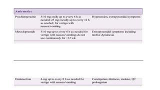
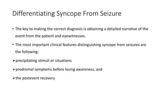
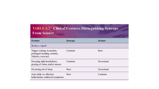
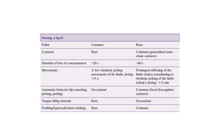
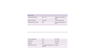
Dizziness, Vertigo, and Hearing Loss
Dr. Zeleke W/Y NR3
INTRODUCTION
• Dizziness is an imprecise term used to describe a variety of symptoms
including but not limited to:
vertigo
light-headedness
faintness
giddiness,
• disequilibrium, confusion, etc.
Contd....
• It affects nearly one-quarter of the general population and is a
common complaint in the emergency room and in the office of the
neurologist, otolaryngologist, and internist.
Contd....
• Vertigo is an illusion of motion, either the environment or the self.
• Most commonly rotatory but may be translational or tilting.
• Presyncope is the sensation encountered before loss of
consciousness.
• Dysequilibrium is not a sensation of motion but a feeling of imbalance
or unsteadiness.
Contd....
• Other or nonspecific dizziness include those whose symptoms do not
easily fit into one of the aforementioned categories or fall into more
than one category.
Contd....
• Rather than using the qualitative description of dizziness to categorize
the subtype, it is often more helpful to use characteristics such as
onset, duration, triggers, history of prior episodes, and associated
symptoms in evaluating these patients.
VERTIGO
Neuroanatomy
• Vertigo primarily results from disorders of the vestibular system,
which includes the vestibular labyrinth, vestibular nerve, vestibular
nuclei in the brainstem, vestibular portions of the cerebellum,
connections between these structures.
• It only rarely higher in the cerebrum.
Contd....
• The vestibular labyrinth:
• located in the temporal bones
• It is composed of the three orthogonally oriented semicircular canals
(anterior, posterior, and lateral) and
• the vestibule, which contains the otolith organs, the utricle and
saccule, which are also angled at approximately 90° to each other.
Contd....
• The SCC responds to angular acceleration and the vestibule responds
to linear acceleration including translation or tilt.
• When the head is rotated, endolymphatic fluid in the semicircular
canals lags behind, leading to a deflection of the gelatinous cupula
within the canal, which activates or inhibits the firing of hair cells.
Contd....
• Activation on one side is paired with inhibition in the complementary
canal on the other.
• The otolith organs, the utricle and saccule, contain hair cells on which
calcium carbonate crystals, the otoconia, rest.
• Translational motion or tilt (via gravity) will activate or inhibit these
cells.
Contd....
• From the vestibular labyrinth, neurons travel centrally through the
vestibular portion of the eighth cranial nerve into the brainstem to
the vestibular nuclei and then project on to the cerebellum, ocular
motor nuclei, spinal cord, and, via some less well-understood
pathways, to the cerebrum.
Contd....
• Integration of the combinations of activations and inhibitions of the
various components of the vestibular system of both ears, along with
visual input and proprioceptive input, detects motion, rotation,
translation, and tilt and affects eye movements and posture.
Contd....
• Vertigo can result from disorders of the peripheral vestibular system
(labyrinth or nerve) or central vestibular system (brainstem,
cerebellum, connections, and rarely, cerebrum), and this localization
is the natural next step in the evaluation of vertigo.
Diagnosis
• In the patient with the acute first presentation of vertigo, the most
significant concern is evaluating for stroke (ischemic or hemorrhagic)
and differentiating from vestibular neuritis.
• A history of vascular risk factors and other neurologic symptoms,
headache, and complaints related to the brainstem is particularly
important.
Contd....
• However, the lack of those symptoms does not exclude an ischemic
etiology.
• Patients presenting with isolated vertigo have a threefold increased
risk of stroke compared to the general population that increases with
the presence of multiple vascular risk factors.
Contd....
• In the patient presenting with recurrent attacks of vertigo, the major
differential includes BPPV, Ménière syndrome, and migraines.
• Vertebral artery compression from neck rotation is a very rare case of
episodic dizziness or vertigo.
Focused History
• Loss of vestibular function affects eye movements and image
stabilization, balance, and spatial orientation.
• In addition to vertigo, patients may feel tilted, the world jiggling while
walking (oscillopsia), spatially disoriented, imbalanced, and, rarely,
suffer from drop attacks, where they may feel pulled or pushed to the
ground.
Contd....
• These drop attacks, known as Tumarkin crises, are most commonly
seen in Ménière syndrome but also may occur in other vestibular
conditions.
Focused Examination
• The examination of the patient with vertigo includes all components of
the neurologic examination with special attention to certain aspects.
• The ear must be examined and hearing must be tested.
• Examination of the eyes is particularly important.
• Start by observing for nystagmus in primary gaze and with eye
movements in all directions.
Contd....
• Reobserve after removing the ability to fixate by the use of Frenzel
lenses, goggles with magnifying lenses that allow you to see eye
movements but does not allow the patient to fixate.
• If Frenzel lenses are unavailable, fixation can be eliminated during
ophthalmoscopic examination of one eye by covering the eye not
being examined with your free hand and observing for nystagmus of
the optic disc.
Contd....
• Remember that this movement is in the opposite direction of the
movement of the front of the eye.
• Pay attention to the type of motion (horizontal, vertical, torsional,
mixed), the effect of fixation, and the effect of gaze.
• Pendular nystagmus is sinusoidal, whereas jerk nystagmus, the more
commonly observed nystagmus, is composed of slow drift in one
direction and a rapid correction back.
Contd....
• It is caused by an imbalance in vestibular input, either peripheral or
central.
• Jerk nystagmus is named for the direction of the fast phase and can
be further categorized by its trajectory and the conditions under
which it is seen.
Subtypes of Nystagmus
• Jerk Nystagmus
• Gaze-evoked: nystagmus at the extremes of gaze beating in the
direction of gaze
• Physiologic: fine nystagmus; usually fatigues
• Drug/medication: often seen with sedatives and anticonvulsants
• Brainstem/cerebellar lesions: may be sustained
Contd....
• Rebound nystagmus: After looking eccentrically for approximately 1
min on return to primary gaze, there is nystagmus beating in the
other direction associated with brainstem or cerebellar lesions.
• Bruns nystagmus: Slow, large-amplitude nystagmus in one direction
and rapid, small-amplitude nystagmus in the other direction suggest a
cerebellopontine angle lesion on the side of the slow, large-amplitude
nystagmus.
Horizontal Nystagmus
• Peripheral nystagmus: Nystagmus only beats in one direction, away
from the affected side, obeys Alexander law, usually mixed with
torsion, and inhibited by fixation.
• Central nystagmus: Nystagmus may change directions, may be purely
horizontal, does not obey Alexander law, and not inhibited by fixation.
Contd....
• Periodic alternating nystagmus: nystagmus alternating directions
every 1-2 min associated with lesions at the cervicomedullary
junction or in the cerebellum
• Dissociated nystagmus: nystagmus differing between the eyes seen
with internuclear ophthalmoplegia or mimicked by myasthenia gravis
Contd....
• Downbeat nystagmus: Nystagmus usually increases on down and
lateral gaze seen with involvement of the dorsal medulla or the
cerebellar flocculus or projections associated with lesions at the
cervicomedullary junction, medications (lithium, carbamazepine,
phenytoin), alcohol, hypomagnesemia, thiamine deficiency,
paraneoplastic syndromes, cerebellar degenerations, and other.
Contd....
• Upbeat nystagmus: associated with brainstem and cerebellar lesions,
most commonly the medulla.
• Congenital nystagmus: often a mixture of jerk and pendular
nystagmus
• Convergence-retraction nystagmus: part of Parinaud dorsal midbrain
syndrome; convergence and retraction of the eyes
Contd....
• Positional nystagmus: seen with specific head motions
• Pendular Nystagmus
• Acquired: seen with brainstem and cerebellar lesions
• Congenital: often a mixture of jerk and pendular nystagmus
Contd....
• Spasmus nutans: infant onset nystagmus is asymmetric and rapid,
often associated with head nodding and head turning; usually
resolves
• Associated with visual loss
• Seesaw nystagmus: opposite conjugate vertical and torsional
movements associated with mesencephalic or parasellar lesions
Contd....
• Oculopalatal myoclonus: rhythmic 2-3-Hz movements seen late after
lesion of Mollaret triangle
• Oculomasticatory myorhythmia: rhythmic movements of eye
convergence and contraction of masticatory or other muscles seen in
20% of patients with Whipple disease
Contd....
• In addition to nystagmus, unilateral peripheral vestibular dysfunction
leads to a subtle skew deviation of vertical eye position with the
ipsilateral eye lower in the orbit relative to the contralateral eye and
conjugate torsion of the eyes to the ipsilateral side.
• In addition to the ocular motor findings, the patient usually has a
small head roll or tilt to the affected side and a tendency to fall
toward the same side.
Contd....
• They may perceive vertical to be slightly tilted.
• Testing for past pointing will reveal deviation to the ipsilateral side.
Tandem gait and Romberg test will be impaired but are not specific.
• The Fukuda step test, marching in place with the eyes closed, will
show deviation toward the side of the vestibular lesion.
Head Impulse Test
• The head thrust or head impulse test evaluates for loss of vestibular
input from either the vestibular labyrinth or vestibular nerve to the
vestibulo-ocular reflex.
• It is performed by asking the patient to focus on a target in front of
him or her.
• The head is then rapidly rotated a small amount (approximately 15°).
Contd....
• In normal circumstances, the eyes remain focused on the target.
• A vestibular lesion on the side to which the patient is being rotated
will lead to a loss of fixation and the eyes will turn with the head and
require a saccade to refixate.
Contd....
• The input from the horizontal canal is most commonly tested by
rotating the head in the horizontal plane.
• The other canals may also be tested by rotating the head along the
plane of the canal in question.
• The head impulse test is generally normal in those with central causes
of vertigo.
Contd....
• In patients presenting with an acute vestibular syndrome, the use of
the head impulse test along with the evaluation of nystagmus and the
presence of skew (HINTS testing) can be used to distinguish an acute
peripheral vestibular lesion from an acute central lesion such as
cerebral infarct with more accuracy than MRI.
Contd....
• Dynamic visual acuity also evaluates this reflex.
• It is performed by testing vision while stationary and then while
shaking the head at approximately 2 Hz.
• Most can read no more than two to four lines worse on the eye chart
with normal function of at least one labyrinth.
The head thrust test
Causes of Peripheral Vertigo
• BPPV: Brief, recurrent, positional nystagmus only in provoking
position
• Vestibular neuritis: Acute, single episode, viral prodrome
• Ramsay Hunt syndrome (herpes zoster oticus): Acute, single episode,
vesicles in/near the ear, facial nerve palsy, deafness
Contd....
• Ménière syndrome: Recurrent, last minutes to hours, unilateral ear
symptoms of fullness, hearing loss, tinnitus
• Trauma: History of trauma
• Perilymphatic fistula: Episodic, associated with Valsalva, loud sounds
(Tullio phenomenon), history of trauma
Contd....
• Superior canal dehiscence: Episodic, associated with Valsalva, loud
sounds
• Cogan syndrome: Ménière-like syndrome with interstitial keratitis
• Acoustic neuroma: Rare vertigo, more imbalance, unilateral hearing
loss or tinnitus
Contd....
• Medications: Aminoglycoside exposure
• Otitis: Evidence of otitis
Contd....
• Labyrinth ischemia: Presence of vascular risk factors, sudden vertigo,
and hearing loss
• Recurrent vestibulopathy: Recurrent attacks but without the ear
symptoms to suggest Ménière syndrome
Causes of Central Vertigo
• Migraine-associated vertigo: Migraine history
• Cerebral ischemia/hemorrhage: Vascular risk factors, other neurologic
symptoms and signs, more prominent dysmetria or ataxia
• Multiple sclerosis: History or presence of other neurologic symptoms
or signs
• Tumor: Presence of other neurologic symptoms or signs
Contd....
• Craniocervical junction abnormalities (Arnold-Chiari, basilar
impression, etc): Associated with headaches, neck pain, other lower
cranial nerve involvement
• Episodic ataxia type 2: Episodic vertigo and ataxia lasting hours to
days
Contd....
• Cerebellar or spinocerebellar degenerations: More commonly ataxia
and not vertigo
• Mal de débarquement: After boat travel (or other), sensation of
motion persists
Treatment
• The treatment of vertigo is primarily directed at the causes.
• Nonspecific treatments for attacks of vertigo, some off-label, include
antihistamines, anticholinergics, benzodiazepines, and antiemetics for
nausea and vomiting.
Contd....
• Symptomatic medications should be stopped after severe symptoms
remit.
• Vestibular rehabilitation is thought to benefit by promoting
compensation, facilitating strategic substitution, limiting inactivity,
and in the case of BPPV ,repositioning otolith debris.
Syncope
Syncope
• Syncope is defined as a transient loss of consciousness due to
decreased cerebral blood flow.
• Seizure is defined as a transient alteration in brain function due to
abnormal electrical cerebral activity.
• When evaluating any patient with a history of transient loss of
consciousness, clinicians should take a very careful history of the
event itself.
PATHOBIOLOGY
• Syncope is defined as a transient loss of consciousness due to
insufficient blood flow to the brain, resulting in a loss of postural
tone.
• The underlying basic mechanisms resulting in decreased cerebral
blood flow are reduced cardiac output and/or low peripheral vascular
resistance (blood pressure).
Contd....
• A loss of consciousness in supine individuals occurs within 6 to 8
seconds after cardiac asystole.
• Whereas in upright individuals, tilt table recordings demonstrate loss
of consciousness if systolic blood pressure drops below 60 mm Hg.
Causes of syncope
Neurally Mediated Reflex Syncope
• The most common type of syncope.
• account for over half of cases.
• It is reflex syncope, also known as neurocardiogenic syncope.
Contd....
• An external factor or set of circumstances (apprehension of pain,
cough, head turning, etc) leads to bradycardia, hypotension, or a
mixture of both phenomena.
• Reflex syncope is divided into two main categories: vasovagal syncope
and carotid sinus syncope
Vasovagal Syncope
• It is the most common type of reflex syncope
• It is caused by a brief loss of neurally mediated circulatory control and
is generally associated with a benign prognosis.
• It an be subdivided into three major categories: postural, central, and
situational.
Contd....
• In the postural form of vasovagal syncope, a common patient
narrative might describe a young person attending a hot and crowded
school assembly or concert, standing for a long period of time and
having skipped a recent meal and ingested some alcohol.
Contd....
• In cases of centrally mediated vasovagal syncope, the trigger may
involve sudden pain (commonly in the setting of venipuncture),
apprehension of pain, or emotional shock immediately preceding the
fainting episode.
Contd....
• Less commonly, syncope occurs in specific situations or appears to be
temporally related to specific triggers.
• Older men may report symptoms soon after arising from bed and
emptying a distended bladder.
Contd....
• Some young people describe recurrent syncope after exercise.
• Others describe syncope after coughing, laughing, or sneezing.
• It is not unusual for a history of vasovagal syncope to run in families.
Carotid Sinus Syndrome
• A more unusual type of reflex syncope is carotid sinus syndrome, which occurs when
normal stimulation of an unusually sensitive carotid sinus leads to hypotension and/or
bradycardia.
• This diagnosis should be suspected in older individuals who report unexplained falls or
syncope with almost no prodromal symptoms; occasionally, a history of head turning,
wearing a tight-collared shirt, or shaving preceding syncope is elicited, but in general, this
diagnosis is often difficult to make and is considered after extensive diagnostic testing
has been inconclusive or negative.
Cardiac Syncope
• The most dangerous type of syncope is cardiac syncope, when a
transient loss of consciousness is caused by either cardiac arrhythmia
or structural heart disease.
• In a large population-based study of syncope, cardiac syncope was
associated with a twofold increased risk of dying, regardless of
etiology, as compared to individuals without a history of syncope.
• Arrhythmias are the most common cause of cardiac syncope, and both
• tachyarrhythmias and bradyarrhythmias have been implicated. The most
• common arrhythmia associated with syncope is sinus node dysfunction. Other
• arrhythmias include atrioventricular conduction system disease,
• supraventricular tachyarrhythmias, and ventricular tachyarrhythmias.
• Mechanical or structural causes of cardiac syncope include prior
• myocardial infarction, cardiomyopathy, aortic stenosis, mitral stenosis,
• pericardial tamponade, myxoma, aortic dissection, and pulmonary emboli.
• Because of the high morbidity associated with cardiac syncope, it is
• imperative to consider this diagnosis when evaluating an individual with
• syncope. The clinical features most predictive of cardiac syncope include
• age older than 60 years, male gender, known structural heart disease, fewer
• events (less than three events by history), syncope while supine, and syncope
• during effort/exercise.
Differentiating Syncope From Seizure
• The key to making the correct diagnosis is obtaining a detailed narrative of the
event from the patient and eyewitnesses.
• The most important clinical features distinguishing syncope from seizures are
the following:
precipitating stimuli or situations
prodromal symptoms before losing awareness, and
the postevent recovery
• Clinical Features of Syncope
• Syncope typically occurs when patients are upright (standing or sitting) and
• can be triggered by exercise, coughing, bearing down (Valsalva maneuver),
• venipuncture, prolonged standing, or pain. Presyncope begins with a sense of
• unwellness accompanied by light-headedness and nausea. Patients may feel
• weak and unsteady on their feet, followed by a decreased awareness or
• detachment from their environment. Immediately before losing
• consciousness, patients experience a “graying of the vision” and/or
• “muffling” of ambient sounds.
• Eyewitnesses will often note that the patients
• appear pale or ashen, diaphoretic, and tachypneic. Sometimes, an
attack can
• be aborted if the patients lie down quickly or lower the head below
the level
• of the heart. Once patients lose consciousness, they will lose tone in
the
• muscles of their trunk and legs and they will limply collapse to the
floor.
• On
• the ground, the limbs are flaccid and patients continue to appear pale
and
• sweaty. The period of unconsciousness is generally brief, lasting only
• seconds up to 1 to 2 minutes. If the degree of decreased cerebral perfusion is
• profound enough, patients may display a few jerking movements of the limbs
• (“convulsive syncope”), but this is also typically very brief (lasting only a
• few seconds). Generally, the pulse and blood pressure quickly return to
• normal with the patients in the recumbent position, and they rapidly regain
• consciousness and quickly become oriented and aware of their surroundings.
• Patients will often realize that they have fainted and remember
details of the
• event up until the moment of losing consciousness. They might
experience
• mild fatigue or brief disorientation after the event, but this should not
last
• more than a few minutes.
Clinical Features of Seizures
• Epileptic seizures are defined as transient alterations in brain function
due to abnormal electrocerebral activity.
• The clinical manifestations of seizures vary widely, depending on brain
volume involvement and neuroanatomic location of electrical
activation.
Contd....
• Seizures can occur day or night, regardless of whether the patient is awake or
asleep, and usually are not triggered by a precipitating stimulus or environment.
• Sometimes, a seizure may begin with the patients reporting an “aura” or warning.
• These subjective experiences might be described as a sense of déjà vu (a sense of
reliving a familiar experience), a noxious smell, sudden anxiety, or tingling over
one side of the body.
Contd....
• Eyewitnesses might find the patients unresponsive to their direct
questions, staring into space, or stopping what they were previously
doing (behavioral arrest).
• Some patients exhibit oral or manual automatisms (lip smacking,
chewing, rubbing of their hands, picking at their clothes).
Contd....
• If a focal seizure then secondarily generalizes (electrical activity
starting off in one area of the brain but then spreading to both
hemispheres of the brain), the patients’ head and eyes may suddenly
and forcibly turn to one side, with stiffening of the limbs, frothing at
the mouth, cyanosis of the lips, followed by rhythmic jerking of the
limbs.
Contd....
• Most secondarily generalized seizures last between 1 and 2 minutes
in duration and rarely over 5 minutes in duration, but afterward, the
patients may be confused and disoriented for minutes to hours.
• Patients may report lateral tongue biting and urinary incontinence
after some seizures.
Clinical Features of Other Spells Potentially
Mistaken for Syncope
• Vertebrobasilar Transient Ischemic Attack
• Transient ischemic attack (TIA) of the vertebrobasilar system is an
extremely rare cause of brief loss of consciousness.
• The underlying pathophysiology is believed to be due to temporary
ischemia of the brainstem reticular activating system.
Contd....
• Almost invariably, TIAs of the vertebrobasilar system are associated
with focal neurologic symptoms due to ischemia of the brainstem,
cerebellum, and/or occipital lobe.
• Therefore, patients and family members will typically describe
associated dysarthria, diplopia, dysphagia, hemianopsia, ataxia,
unilateral weakness, or numbness.
Hypoglycemia
• Hypoglycemia may cause feelings of light-headedness or dizziness and
rarely results in a brief loss of consciousness.
• A more typical presentation of hypoglycemia is a slow and insidious
onset of delirium that may last minutes to hours and be associated
with diaphoresis, hunger, tremulousness, anxiety, and palpitations.
• If severe enough, hypoglycemia can progress to coma (otherwise,
prolonged unresponsiveness) and/or generalized tonic-clonic
seizures.
Basilar Migraine
• Basilar migraine may cause confusion but rarely leads to loss of
consciousness.
• It is associated with headache, ataxia, and positive visual
phenomenon developing over minutes to hours and therefore is
unlikely to be confused with syncope.
Subclavian Steal Syndrome
• It is characterized by a reversal of blood flow in the vertebral artery,
away from the brain, in order to supply blood flow to an ischemic
arm.
• In this syndrome, exercising the affected arm leads to dizziness,
vertigo, dysarthria, dysphagia, ataxia, unilateral weakness, and
sometimes loss of consciousness.
• Supranuclear Causes of Diplopia
• INO due to lesion of the medial longitudinal fasciculus (MLF) is
• characterized by horizontal diplopia that is present in contralateral gaze only.
• Often, there is nystagmus of the abducting (normal) eye and slowed adducting
• saccade of the affected eye. Sometimes, this is the only finding. Adduction of
• the affected eye is often better during convergence because this does not use
• the MLF. INO localizes to the brainstem as is a common demyelinating
• syndrome. It can also result from small vessel stroke, sometimes, below the
• level of detection of diffusion weighted magnetic resonance imaging.
• Skew deviation, a supranuclear disorder, is characterized by vertical
• diplopia with tilting of images in both eyes. Typically, it is associated with
• other vestibular or cerebellar symptoms and signs. Stroke and tumor are
• common causes.
• Convergence insufficiency is trouble bringing the eyes in at near leading
• to horizontal diplopia when reading and is common in Parkinson disease and
• after concussion. Divergence insufficiency is trouble bringing the eyes apart
• leading to horizontal diplopia at distance. Both have similar symptoms in all
• directions of gaze, which distinguishes them from sixth nerve palsy and INO.
Third Nerve Palsy
• A nonnuclear, third nerve palsy in isolation affects the superior rectus,
inferior rectus, medical rectus, and inferior oblique to cause
oblique diplopia in primary gaze
vertical diplopia in up and down gaze
horizontal diplopia in contralateral gaze, and
no diplopia in ipsilateral gaze
Contd....
• Additional localizing signs are a larger ipsilateral pupil and ptosis.
• A nuclear third nerve palsy typically causes:
bilateral ptosis and bilateral upgaze deficits
typical ipsilateral cranial nerve III signs
Contd....
• Aneurysm compressing the ipsilateral third nerve is a can’t miss
diagnosis, which requires emergent angiographic imaging to exclude
it.
• Uncal herniation, typically associated with altered mental status,
causes pupillary dilation through third nerve compression.
Contd....
• Skull base tumors can also cause third nerve palsy through
compression.
• In chronic compression, there can be aberrant regeneration, which is
demonstrated on exam when one third nerve action (eg lid raising),
occurs when another (eg, downgaze) is attempted.
Contd....
• Brainstem parenchymal events such as stroke, demyelination, and
tumor can cause third nerve palsy accompanying:
contralateral movement disorder (Benedikt syndrome)
contralateral ataxia (Claude syndrome), or
contralateral hemiparesis (Weber syndrome)
Fourth Nerve Palsy
• A nonnuclear fourth nerve palsy in isolation affects the ipsilateral
superior oblique muscle to cause vertical diplopia that is worse in
down gaze, contralateral gaze, and ipsilateral head tilt.
• Due to the torsional action of the superior oblique, the image in the
affected eye can appear tilted.
Contd....
• Patients may adopt a compensatory head tilt.
• A nuclear fourth nerve palsy causes contralateral superior oblique
dysfunction.
• Brainstem causes of fourth nerve palsy can be accompanied by a
contralateral Horner syndrome.
Contd....
• Fascicular causes include microvascular, traumatic, and compressive.
• Fourth nerve palsies are a common childhood strabismus pattern that
can decompensate in adulthood to cause new-onset diplopia.
Sixth Nerve Palsy
• A nonnuclear sixth nerve palsy in isolation affects the lateral rectus
muscle to cause horizontal diplopia in ipsilateral gaze that resolves in
contralateral gaze.
• A nuclear sixth nerve palsy causes an ipsilateral gaze palsy (ie,
affecting both eyes and therefore without diplopia).
Contd....
• Sixth nerve palsy is a false localizing sign of elevated ICP, for example,
due to tumor, venous sinus thrombosis or meningitis, and intracranial
hypotension as might occur from cerebrospinal fluid leak.
Contd....
• As with third and fourth nerve palsies, it can be caused by
microvascular disease, compression, trauma, and brainstem events.
• The sixth nerve floats freely in the cavernous sinus and may be
affected in isolation by disease in this region.
• It is affected in petrous apicitis in Gradenigo syndrome.
Cavernous Sinus Syndrome
• Complete or partial involvement of cranial nerves III, IV , VI,
sympathetic pathways (Horner syndrome), and V1 and V2 localizes to
the cavernous sinus.
• Any process that elevates venous pressure in the cavernous sinus can
transmit this pressure to the orbit to cause significant orbital signs
including proptosis and red eyes.
Contd....
• Vision can be affected if the process extends superiorly to the optic
chiasm or if blood flow to the eye is affected.
Descriptive terms for involuntary movements by
category
Explanation of Descriptive Terms for
Involuntary Movements by Category
• Voluntariness
• Voluntary: Completely within the control of the patient
• Example: Lifting a cup to drink
Contd....
• Semivoluntary: Partially within the control of the patient; may be
suppressed if even temporarily; may be associated with an
uncontrollable urge to move and relief after movement
• Phenomenology: Akathisia
Contd....
• Involuntary: Completely outside of the patient’s control;
insuppressible
• Phenomenology: Myoclonus
Rhythmicity
• Rhythmic
Explanation: Occurring at a regular frequency
Phenomenology: Tremor
Arrhythmic
Explanation: occurring at an irregular frequency
Phenomenology: Dystonia
Pattern
• Stereotyped
Explanation: Patterned and somewhat predictable
Phenomenology: Tics
• Nonstereotyped
Explantion: Unpatterned and unpredictable
Phenomenology: Chorea
Descriptive Terms
Phenomenology and their Descriptors
• Myoclonus Involuntary, jerky
• Dystonia Involuntary, sustained
• Chorea Involuntary, nonstereotyped, flowing
• Athetosis Involuntary, nonstereotyped, flowing
Contd....
• Ballism : Involuntary, nonstereotyped, ballistic
• Tics: Semivoluntary, stereotyped
• Stereotypies: Semivoluntary, stereotyped
• Tremor: Involuntary, rhythmic, oscillatory
• Akathisia: Semivoluntary, stereotyped or nonstereotyped, flowing
Hyperekplexia
• Hyperekplexia (exaggerated startle) consists of dramatic, complex
motor responses to sudden tactile or auditory stimuli.
• The reaction can consist of a blink; facial contortion; abduction of the
arms; and flexion of the neck, trunk, and arms.
Contd....
• Sometimes, instead of movements, the body becomes stiff and
immobile.
• When it is severe, the patient’s movements must be curtailed
because a sudden attack may lead to injury from falling.
• The etiology may be hereditary or sporadic.
Red Flags for Functional/Psychogenic
Movements
• Inconsistency: Movements that change quality (eg, change in
direction or frequency)
• Combination of several hyperkinesias that do not ordinarily occur
together: Unusual combinations of movements that do not typically
co-occur in organic disease
Contd....
• Fixed postures: Postures that are sustained for prolonged periods of time
• Deliberate slowness: Practiced slowness that resolves when the patient is
unaware that they are still being observed
• Effortfulness: patient demonstrates extraordinary effort during tasks that
should not be affected (cognitive tasks, tasks with unaffected body parts).
Contd....
• Suggestibility: the movement is reproduced when the examiner
performs a maneuver that he or she suggests will acutely bring on the
movement.
• Distractibility: the movement stops when the examiner draws the
patient’s attention away from the affected body part to perform a
task that requires considerable effort and concentration.
Contd....
• Entrainment (in tremor disorders): tremor frequency changes to
match the frequency of tapping with the opposite limb.
• Give-way weakness: during strength testing, the patient initially
provides resistance against the examiner’s force but then suddenly
“gives way” and provides no further muscular resistance.
Contd....
• Nonanatomic sensory impairment: sensory deficits that do not follow
anatomic patterns/boundaries
• Marked fatigue and exhaustion: by history, the amount of fatigue is
out of proportion to the severity of the movements.
NEUROANATOMY
• Most involuntary movements are the result of central nervous system
disorders and, more specifically, lesions involving the basal ganglia or
cerebellum.
• E.g. the motor features of PD are related to a lesion in the SNpc
Contd....
• chorea is related to disorders of the caudate nucleus but sometimes
involving other structures, and
• ballism is most often related to lesions of the subthalamic nucleus.
• Ataxia and intention tremor are related to lesions of the cerebellum
Contd....
• Despite central nervous system predominance in movement disorders
pathophysiology, abnormal movements attributed to a peripheral
etiology do also occur (eg, hemifacial spasm, painful legs–moving
toes, jumpy stump).
Contd....
• The neural site of origin of myoclonus spans central and peripheral
localizations, including the cerebral cortex (cortical myoclonus), brain
stem (reticular reflex myoclonus), spinal cord (propriospinal
myoclonus), or peripheral nerve (eg, hemifacial spasm).
Clinical, epidemiologic, and pathophysiologic features of
cerebral palsy
• CP is a developmental, nonprogressive disorder manifested by
abnormal movements (chorea, athetosis, dystonia), spasticity, and/ or
ataxia accompanied by disturbances of cognition, behavior,
communication, and other neurologic and musculoskeletal problems.
Contd....
• CP is attributed to damage in the developing fetal or infant brain, such
as perinatal hypoxia–ischemia, intracranial hemorrhage or cerebral
infarction, neonatal hyperbilirubinemia (kernicterus), and brain
maldevelopment.
• Prevalence: 1.7 to 3.1 in 1000 live births, higher in developing
countries; the most common cause of disability in early childhood.
Contd....
• Dyskinetic CP is the third most common form of CP (15%), after
spastic (hemiplegic > quadriplegic > diplegic) and ataxic CP.
• Dystonia, choreoathetosis: exacerbated by action
• 70% have lesions in the basal ganglia or thalamus
Contd....
• Exclude CP mimickers (e.g., dopa responsive dystonia (DRD),
sepiapterin reductase deficiency (SRD), beta-propeller protein–
associated neurodegeneration (BPAN), congenital disorders of
glycosylation (CDG), ADCY5-related movement disorders
Tics
• Simple phonic tics typically consist of:
sniffing
throat clearing
grunting
squeaking
screaming
coughing, blowing, and sucking sounds
Contd....
• Complex phonic tics include:
linguistically meaningful utterances and verbalizations, such as
shouting of obscenities or profanities (coprolalia),
repetition of someone else’s words or phrases (echolalia), and
repetition of one’s own utterances, particularly the last syllable, word
or phrase in a sentence (palilalia).
Contd....
• Some TS patients also manifest sudden and transient cessation of all
motor activity (blocking tics) without alteration of consciousness.
Contd....
• Suppressibility, although characteristic and common in tics, is not
unique or specific for tics, and this phenomenon has been well
documented in other hyperkinetic movement disorders.
• Using functional MRI, Peterson and colleagues (1998a) showed
decreased neuronal activity during periods of suppression in the
ventral globus pallidus (GP), putamen, and thalamus.
Contd....
• There was increased activity in the right caudate nucleus, right frontal
cortex, and other cortical areas that are normally involved in the
inhibition of unwanted impulses (prefrontal, parietal, temporal, and
cingulate cortices).
Contd....
• Besides temporary suppressibility, tics are characterized by
suggestibility and exacerbation with stress, excitement, boredom,
fatigue, and exposure to heat.
• Emotional stress associated with life events or other stresses have
been documented to potentially markedly exacerbate tics, but onset
of TS is not necessarily related to stressful life events.
Contd....
• Tics may also increase during relaxation after a period of stress.
• In contrast to other hyperkinetic movement disorders that are usually
completely suppressed during sleep, motor and phonic tics may
persist during all stages of sleep.
Contd....
• In addition, patients with TS often have disturbances of sleep, such as
increased sleep fragmentation, higher frequency of arousals,
decreased rapid eye movement (REM) sleep, and enuresis.
• Many patients note a reduction in their tics when they are distracted
while concentrating on mental or physical tasks (such as when playing
a video game or during an orgasm).
Contd....
• Other patients experience increased frequency and intensity of their
tics when distracted, especially when they no longer have the need to
suppress the tics.
• Tics are also typically exacerbated by dopaminergic drugs and by
stimulants, including methylphenidate and cocaine.
Contd....
• Finally, it should be noted that a broad spectrum of movements may
be present in patients with TS that can be confused with tics, such as
akathisia, chorea, dystonia, compulsive movements, and fidgeting, as
part of hyperactivity associated with ADHD.
ANATOMY OF THE CEREBELLUM
• The cerebellum is tucked underneath the cerebral cortex and next to
the brainstem and has extensive connections to both of those
structures.
• It has a cortex that branches and folds, central white matter, and deep
nuclei.
Contd....
• The gray matter constituting the cortex is divided into three layers:
the external molecular layer, which contains the outer stellate
cells and inner basket cells
the middle layer, which contains the Purkinje cells
the internal granular layer, which contains granular cells
Contd....
• The deep cerebellar nuclei (the dentate nucleus, interposed nuclei,
and fastigial nucleus) are the only output cells of the cerebellar cortex
and send their excitatory signal to numerous brain regions.
Contd....
• The cerebellar circuitry is designed to modify signals from the brain,
process those signals,and then send the processed signals back to
different brain areas.
• The signal begins in the cerebral cortex and then synapses on neurons
in the pontine nuclei.
Contd....
• The axons from the contralateral pontine nuclei, called mossy fibers
because of the appearance of their synaptic terminals, then synapse
on the granular cells in the inner layer of the cerebellar cortex gray
matter.
Contd....
• The axons of the granular cells are parallel fibers that ascend to the
molecular layer of the cerebellum, bifurcate, and form T-shaped
branches that then synapse on many Purkinje cells.
• Each Purkinje cell receives input from many parallel fibers, and each
parallel fiber modulates tens of thousands of Purkinje cells.
• The Purkinje cells also receive direct modulatory input on their
dendritic shaft from the inferior olive’s climbing fibers, thus allowing
the climbing fibers to modulate the parallel fiber–to–Purkinje
connection.
• The Purkinje cells then receive further inhibitory input from the
basket and stellate interneurons in the molecular layer that shape the
spike activity of the Purkinje cell output.
• The GABA-ergic Purkinje cells then inhibit the deep cerebellar nuclei,
which in turn also receive excitatory inputs from the mossy and
climbing fibers, thus creating a loop in which the Purkinje cells
modulate the excitatory outputs from the deep cerebellar nuclei.
• This circuitry is repeated in all parts of the cerebellum, which can be
divided into different lobes based on function.
• The vermis and paravermis make up the central portion of the
cerebellum and are also referred to as the spinocerebellum.
• The vermis is responsible for axial function, while the paravermis is
responsible primarily for limb function.
Contd....
• The spinocerebellum receives sensory input from the trigeminal
nucleus and the dorsal column of the spinal cord and
• then connects to:
• (1) the fastigial nuclei to modulate eye movements and
• (2) the rubrospinal and vestibulospinal tract to modulate muscle tone.
• The cerebrocerebellar hemispheres are located on each side of the
vermis and paravermis and are responsible for motor planning and
timing (primarily anterior lobe) as well as cognition (primarily
posterior lobe).
Contd....
• The cerebrocerebellum receives input from the ipsilateral primary
and supplementary motor cortex and the contralateral inferior olivary
nucleus and connects to the cerebral premotor and motor cortex and
the red nucleus.
• Finally, tucked into the underside of the cerebellum is the
flocculonodular lobe, which is also called the vestibulocerebellum and
is responsible for the vestibular system.
• It receives input from the ipsilateral vestibular nuclei and pretectal
area of the midbrain and visual cortex and then connects directly
back to the vestibular nucleus.
Contd....
• The vasculature of the cerebellum originates from the vertebrobasilar
anterior system and is divided such that the three primary vessels
cover different portions of the cerebellar lobes.
• The SCA supplies the superior area of the vermis and the
superomedial cortex.
• The AICA supplies the anterior inferior cerebellum, flocculus, and
MCP.
Contd....
• The PICA supplies the cerebellar nuclei, the inferior surface of the
vermis, and the undersurface area of the cerebellar hemisphere.
Contd....
• The SCA, AICA, and PICA all also provide blood supply to different
areas of the brainstem and have additional branches within the
cerebellum, which explains the variation in examination and MRI
findings following strokes of these vessels.
Cerebellar Signs and Symptoms on Bedside
Examination
• The localization of a patient’s symptoms to the cerebellum relies on
identification of characteristic changes in balance, gait, speech,
swallowing, eye, and appendicular movements.
BALANCE AND GAIT
• Most individuals with cerebellar ataxia present with balance and gait
difficulties.
• Patients describe falls and clumsiness but often describe the initial
symptoms as being difficulty maintaining balance on uneven ground,
when going up or (primarily) down stairs, or when running.
Contd....
• Examination usually reveals a wide-based gait with some titubation
observed, specifically on turns.
• Because this same gait can be observed in sensory or vestibular-
based ataxia, the remainder of the examination allows for cerebellar
localization.
SPEECH AND SWALLOWING
• Speech, swallowing, and eye movement abnormalities are especially
important for cerebellar localization,as these findings indicate that
the lesion is above the spinal cord.
• Although speech abnormalities vary slightly among the different
ataxias, most individuals have a scanning-type speech,with difficulties
with prosody, vocal modulation, articulation, and phonation.
Contd....
• This pattern is different from that heard in individuals with Parkinson disease
in that hypophonia is not prominent in the cerebellar ataxias.
• Dysphagia is also very common in individuals with cerebellar ataxia, although
its prevalence varies widely across the different etiologies of cerebellar ataxia.
• Individuals often report coughing while ingesting food and liquid and getting
food or pills stuck in their throats.
EYE MOVEMENTS AND VISION
• Eye movement changes are often the best way to localize the
patient’s ataxia to the cerebellum.
Patients describe the
world moving around (oscillopsia)
delayed focusing when they move their eyes
Contd....
difficulty looking up, especially when lying down (eg, individuals who
bench press weights will have difficulty looking at the bar)
difficulty with reading signs when in a moving car; or
challenges looking down long grocery store aisles
• On examination, these symptoms manifest as down beat
nystagmus,saccadic intrusions on smooth pursuit, square-wave jerks,
and hypermetric and hypometric saccades.
• Downbeat nystagmus is the most classic examination finding and
localizes to changes in the bilateral flocculus lobe of the cerebellum as
well as the craniocervical junction and bilateral medial longitudinal
fasciculi, although, when combined with other cerebellar findings,the
flocculus localization seems the most likely for these patients.
• However, individuals without downbeat nystagmus can have
cerebellar localization, with hypermetric and hypometric saccades
also observed in most individuals with cerebellar ataxia.
• These saccadic eye movement changes are essentially the eye
manifestations of the undershoot and overshoot observed in the
appendicular examination, as described subsequently.
Contd....
• Individuals with a pure cerebellar ataxia have a normal vestibular
system and therefore a normal head impulse test, but many
neurodegenerative ataxias also feature vestibular involvement, so
assessing the vestibular system facilitates the differential diagnosis.
APPENDICULAR ATAXIA
• Individuals with appendicular ataxia describe
significant clumsiness, manifesting as knocking over drinks at the
dinner table, hitting the wrong letter on a computer or phone
keyboard, and
having difficulty with activities of daily living such as cutting food,
fastening buttons, and pulling up zippers.
• It is important to determine whether this clumsiness is due to
cerebellar lesions or an alternative localization, most often
extrapyramidal lesions.
Contd....
• Sometimes it is possible to distinguish between the two in the history
by determining whether patients experience difficulty meeting their
target (eg, grabbing the fork and knife, finding the button) versus a
feeling that their fingers will not do what their brain is asking.
Contd....
• On examination, patients demonstrate past pointing on the finger
chase test, dysdiadochokinesia, and dysmetria on the finger-to-nose
test, although often a finger-to-chin test is better to ensure that
patients do not hit themselves in the eye.
• Tremors are also commonly noted in individuals with ataxia, although
sometimes what is described as tremor is nonrhythmic and really a
combination of truncal ataxia and dysmetria.
EXTRAPYRAMIDAL FINDINGS AND MIMICS
• Muscle cramps and stiffness as well as slowness and clumsiness are
also common concerns in individuals with cerebellar ataxia.
• Examination of muscle tone reveals both a pyramidal and
extrapyramidal localization depending on the etiology of the
cerebellar degeneration, with spasticity in some patients and rigidity
in others; in some patients, the bedside examination reveals a clear
muscle tightness, but whether it is rigidity or
• spasticity is difficult to determine. Individuals with cerebellar ataxia
without
• extrapyramidal involvement often demonstrate pauses and
clumsiness on finger
• tapping,handopeningandclosing,andpronationandsupinationbutdono
thave
• a decrementing bradykinesia ( VIDEO 9-3).
• Many of the genetic ataxias do have extrapyramidal involvement and
therefore feature true decrementing bradykinesia, although such
findings also point toward MSA as a possible diagnosis.
AUTONOMIC NERVOUS SYSTEM
• Autonomic nervous system involvement is very common in the other
neurodegenerative ataxias.
• Patients commonly report urinary urgency, frequency, and
incontinence; lightheadedness when standing; and sometimes sexual
dysfunction.
• In one survey, lower urinary tract symptoms were reported in 8% to
50% of men with a variety of genetic spinocerebellarataxias; also
reported were clear changes in urodynamic studies performed on a
smaller subset of the same cohort.
Contd....
• Another analysis of nine individuals with spinocerebellar ataxia (SCA)
type 2 identified autonomic nervous system symptoms and
neurophysiologic study changes in all the individuals, ranging from
urinary tract to cardiovascular system to gastrointestinal dysfunction.
Contd....
• Although such detailed analysis is not available for all the SCAs and
neurodegenerative ataxias, these findings show that autonomic
nervous system dysfunction is part of the overall clinical picture and
should be addressed as part of patient care.
COGNITION
• The cerebellum plays a critical role in cognition, and individuals with
cerebellar neurodegeneration often report cognitive difficulties.
• Individuals with what is known as cerebellar cognitive affective
syndrome present with challenges in executive function, linguistic
processing, and spatial cognition.
• These cognitive changes were once thought to be related to the
cerebellar connections to the cortex, but more recent research has
demonstrated that the relevant processes occur in the cerebellum
itself, particularly the more posterior part of the cerebellum,
• with the anterior cerebellumbeing more related to motor processes.
• Cerebellar cognitive affective syndrome has been identified in
individuals with acute (eg, stroke) and more subacute (eg,
neurodegeneration, tumors) disorders of cerebellar processes and
can be measured using a validated scale that is specific to the
cerebellar deficits.
Contd....
• The recognition that these cognitive changes are part of the disease
process is a critical part of counseling for patients and their families
and is an area of intensive research, both to better understand the
disorder and to identify more effective treatments.
PSYCHIATRICCHANGES
• Psychiatric changes associated with cerebellar disease may cause
significant challenges for patients and their families and are found in
almost all patients.
• Although depression can certainly be part of the disease course,
anxiety and irritability coupled with disinhibition are more common.
Contd....
• Patients often report feeling particularly anxious in crowds of people
or whenever they need to walk and maneuver to get somewhere
without falling.
• The irritability and disinhibition are most often noticed by the spouse
or care partner of the individual with ataxia and are often described
as “going from an emotional 0 to 60” in the snap of a finger.
Contd....
• The individual with ataxia is sometimes not aware of this change, but
it may cause significant difficulties in their personal relationships.

Dizziness.pptx

  • 1.
    Dizziness, Vertigo, andHearing Loss Dr. Zeleke W/Y NR3
  • 2.
    INTRODUCTION • Dizziness isan imprecise term used to describe a variety of symptoms including but not limited to: vertigo light-headedness faintness giddiness, • disequilibrium, confusion, etc.
  • 3.
    Contd.... • It affectsnearly one-quarter of the general population and is a common complaint in the emergency room and in the office of the neurologist, otolaryngologist, and internist.
  • 4.
    Contd.... • Vertigo isan illusion of motion, either the environment or the self. • Most commonly rotatory but may be translational or tilting. • Presyncope is the sensation encountered before loss of consciousness. • Dysequilibrium is not a sensation of motion but a feeling of imbalance or unsteadiness.
  • 5.
    Contd.... • Other ornonspecific dizziness include those whose symptoms do not easily fit into one of the aforementioned categories or fall into more than one category.
  • 6.
    Contd.... • Rather thanusing the qualitative description of dizziness to categorize the subtype, it is often more helpful to use characteristics such as onset, duration, triggers, history of prior episodes, and associated symptoms in evaluating these patients.
  • 8.
    VERTIGO Neuroanatomy • Vertigo primarilyresults from disorders of the vestibular system, which includes the vestibular labyrinth, vestibular nerve, vestibular nuclei in the brainstem, vestibular portions of the cerebellum, connections between these structures. • It only rarely higher in the cerebrum.
  • 9.
    Contd.... • The vestibularlabyrinth: • located in the temporal bones • It is composed of the three orthogonally oriented semicircular canals (anterior, posterior, and lateral) and • the vestibule, which contains the otolith organs, the utricle and saccule, which are also angled at approximately 90° to each other.
  • 10.
    Contd.... • The SCCresponds to angular acceleration and the vestibule responds to linear acceleration including translation or tilt. • When the head is rotated, endolymphatic fluid in the semicircular canals lags behind, leading to a deflection of the gelatinous cupula within the canal, which activates or inhibits the firing of hair cells.
  • 11.
    Contd.... • Activation onone side is paired with inhibition in the complementary canal on the other. • The otolith organs, the utricle and saccule, contain hair cells on which calcium carbonate crystals, the otoconia, rest. • Translational motion or tilt (via gravity) will activate or inhibit these cells.
  • 12.
    Contd.... • From thevestibular labyrinth, neurons travel centrally through the vestibular portion of the eighth cranial nerve into the brainstem to the vestibular nuclei and then project on to the cerebellum, ocular motor nuclei, spinal cord, and, via some less well-understood pathways, to the cerebrum.
  • 13.
    Contd.... • Integration ofthe combinations of activations and inhibitions of the various components of the vestibular system of both ears, along with visual input and proprioceptive input, detects motion, rotation, translation, and tilt and affects eye movements and posture.
  • 14.
    Contd.... • Vertigo canresult from disorders of the peripheral vestibular system (labyrinth or nerve) or central vestibular system (brainstem, cerebellum, connections, and rarely, cerebrum), and this localization is the natural next step in the evaluation of vertigo.
  • 16.
    Diagnosis • In thepatient with the acute first presentation of vertigo, the most significant concern is evaluating for stroke (ischemic or hemorrhagic) and differentiating from vestibular neuritis. • A history of vascular risk factors and other neurologic symptoms, headache, and complaints related to the brainstem is particularly important.
  • 17.
    Contd.... • However, thelack of those symptoms does not exclude an ischemic etiology. • Patients presenting with isolated vertigo have a threefold increased risk of stroke compared to the general population that increases with the presence of multiple vascular risk factors.
  • 18.
    Contd.... • In thepatient presenting with recurrent attacks of vertigo, the major differential includes BPPV, Ménière syndrome, and migraines. • Vertebral artery compression from neck rotation is a very rare case of episodic dizziness or vertigo.
  • 19.
    Focused History • Lossof vestibular function affects eye movements and image stabilization, balance, and spatial orientation. • In addition to vertigo, patients may feel tilted, the world jiggling while walking (oscillopsia), spatially disoriented, imbalanced, and, rarely, suffer from drop attacks, where they may feel pulled or pushed to the ground.
  • 20.
    Contd.... • These dropattacks, known as Tumarkin crises, are most commonly seen in Ménière syndrome but also may occur in other vestibular conditions.
  • 21.
    Focused Examination • Theexamination of the patient with vertigo includes all components of the neurologic examination with special attention to certain aspects. • The ear must be examined and hearing must be tested. • Examination of the eyes is particularly important. • Start by observing for nystagmus in primary gaze and with eye movements in all directions.
  • 22.
    Contd.... • Reobserve afterremoving the ability to fixate by the use of Frenzel lenses, goggles with magnifying lenses that allow you to see eye movements but does not allow the patient to fixate. • If Frenzel lenses are unavailable, fixation can be eliminated during ophthalmoscopic examination of one eye by covering the eye not being examined with your free hand and observing for nystagmus of the optic disc.
  • 23.
    Contd.... • Remember thatthis movement is in the opposite direction of the movement of the front of the eye. • Pay attention to the type of motion (horizontal, vertical, torsional, mixed), the effect of fixation, and the effect of gaze. • Pendular nystagmus is sinusoidal, whereas jerk nystagmus, the more commonly observed nystagmus, is composed of slow drift in one direction and a rapid correction back.
  • 24.
    Contd.... • It iscaused by an imbalance in vestibular input, either peripheral or central. • Jerk nystagmus is named for the direction of the fast phase and can be further categorized by its trajectory and the conditions under which it is seen.
  • 26.
    Subtypes of Nystagmus •Jerk Nystagmus • Gaze-evoked: nystagmus at the extremes of gaze beating in the direction of gaze • Physiologic: fine nystagmus; usually fatigues • Drug/medication: often seen with sedatives and anticonvulsants • Brainstem/cerebellar lesions: may be sustained
  • 27.
    Contd.... • Rebound nystagmus:After looking eccentrically for approximately 1 min on return to primary gaze, there is nystagmus beating in the other direction associated with brainstem or cerebellar lesions. • Bruns nystagmus: Slow, large-amplitude nystagmus in one direction and rapid, small-amplitude nystagmus in the other direction suggest a cerebellopontine angle lesion on the side of the slow, large-amplitude nystagmus.
  • 28.
    Horizontal Nystagmus • Peripheralnystagmus: Nystagmus only beats in one direction, away from the affected side, obeys Alexander law, usually mixed with torsion, and inhibited by fixation. • Central nystagmus: Nystagmus may change directions, may be purely horizontal, does not obey Alexander law, and not inhibited by fixation.
  • 29.
    Contd.... • Periodic alternatingnystagmus: nystagmus alternating directions every 1-2 min associated with lesions at the cervicomedullary junction or in the cerebellum • Dissociated nystagmus: nystagmus differing between the eyes seen with internuclear ophthalmoplegia or mimicked by myasthenia gravis
  • 30.
    Contd.... • Downbeat nystagmus:Nystagmus usually increases on down and lateral gaze seen with involvement of the dorsal medulla or the cerebellar flocculus or projections associated with lesions at the cervicomedullary junction, medications (lithium, carbamazepine, phenytoin), alcohol, hypomagnesemia, thiamine deficiency, paraneoplastic syndromes, cerebellar degenerations, and other.
  • 31.
    Contd.... • Upbeat nystagmus:associated with brainstem and cerebellar lesions, most commonly the medulla. • Congenital nystagmus: often a mixture of jerk and pendular nystagmus • Convergence-retraction nystagmus: part of Parinaud dorsal midbrain syndrome; convergence and retraction of the eyes
  • 32.
    Contd.... • Positional nystagmus:seen with specific head motions • Pendular Nystagmus • Acquired: seen with brainstem and cerebellar lesions • Congenital: often a mixture of jerk and pendular nystagmus
  • 33.
    Contd.... • Spasmus nutans:infant onset nystagmus is asymmetric and rapid, often associated with head nodding and head turning; usually resolves • Associated with visual loss • Seesaw nystagmus: opposite conjugate vertical and torsional movements associated with mesencephalic or parasellar lesions
  • 34.
    Contd.... • Oculopalatal myoclonus:rhythmic 2-3-Hz movements seen late after lesion of Mollaret triangle • Oculomasticatory myorhythmia: rhythmic movements of eye convergence and contraction of masticatory or other muscles seen in 20% of patients with Whipple disease
  • 35.
    Contd.... • In additionto nystagmus, unilateral peripheral vestibular dysfunction leads to a subtle skew deviation of vertical eye position with the ipsilateral eye lower in the orbit relative to the contralateral eye and conjugate torsion of the eyes to the ipsilateral side. • In addition to the ocular motor findings, the patient usually has a small head roll or tilt to the affected side and a tendency to fall toward the same side.
  • 36.
    Contd.... • They mayperceive vertical to be slightly tilted. • Testing for past pointing will reveal deviation to the ipsilateral side. Tandem gait and Romberg test will be impaired but are not specific. • The Fukuda step test, marching in place with the eyes closed, will show deviation toward the side of the vestibular lesion.
  • 37.
    Head Impulse Test •The head thrust or head impulse test evaluates for loss of vestibular input from either the vestibular labyrinth or vestibular nerve to the vestibulo-ocular reflex. • It is performed by asking the patient to focus on a target in front of him or her. • The head is then rapidly rotated a small amount (approximately 15°).
  • 38.
    Contd.... • In normalcircumstances, the eyes remain focused on the target. • A vestibular lesion on the side to which the patient is being rotated will lead to a loss of fixation and the eyes will turn with the head and require a saccade to refixate.
  • 39.
    Contd.... • The inputfrom the horizontal canal is most commonly tested by rotating the head in the horizontal plane. • The other canals may also be tested by rotating the head along the plane of the canal in question. • The head impulse test is generally normal in those with central causes of vertigo.
  • 40.
    Contd.... • In patientspresenting with an acute vestibular syndrome, the use of the head impulse test along with the evaluation of nystagmus and the presence of skew (HINTS testing) can be used to distinguish an acute peripheral vestibular lesion from an acute central lesion such as cerebral infarct with more accuracy than MRI.
  • 41.
    Contd.... • Dynamic visualacuity also evaluates this reflex. • It is performed by testing vision while stationary and then while shaking the head at approximately 2 Hz. • Most can read no more than two to four lines worse on the eye chart with normal function of at least one labyrinth.
  • 42.
  • 43.
    Causes of PeripheralVertigo • BPPV: Brief, recurrent, positional nystagmus only in provoking position • Vestibular neuritis: Acute, single episode, viral prodrome • Ramsay Hunt syndrome (herpes zoster oticus): Acute, single episode, vesicles in/near the ear, facial nerve palsy, deafness
  • 44.
    Contd.... • Ménière syndrome:Recurrent, last minutes to hours, unilateral ear symptoms of fullness, hearing loss, tinnitus • Trauma: History of trauma • Perilymphatic fistula: Episodic, associated with Valsalva, loud sounds (Tullio phenomenon), history of trauma
  • 45.
    Contd.... • Superior canaldehiscence: Episodic, associated with Valsalva, loud sounds • Cogan syndrome: Ménière-like syndrome with interstitial keratitis • Acoustic neuroma: Rare vertigo, more imbalance, unilateral hearing loss or tinnitus
  • 46.
    Contd.... • Medications: Aminoglycosideexposure • Otitis: Evidence of otitis
  • 47.
    Contd.... • Labyrinth ischemia:Presence of vascular risk factors, sudden vertigo, and hearing loss • Recurrent vestibulopathy: Recurrent attacks but without the ear symptoms to suggest Ménière syndrome
  • 48.
    Causes of CentralVertigo • Migraine-associated vertigo: Migraine history • Cerebral ischemia/hemorrhage: Vascular risk factors, other neurologic symptoms and signs, more prominent dysmetria or ataxia • Multiple sclerosis: History or presence of other neurologic symptoms or signs • Tumor: Presence of other neurologic symptoms or signs
  • 49.
    Contd.... • Craniocervical junctionabnormalities (Arnold-Chiari, basilar impression, etc): Associated with headaches, neck pain, other lower cranial nerve involvement • Episodic ataxia type 2: Episodic vertigo and ataxia lasting hours to days
  • 50.
    Contd.... • Cerebellar orspinocerebellar degenerations: More commonly ataxia and not vertigo • Mal de débarquement: After boat travel (or other), sensation of motion persists
  • 51.
    Treatment • The treatmentof vertigo is primarily directed at the causes. • Nonspecific treatments for attacks of vertigo, some off-label, include antihistamines, anticholinergics, benzodiazepines, and antiemetics for nausea and vomiting.
  • 52.
    Contd.... • Symptomatic medicationsshould be stopped after severe symptoms remit. • Vestibular rehabilitation is thought to benefit by promoting compensation, facilitating strategic substitution, limiting inactivity, and in the case of BPPV ,repositioning otolith debris.
  • 56.
  • 57.
    Syncope • Syncope isdefined as a transient loss of consciousness due to decreased cerebral blood flow. • Seizure is defined as a transient alteration in brain function due to abnormal electrical cerebral activity. • When evaluating any patient with a history of transient loss of consciousness, clinicians should take a very careful history of the event itself.
  • 58.
    PATHOBIOLOGY • Syncope isdefined as a transient loss of consciousness due to insufficient blood flow to the brain, resulting in a loss of postural tone. • The underlying basic mechanisms resulting in decreased cerebral blood flow are reduced cardiac output and/or low peripheral vascular resistance (blood pressure).
  • 59.
    Contd.... • A lossof consciousness in supine individuals occurs within 6 to 8 seconds after cardiac asystole. • Whereas in upright individuals, tilt table recordings demonstrate loss of consciousness if systolic blood pressure drops below 60 mm Hg.
  • 60.
  • 61.
    Neurally Mediated ReflexSyncope • The most common type of syncope. • account for over half of cases. • It is reflex syncope, also known as neurocardiogenic syncope.
  • 62.
    Contd.... • An externalfactor or set of circumstances (apprehension of pain, cough, head turning, etc) leads to bradycardia, hypotension, or a mixture of both phenomena. • Reflex syncope is divided into two main categories: vasovagal syncope and carotid sinus syncope
  • 63.
    Vasovagal Syncope • Itis the most common type of reflex syncope • It is caused by a brief loss of neurally mediated circulatory control and is generally associated with a benign prognosis. • It an be subdivided into three major categories: postural, central, and situational.
  • 64.
    Contd.... • In thepostural form of vasovagal syncope, a common patient narrative might describe a young person attending a hot and crowded school assembly or concert, standing for a long period of time and having skipped a recent meal and ingested some alcohol.
  • 65.
    Contd.... • In casesof centrally mediated vasovagal syncope, the trigger may involve sudden pain (commonly in the setting of venipuncture), apprehension of pain, or emotional shock immediately preceding the fainting episode.
  • 66.
    Contd.... • Less commonly,syncope occurs in specific situations or appears to be temporally related to specific triggers. • Older men may report symptoms soon after arising from bed and emptying a distended bladder.
  • 67.
    Contd.... • Some youngpeople describe recurrent syncope after exercise. • Others describe syncope after coughing, laughing, or sneezing. • It is not unusual for a history of vasovagal syncope to run in families.
  • 68.
    Carotid Sinus Syndrome •A more unusual type of reflex syncope is carotid sinus syndrome, which occurs when normal stimulation of an unusually sensitive carotid sinus leads to hypotension and/or bradycardia. • This diagnosis should be suspected in older individuals who report unexplained falls or syncope with almost no prodromal symptoms; occasionally, a history of head turning, wearing a tight-collared shirt, or shaving preceding syncope is elicited, but in general, this diagnosis is often difficult to make and is considered after extensive diagnostic testing has been inconclusive or negative.
  • 69.
    Cardiac Syncope • Themost dangerous type of syncope is cardiac syncope, when a transient loss of consciousness is caused by either cardiac arrhythmia or structural heart disease. • In a large population-based study of syncope, cardiac syncope was associated with a twofold increased risk of dying, regardless of etiology, as compared to individuals without a history of syncope.
  • 70.
    • Arrhythmias arethe most common cause of cardiac syncope, and both • tachyarrhythmias and bradyarrhythmias have been implicated. The most • common arrhythmia associated with syncope is sinus node dysfunction. Other • arrhythmias include atrioventricular conduction system disease, • supraventricular tachyarrhythmias, and ventricular tachyarrhythmias. • Mechanical or structural causes of cardiac syncope include prior • myocardial infarction, cardiomyopathy, aortic stenosis, mitral stenosis,
  • 71.
    • pericardial tamponade,myxoma, aortic dissection, and pulmonary emboli. • Because of the high morbidity associated with cardiac syncope, it is • imperative to consider this diagnosis when evaluating an individual with • syncope. The clinical features most predictive of cardiac syncope include • age older than 60 years, male gender, known structural heart disease, fewer • events (less than three events by history), syncope while supine, and syncope • during effort/exercise.
  • 72.
    Differentiating Syncope FromSeizure • The key to making the correct diagnosis is obtaining a detailed narrative of the event from the patient and eyewitnesses. • The most important clinical features distinguishing syncope from seizures are the following: precipitating stimuli or situations prodromal symptoms before losing awareness, and the postevent recovery
  • 76.
    • Clinical Featuresof Syncope • Syncope typically occurs when patients are upright (standing or sitting) and • can be triggered by exercise, coughing, bearing down (Valsalva maneuver), • venipuncture, prolonged standing, or pain. Presyncope begins with a sense of • unwellness accompanied by light-headedness and nausea. Patients may feel • weak and unsteady on their feet, followed by a decreased awareness or • detachment from their environment. Immediately before losing • consciousness, patients experience a “graying of the vision” and/or • “muffling” of ambient sounds.
  • 77.
    • Eyewitnesses willoften note that the patients • appear pale or ashen, diaphoretic, and tachypneic. Sometimes, an attack can • be aborted if the patients lie down quickly or lower the head below the level • of the heart. Once patients lose consciousness, they will lose tone in the • muscles of their trunk and legs and they will limply collapse to the floor.
  • 78.
    • On • theground, the limbs are flaccid and patients continue to appear pale and • sweaty. The period of unconsciousness is generally brief, lasting only
  • 79.
    • seconds upto 1 to 2 minutes. If the degree of decreased cerebral perfusion is • profound enough, patients may display a few jerking movements of the limbs • (“convulsive syncope”), but this is also typically very brief (lasting only a • few seconds). Generally, the pulse and blood pressure quickly return to • normal with the patients in the recumbent position, and they rapidly regain • consciousness and quickly become oriented and aware of their surroundings.
  • 80.
    • Patients willoften realize that they have fainted and remember details of the • event up until the moment of losing consciousness. They might experience • mild fatigue or brief disorientation after the event, but this should not last • more than a few minutes.
  • 81.
    Clinical Features ofSeizures • Epileptic seizures are defined as transient alterations in brain function due to abnormal electrocerebral activity. • The clinical manifestations of seizures vary widely, depending on brain volume involvement and neuroanatomic location of electrical activation.
  • 82.
    Contd.... • Seizures canoccur day or night, regardless of whether the patient is awake or asleep, and usually are not triggered by a precipitating stimulus or environment. • Sometimes, a seizure may begin with the patients reporting an “aura” or warning. • These subjective experiences might be described as a sense of déjà vu (a sense of reliving a familiar experience), a noxious smell, sudden anxiety, or tingling over one side of the body.
  • 83.
    Contd.... • Eyewitnesses mightfind the patients unresponsive to their direct questions, staring into space, or stopping what they were previously doing (behavioral arrest). • Some patients exhibit oral or manual automatisms (lip smacking, chewing, rubbing of their hands, picking at their clothes).
  • 84.
    Contd.... • If afocal seizure then secondarily generalizes (electrical activity starting off in one area of the brain but then spreading to both hemispheres of the brain), the patients’ head and eyes may suddenly and forcibly turn to one side, with stiffening of the limbs, frothing at the mouth, cyanosis of the lips, followed by rhythmic jerking of the limbs.
  • 85.
    Contd.... • Most secondarilygeneralized seizures last between 1 and 2 minutes in duration and rarely over 5 minutes in duration, but afterward, the patients may be confused and disoriented for minutes to hours. • Patients may report lateral tongue biting and urinary incontinence after some seizures.
  • 86.
    Clinical Features ofOther Spells Potentially Mistaken for Syncope • Vertebrobasilar Transient Ischemic Attack • Transient ischemic attack (TIA) of the vertebrobasilar system is an extremely rare cause of brief loss of consciousness. • The underlying pathophysiology is believed to be due to temporary ischemia of the brainstem reticular activating system.
  • 87.
    Contd.... • Almost invariably,TIAs of the vertebrobasilar system are associated with focal neurologic symptoms due to ischemia of the brainstem, cerebellum, and/or occipital lobe. • Therefore, patients and family members will typically describe associated dysarthria, diplopia, dysphagia, hemianopsia, ataxia, unilateral weakness, or numbness.
  • 88.
    Hypoglycemia • Hypoglycemia maycause feelings of light-headedness or dizziness and rarely results in a brief loss of consciousness. • A more typical presentation of hypoglycemia is a slow and insidious onset of delirium that may last minutes to hours and be associated with diaphoresis, hunger, tremulousness, anxiety, and palpitations. • If severe enough, hypoglycemia can progress to coma (otherwise, prolonged unresponsiveness) and/or generalized tonic-clonic seizures.
  • 89.
    Basilar Migraine • Basilarmigraine may cause confusion but rarely leads to loss of consciousness. • It is associated with headache, ataxia, and positive visual phenomenon developing over minutes to hours and therefore is unlikely to be confused with syncope.
  • 90.
    Subclavian Steal Syndrome •It is characterized by a reversal of blood flow in the vertebral artery, away from the brain, in order to supply blood flow to an ischemic arm. • In this syndrome, exercising the affected arm leads to dizziness, vertigo, dysarthria, dysphagia, ataxia, unilateral weakness, and sometimes loss of consciousness.
  • 91.
    • Supranuclear Causesof Diplopia • INO due to lesion of the medial longitudinal fasciculus (MLF) is • characterized by horizontal diplopia that is present in contralateral gaze only. • Often, there is nystagmus of the abducting (normal) eye and slowed adducting • saccade of the affected eye. Sometimes, this is the only finding. Adduction of • the affected eye is often better during convergence because this does not use • the MLF. INO localizes to the brainstem as is a common demyelinating • syndrome. It can also result from small vessel stroke, sometimes, below the • level of detection of diffusion weighted magnetic resonance imaging.
  • 92.
    • Skew deviation,a supranuclear disorder, is characterized by vertical • diplopia with tilting of images in both eyes. Typically, it is associated with • other vestibular or cerebellar symptoms and signs. Stroke and tumor are • common causes. • Convergence insufficiency is trouble bringing the eyes in at near leading • to horizontal diplopia when reading and is common in Parkinson disease and • after concussion. Divergence insufficiency is trouble bringing the eyes apart • leading to horizontal diplopia at distance. Both have similar symptoms in all • directions of gaze, which distinguishes them from sixth nerve palsy and INO.
  • 93.
    Third Nerve Palsy •A nonnuclear, third nerve palsy in isolation affects the superior rectus, inferior rectus, medical rectus, and inferior oblique to cause oblique diplopia in primary gaze vertical diplopia in up and down gaze horizontal diplopia in contralateral gaze, and no diplopia in ipsilateral gaze
  • 94.
    Contd.... • Additional localizingsigns are a larger ipsilateral pupil and ptosis. • A nuclear third nerve palsy typically causes: bilateral ptosis and bilateral upgaze deficits typical ipsilateral cranial nerve III signs
  • 95.
    Contd.... • Aneurysm compressingthe ipsilateral third nerve is a can’t miss diagnosis, which requires emergent angiographic imaging to exclude it. • Uncal herniation, typically associated with altered mental status, causes pupillary dilation through third nerve compression.
  • 96.
    Contd.... • Skull basetumors can also cause third nerve palsy through compression. • In chronic compression, there can be aberrant regeneration, which is demonstrated on exam when one third nerve action (eg lid raising), occurs when another (eg, downgaze) is attempted.
  • 97.
    Contd.... • Brainstem parenchymalevents such as stroke, demyelination, and tumor can cause third nerve palsy accompanying: contralateral movement disorder (Benedikt syndrome) contralateral ataxia (Claude syndrome), or contralateral hemiparesis (Weber syndrome)
  • 98.
    Fourth Nerve Palsy •A nonnuclear fourth nerve palsy in isolation affects the ipsilateral superior oblique muscle to cause vertical diplopia that is worse in down gaze, contralateral gaze, and ipsilateral head tilt. • Due to the torsional action of the superior oblique, the image in the affected eye can appear tilted.
  • 99.
    Contd.... • Patients mayadopt a compensatory head tilt. • A nuclear fourth nerve palsy causes contralateral superior oblique dysfunction. • Brainstem causes of fourth nerve palsy can be accompanied by a contralateral Horner syndrome.
  • 100.
    Contd.... • Fascicular causesinclude microvascular, traumatic, and compressive. • Fourth nerve palsies are a common childhood strabismus pattern that can decompensate in adulthood to cause new-onset diplopia.
  • 101.
    Sixth Nerve Palsy •A nonnuclear sixth nerve palsy in isolation affects the lateral rectus muscle to cause horizontal diplopia in ipsilateral gaze that resolves in contralateral gaze. • A nuclear sixth nerve palsy causes an ipsilateral gaze palsy (ie, affecting both eyes and therefore without diplopia).
  • 102.
    Contd.... • Sixth nervepalsy is a false localizing sign of elevated ICP, for example, due to tumor, venous sinus thrombosis or meningitis, and intracranial hypotension as might occur from cerebrospinal fluid leak.
  • 103.
    Contd.... • As withthird and fourth nerve palsies, it can be caused by microvascular disease, compression, trauma, and brainstem events. • The sixth nerve floats freely in the cavernous sinus and may be affected in isolation by disease in this region. • It is affected in petrous apicitis in Gradenigo syndrome.
  • 104.
    Cavernous Sinus Syndrome •Complete or partial involvement of cranial nerves III, IV , VI, sympathetic pathways (Horner syndrome), and V1 and V2 localizes to the cavernous sinus. • Any process that elevates venous pressure in the cavernous sinus can transmit this pressure to the orbit to cause significant orbital signs including proptosis and red eyes.
  • 105.
    Contd.... • Vision canbe affected if the process extends superiorly to the optic chiasm or if blood flow to the eye is affected.
  • 106.
    Descriptive terms forinvoluntary movements by category
  • 107.
    Explanation of DescriptiveTerms for Involuntary Movements by Category • Voluntariness • Voluntary: Completely within the control of the patient • Example: Lifting a cup to drink
  • 108.
    Contd.... • Semivoluntary: Partiallywithin the control of the patient; may be suppressed if even temporarily; may be associated with an uncontrollable urge to move and relief after movement • Phenomenology: Akathisia
  • 109.
    Contd.... • Involuntary: Completelyoutside of the patient’s control; insuppressible • Phenomenology: Myoclonus
  • 110.
    Rhythmicity • Rhythmic Explanation: Occurringat a regular frequency Phenomenology: Tremor Arrhythmic Explanation: occurring at an irregular frequency Phenomenology: Dystonia
  • 111.
    Pattern • Stereotyped Explanation: Patternedand somewhat predictable Phenomenology: Tics • Nonstereotyped Explantion: Unpatterned and unpredictable Phenomenology: Chorea
  • 112.
    Descriptive Terms Phenomenology andtheir Descriptors • Myoclonus Involuntary, jerky • Dystonia Involuntary, sustained • Chorea Involuntary, nonstereotyped, flowing • Athetosis Involuntary, nonstereotyped, flowing
  • 113.
    Contd.... • Ballism :Involuntary, nonstereotyped, ballistic • Tics: Semivoluntary, stereotyped • Stereotypies: Semivoluntary, stereotyped • Tremor: Involuntary, rhythmic, oscillatory • Akathisia: Semivoluntary, stereotyped or nonstereotyped, flowing
  • 114.
    Hyperekplexia • Hyperekplexia (exaggeratedstartle) consists of dramatic, complex motor responses to sudden tactile or auditory stimuli. • The reaction can consist of a blink; facial contortion; abduction of the arms; and flexion of the neck, trunk, and arms.
  • 115.
    Contd.... • Sometimes, insteadof movements, the body becomes stiff and immobile. • When it is severe, the patient’s movements must be curtailed because a sudden attack may lead to injury from falling. • The etiology may be hereditary or sporadic.
  • 116.
    Red Flags forFunctional/Psychogenic Movements • Inconsistency: Movements that change quality (eg, change in direction or frequency) • Combination of several hyperkinesias that do not ordinarily occur together: Unusual combinations of movements that do not typically co-occur in organic disease
  • 117.
    Contd.... • Fixed postures:Postures that are sustained for prolonged periods of time • Deliberate slowness: Practiced slowness that resolves when the patient is unaware that they are still being observed • Effortfulness: patient demonstrates extraordinary effort during tasks that should not be affected (cognitive tasks, tasks with unaffected body parts).
  • 118.
    Contd.... • Suggestibility: themovement is reproduced when the examiner performs a maneuver that he or she suggests will acutely bring on the movement. • Distractibility: the movement stops when the examiner draws the patient’s attention away from the affected body part to perform a task that requires considerable effort and concentration.
  • 119.
    Contd.... • Entrainment (intremor disorders): tremor frequency changes to match the frequency of tapping with the opposite limb. • Give-way weakness: during strength testing, the patient initially provides resistance against the examiner’s force but then suddenly “gives way” and provides no further muscular resistance.
  • 120.
    Contd.... • Nonanatomic sensoryimpairment: sensory deficits that do not follow anatomic patterns/boundaries • Marked fatigue and exhaustion: by history, the amount of fatigue is out of proportion to the severity of the movements.
  • 121.
    NEUROANATOMY • Most involuntarymovements are the result of central nervous system disorders and, more specifically, lesions involving the basal ganglia or cerebellum. • E.g. the motor features of PD are related to a lesion in the SNpc
  • 122.
    Contd.... • chorea isrelated to disorders of the caudate nucleus but sometimes involving other structures, and • ballism is most often related to lesions of the subthalamic nucleus. • Ataxia and intention tremor are related to lesions of the cerebellum
  • 123.
    Contd.... • Despite centralnervous system predominance in movement disorders pathophysiology, abnormal movements attributed to a peripheral etiology do also occur (eg, hemifacial spasm, painful legs–moving toes, jumpy stump).
  • 124.
    Contd.... • The neuralsite of origin of myoclonus spans central and peripheral localizations, including the cerebral cortex (cortical myoclonus), brain stem (reticular reflex myoclonus), spinal cord (propriospinal myoclonus), or peripheral nerve (eg, hemifacial spasm).
  • 125.
    Clinical, epidemiologic, andpathophysiologic features of cerebral palsy • CP is a developmental, nonprogressive disorder manifested by abnormal movements (chorea, athetosis, dystonia), spasticity, and/ or ataxia accompanied by disturbances of cognition, behavior, communication, and other neurologic and musculoskeletal problems.
  • 126.
    Contd.... • CP isattributed to damage in the developing fetal or infant brain, such as perinatal hypoxia–ischemia, intracranial hemorrhage or cerebral infarction, neonatal hyperbilirubinemia (kernicterus), and brain maldevelopment. • Prevalence: 1.7 to 3.1 in 1000 live births, higher in developing countries; the most common cause of disability in early childhood.
  • 127.
    Contd.... • Dyskinetic CPis the third most common form of CP (15%), after spastic (hemiplegic > quadriplegic > diplegic) and ataxic CP. • Dystonia, choreoathetosis: exacerbated by action • 70% have lesions in the basal ganglia or thalamus
  • 128.
    Contd.... • Exclude CPmimickers (e.g., dopa responsive dystonia (DRD), sepiapterin reductase deficiency (SRD), beta-propeller protein– associated neurodegeneration (BPAN), congenital disorders of glycosylation (CDG), ADCY5-related movement disorders
  • 129.
    Tics • Simple phonictics typically consist of: sniffing throat clearing grunting squeaking screaming coughing, blowing, and sucking sounds
  • 130.
    Contd.... • Complex phonictics include: linguistically meaningful utterances and verbalizations, such as shouting of obscenities or profanities (coprolalia), repetition of someone else’s words or phrases (echolalia), and repetition of one’s own utterances, particularly the last syllable, word or phrase in a sentence (palilalia).
  • 131.
    Contd.... • Some TSpatients also manifest sudden and transient cessation of all motor activity (blocking tics) without alteration of consciousness.
  • 132.
    Contd.... • Suppressibility, althoughcharacteristic and common in tics, is not unique or specific for tics, and this phenomenon has been well documented in other hyperkinetic movement disorders. • Using functional MRI, Peterson and colleagues (1998a) showed decreased neuronal activity during periods of suppression in the ventral globus pallidus (GP), putamen, and thalamus.
  • 133.
    Contd.... • There wasincreased activity in the right caudate nucleus, right frontal cortex, and other cortical areas that are normally involved in the inhibition of unwanted impulses (prefrontal, parietal, temporal, and cingulate cortices).
  • 134.
    Contd.... • Besides temporarysuppressibility, tics are characterized by suggestibility and exacerbation with stress, excitement, boredom, fatigue, and exposure to heat. • Emotional stress associated with life events or other stresses have been documented to potentially markedly exacerbate tics, but onset of TS is not necessarily related to stressful life events.
  • 135.
    Contd.... • Tics mayalso increase during relaxation after a period of stress. • In contrast to other hyperkinetic movement disorders that are usually completely suppressed during sleep, motor and phonic tics may persist during all stages of sleep.
  • 136.
    Contd.... • In addition,patients with TS often have disturbances of sleep, such as increased sleep fragmentation, higher frequency of arousals, decreased rapid eye movement (REM) sleep, and enuresis. • Many patients note a reduction in their tics when they are distracted while concentrating on mental or physical tasks (such as when playing a video game or during an orgasm).
  • 137.
    Contd.... • Other patientsexperience increased frequency and intensity of their tics when distracted, especially when they no longer have the need to suppress the tics. • Tics are also typically exacerbated by dopaminergic drugs and by stimulants, including methylphenidate and cocaine.
  • 138.
    Contd.... • Finally, itshould be noted that a broad spectrum of movements may be present in patients with TS that can be confused with tics, such as akathisia, chorea, dystonia, compulsive movements, and fidgeting, as part of hyperactivity associated with ADHD.
  • 139.
    ANATOMY OF THECEREBELLUM • The cerebellum is tucked underneath the cerebral cortex and next to the brainstem and has extensive connections to both of those structures. • It has a cortex that branches and folds, central white matter, and deep nuclei.
  • 140.
    Contd.... • The graymatter constituting the cortex is divided into three layers: the external molecular layer, which contains the outer stellate cells and inner basket cells the middle layer, which contains the Purkinje cells the internal granular layer, which contains granular cells
  • 141.
    Contd.... • The deepcerebellar nuclei (the dentate nucleus, interposed nuclei, and fastigial nucleus) are the only output cells of the cerebellar cortex and send their excitatory signal to numerous brain regions.
  • 142.
    Contd.... • The cerebellarcircuitry is designed to modify signals from the brain, process those signals,and then send the processed signals back to different brain areas. • The signal begins in the cerebral cortex and then synapses on neurons in the pontine nuclei.
  • 143.
    Contd.... • The axonsfrom the contralateral pontine nuclei, called mossy fibers because of the appearance of their synaptic terminals, then synapse on the granular cells in the inner layer of the cerebellar cortex gray matter.
  • 144.
    Contd.... • The axonsof the granular cells are parallel fibers that ascend to the molecular layer of the cerebellum, bifurcate, and form T-shaped branches that then synapse on many Purkinje cells. • Each Purkinje cell receives input from many parallel fibers, and each parallel fiber modulates tens of thousands of Purkinje cells.
  • 145.
    • The Purkinjecells also receive direct modulatory input on their dendritic shaft from the inferior olive’s climbing fibers, thus allowing the climbing fibers to modulate the parallel fiber–to–Purkinje connection. • The Purkinje cells then receive further inhibitory input from the basket and stellate interneurons in the molecular layer that shape the spike activity of the Purkinje cell output.
  • 146.
    • The GABA-ergicPurkinje cells then inhibit the deep cerebellar nuclei, which in turn also receive excitatory inputs from the mossy and climbing fibers, thus creating a loop in which the Purkinje cells modulate the excitatory outputs from the deep cerebellar nuclei.
  • 147.
    • This circuitryis repeated in all parts of the cerebellum, which can be divided into different lobes based on function. • The vermis and paravermis make up the central portion of the cerebellum and are also referred to as the spinocerebellum. • The vermis is responsible for axial function, while the paravermis is responsible primarily for limb function.
  • 148.
    Contd.... • The spinocerebellumreceives sensory input from the trigeminal nucleus and the dorsal column of the spinal cord and • then connects to: • (1) the fastigial nuclei to modulate eye movements and • (2) the rubrospinal and vestibulospinal tract to modulate muscle tone.
  • 149.
    • The cerebrocerebellarhemispheres are located on each side of the vermis and paravermis and are responsible for motor planning and timing (primarily anterior lobe) as well as cognition (primarily posterior lobe).
  • 150.
    Contd.... • The cerebrocerebellumreceives input from the ipsilateral primary and supplementary motor cortex and the contralateral inferior olivary nucleus and connects to the cerebral premotor and motor cortex and the red nucleus.
  • 151.
    • Finally, tuckedinto the underside of the cerebellum is the flocculonodular lobe, which is also called the vestibulocerebellum and is responsible for the vestibular system. • It receives input from the ipsilateral vestibular nuclei and pretectal area of the midbrain and visual cortex and then connects directly back to the vestibular nucleus.
  • 152.
    Contd.... • The vasculatureof the cerebellum originates from the vertebrobasilar anterior system and is divided such that the three primary vessels cover different portions of the cerebellar lobes. • The SCA supplies the superior area of the vermis and the superomedial cortex. • The AICA supplies the anterior inferior cerebellum, flocculus, and MCP.
  • 153.
    Contd.... • The PICAsupplies the cerebellar nuclei, the inferior surface of the vermis, and the undersurface area of the cerebellar hemisphere.
  • 154.
    Contd.... • The SCA,AICA, and PICA all also provide blood supply to different areas of the brainstem and have additional branches within the cerebellum, which explains the variation in examination and MRI findings following strokes of these vessels.
  • 155.
    Cerebellar Signs andSymptoms on Bedside Examination • The localization of a patient’s symptoms to the cerebellum relies on identification of characteristic changes in balance, gait, speech, swallowing, eye, and appendicular movements.
  • 156.
    BALANCE AND GAIT •Most individuals with cerebellar ataxia present with balance and gait difficulties. • Patients describe falls and clumsiness but often describe the initial symptoms as being difficulty maintaining balance on uneven ground, when going up or (primarily) down stairs, or when running.
  • 157.
    Contd.... • Examination usuallyreveals a wide-based gait with some titubation observed, specifically on turns. • Because this same gait can be observed in sensory or vestibular- based ataxia, the remainder of the examination allows for cerebellar localization.
  • 158.
    SPEECH AND SWALLOWING •Speech, swallowing, and eye movement abnormalities are especially important for cerebellar localization,as these findings indicate that the lesion is above the spinal cord. • Although speech abnormalities vary slightly among the different ataxias, most individuals have a scanning-type speech,with difficulties with prosody, vocal modulation, articulation, and phonation.
  • 159.
    Contd.... • This patternis different from that heard in individuals with Parkinson disease in that hypophonia is not prominent in the cerebellar ataxias. • Dysphagia is also very common in individuals with cerebellar ataxia, although its prevalence varies widely across the different etiologies of cerebellar ataxia. • Individuals often report coughing while ingesting food and liquid and getting food or pills stuck in their throats.
  • 160.
    EYE MOVEMENTS ANDVISION • Eye movement changes are often the best way to localize the patient’s ataxia to the cerebellum. Patients describe the world moving around (oscillopsia) delayed focusing when they move their eyes
  • 161.
    Contd.... difficulty looking up,especially when lying down (eg, individuals who bench press weights will have difficulty looking at the bar) difficulty with reading signs when in a moving car; or challenges looking down long grocery store aisles
  • 162.
    • On examination,these symptoms manifest as down beat nystagmus,saccadic intrusions on smooth pursuit, square-wave jerks, and hypermetric and hypometric saccades.
  • 163.
    • Downbeat nystagmusis the most classic examination finding and localizes to changes in the bilateral flocculus lobe of the cerebellum as well as the craniocervical junction and bilateral medial longitudinal fasciculi, although, when combined with other cerebellar findings,the flocculus localization seems the most likely for these patients.
  • 164.
    • However, individualswithout downbeat nystagmus can have cerebellar localization, with hypermetric and hypometric saccades also observed in most individuals with cerebellar ataxia. • These saccadic eye movement changes are essentially the eye manifestations of the undershoot and overshoot observed in the appendicular examination, as described subsequently.
  • 165.
    Contd.... • Individuals witha pure cerebellar ataxia have a normal vestibular system and therefore a normal head impulse test, but many neurodegenerative ataxias also feature vestibular involvement, so assessing the vestibular system facilitates the differential diagnosis.
  • 166.
    APPENDICULAR ATAXIA • Individualswith appendicular ataxia describe significant clumsiness, manifesting as knocking over drinks at the dinner table, hitting the wrong letter on a computer or phone keyboard, and having difficulty with activities of daily living such as cutting food, fastening buttons, and pulling up zippers.
  • 167.
    • It isimportant to determine whether this clumsiness is due to cerebellar lesions or an alternative localization, most often extrapyramidal lesions.
  • 168.
    Contd.... • Sometimes itis possible to distinguish between the two in the history by determining whether patients experience difficulty meeting their target (eg, grabbing the fork and knife, finding the button) versus a feeling that their fingers will not do what their brain is asking.
  • 169.
    Contd.... • On examination,patients demonstrate past pointing on the finger chase test, dysdiadochokinesia, and dysmetria on the finger-to-nose test, although often a finger-to-chin test is better to ensure that patients do not hit themselves in the eye. • Tremors are also commonly noted in individuals with ataxia, although sometimes what is described as tremor is nonrhythmic and really a combination of truncal ataxia and dysmetria.
  • 170.
    EXTRAPYRAMIDAL FINDINGS ANDMIMICS • Muscle cramps and stiffness as well as slowness and clumsiness are also common concerns in individuals with cerebellar ataxia. • Examination of muscle tone reveals both a pyramidal and extrapyramidal localization depending on the etiology of the cerebellar degeneration, with spasticity in some patients and rigidity in others; in some patients, the bedside examination reveals a clear muscle tightness, but whether it is rigidity or
  • 171.
    • spasticity isdifficult to determine. Individuals with cerebellar ataxia without • extrapyramidal involvement often demonstrate pauses and clumsiness on finger • tapping,handopeningandclosing,andpronationandsupinationbutdono thave • a decrementing bradykinesia ( VIDEO 9-3).
  • 172.
    • Many ofthe genetic ataxias do have extrapyramidal involvement and therefore feature true decrementing bradykinesia, although such findings also point toward MSA as a possible diagnosis.
  • 173.
    AUTONOMIC NERVOUS SYSTEM •Autonomic nervous system involvement is very common in the other neurodegenerative ataxias. • Patients commonly report urinary urgency, frequency, and incontinence; lightheadedness when standing; and sometimes sexual dysfunction.
  • 174.
    • In onesurvey, lower urinary tract symptoms were reported in 8% to 50% of men with a variety of genetic spinocerebellarataxias; also reported were clear changes in urodynamic studies performed on a smaller subset of the same cohort.
  • 175.
    Contd.... • Another analysisof nine individuals with spinocerebellar ataxia (SCA) type 2 identified autonomic nervous system symptoms and neurophysiologic study changes in all the individuals, ranging from urinary tract to cardiovascular system to gastrointestinal dysfunction.
  • 176.
    Contd.... • Although suchdetailed analysis is not available for all the SCAs and neurodegenerative ataxias, these findings show that autonomic nervous system dysfunction is part of the overall clinical picture and should be addressed as part of patient care.
  • 177.
    COGNITION • The cerebellumplays a critical role in cognition, and individuals with cerebellar neurodegeneration often report cognitive difficulties. • Individuals with what is known as cerebellar cognitive affective syndrome present with challenges in executive function, linguistic processing, and spatial cognition.
  • 178.
    • These cognitivechanges were once thought to be related to the cerebellar connections to the cortex, but more recent research has demonstrated that the relevant processes occur in the cerebellum itself, particularly the more posterior part of the cerebellum, • with the anterior cerebellumbeing more related to motor processes.
  • 179.
    • Cerebellar cognitiveaffective syndrome has been identified in individuals with acute (eg, stroke) and more subacute (eg, neurodegeneration, tumors) disorders of cerebellar processes and can be measured using a validated scale that is specific to the cerebellar deficits.
  • 180.
    Contd.... • The recognitionthat these cognitive changes are part of the disease process is a critical part of counseling for patients and their families and is an area of intensive research, both to better understand the disorder and to identify more effective treatments.
  • 181.
    PSYCHIATRICCHANGES • Psychiatric changesassociated with cerebellar disease may cause significant challenges for patients and their families and are found in almost all patients. • Although depression can certainly be part of the disease course, anxiety and irritability coupled with disinhibition are more common.
  • 182.
    Contd.... • Patients oftenreport feeling particularly anxious in crowds of people or whenever they need to walk and maneuver to get somewhere without falling. • The irritability and disinhibition are most often noticed by the spouse or care partner of the individual with ataxia and are often described as “going from an emotional 0 to 60” in the snap of a finger.
  • 183.
    Contd.... • The individualwith ataxia is sometimes not aware of this change, but it may cause significant difficulties in their personal relationships.