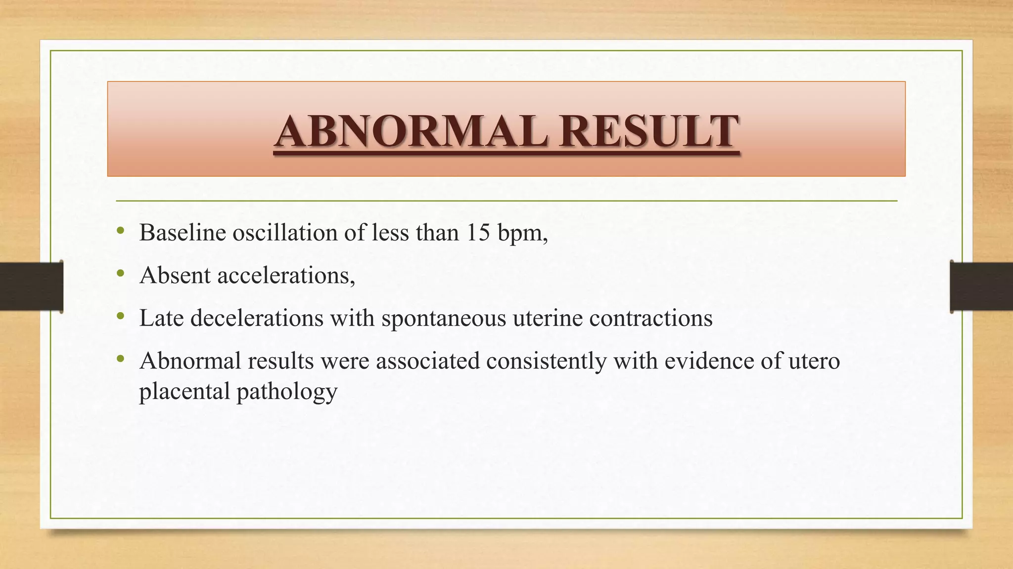
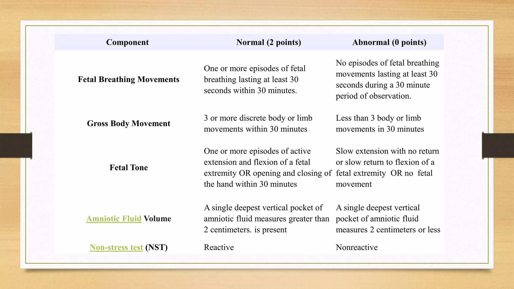
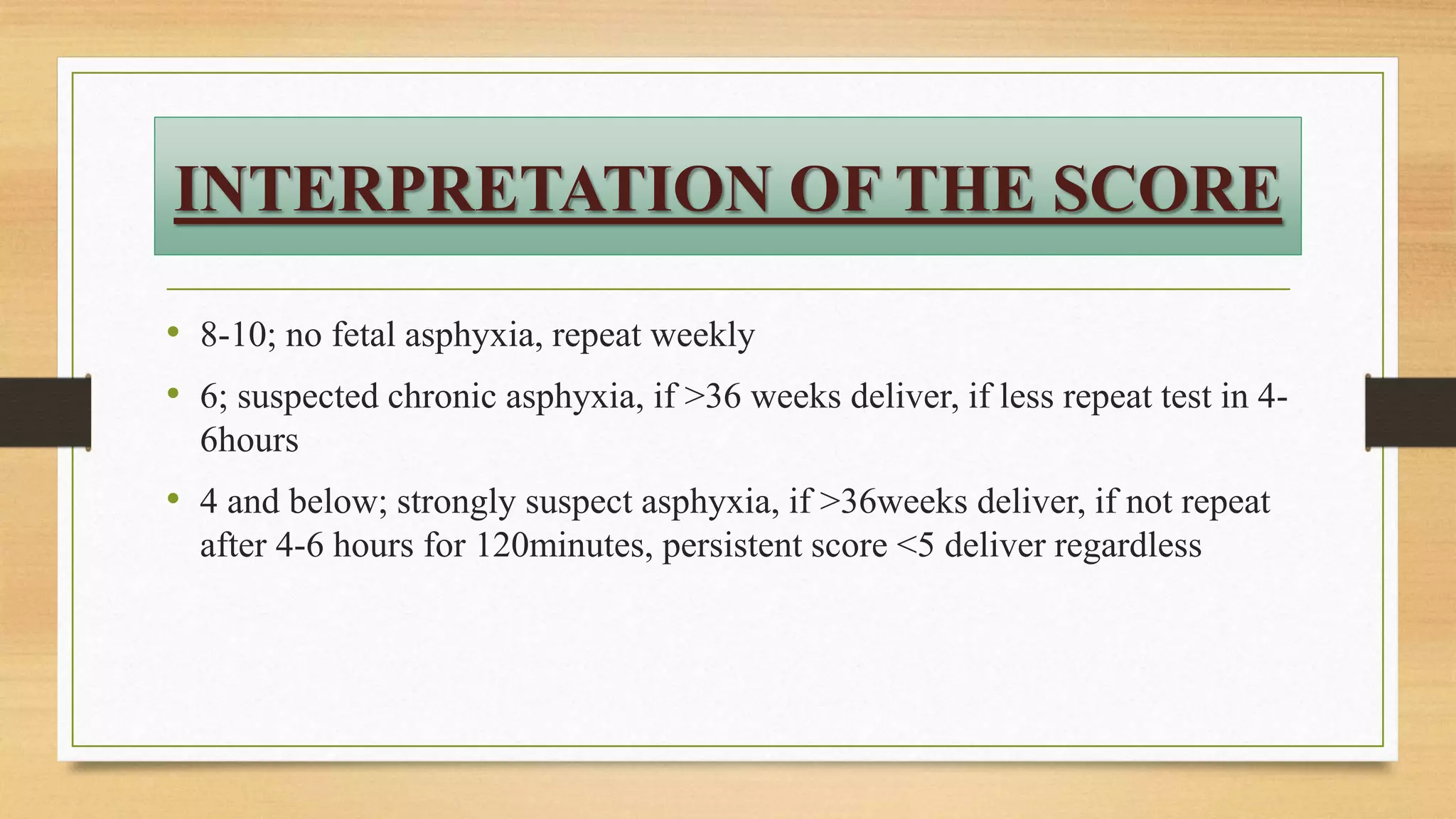
METRO COLLEGE OF
NURSING
BIOPHYSICAL PROFILE
SANDHYA KUMARI
Nursing Tutor (OBG)
MCON, Greater Noida
INTRODUCTION
• It is a technique employed to forecast fetal well-being focus on fetal biophysical findings
that include heart rate, movement, breathing, and amniotic fluid production
• It contains 5 parameters; non stress test (NST), fetal breathing, fetal movements, muscle
tone and amniotic fluid volume
• Modified Biophysical Profile consists of NST and ultrasono graphically determined
amniotic fluid index (AFI)
• Modified BPP is considered abnormal (non reassuring) when the NST is non-reactive
and/or the AFI is < 5.
• The BPP is most common in the third trimester
DEFINITION
• The biophysical profile is a test used to evaluate the well-being of the
fetus. The biophysical profile uses ultrasound and cardiotocography
(CTG), also known electronic fetal heart rate monitoring, to examine the
fetus. There are five components measured during the biophysical
examination. A score of 2 points is given for each component that meets
criteria as listed in the table below. The test is continued until all criteria
are met or 30 minutes have elapsed. The points are then added for a
possible maximum score of 10.
NONSTRESS TEST
• Currently, non stress test is the most widely used primary testing method
for assessment of fetal well-being
• It describes FHR acceleration in response to fetal movement as a sign of
fetal health
• Involved the use of Doppler-detected FHR acceleration coincident with
fetal movements perceived by the mother
• As hypoxia develops, these fetal heart rate accelerations diminish
NORMAL RESULT
• Two or more accelerations that peak at 15 bpm or more above baseline,
each lasting 15 seconds or more, and all occurring within 20 minutes of
beginning the test
• A 40-minute or longer tracing to account for fetal sleep cycles should be
performed before concluding that there was insufficient fetal reactivity
• 1 acceleration was just as reliable as 2 in predicting healthy fetal status
• Also accelerations with or without fetal movements may be accepted
ABNORMAL RESULT
• Baseline oscillation of less than 15 bpm,
• Absent accelerations,
• Late decelerations with spontaneous uterine contractions
• Abnormal results were associated consistently with evidence of utero
placental pathology
EXAMPLE
• IUGR
• Oligohydramnios
• Meconium
• Interval between testing set at 7 days; but more frequent testing is
advocated for women with post-term pregnancy, multifetal gestation, type
1 diabetes mellitus, IUGR, or gestational hypertension
• In these circumstances, twice-weekly tests, with additional testing is
advised
FETAL MOVEMENTS
• Passive unstimulated fetal activity commences as early as 7 weeks’
gestation and becomes more sophisticated and coordinated by the end of
pregnancy
• Between 20 and 30 weeks, general body movements become organized,
and by 36 weeks behavioural states are established in most normal
foetuses
Four fetal behavioural states described
• State 1F is a quiescent state; quiet sleep, with a narrow oscillatory
bandwidth of the FHR
• State 2F includes frequent gross body movements, continuous eye
movements, and wider oscillation of the FHR. It is analogous to REM or
active sleep in the neonate
• State 3F includes continuous eye movements in the absence of body
movements and no heart rate accelerations
• State 4F is one of vigorous body movement with continuous eye
movements and heart rate accelerations. This state corresponds to the
awake state in newborns
CONTINUE
• State 4F is one of vigorous body movement with continuous eye
movements and heart rate accelerations. This state corresponds to the
awake state in newborns
Fetuses spend most of their time in states 1F and 2F (>75% at 38 weeks)
Mean length of the quiet or inactive state for term fetuses was 23 minutes
(up to 75min)
Amniotic fluid volume is another important determinant of fetal activity
Perception of 10 fetal movements in up to 2 hours is considered normal.
FETAL BREATHING
• The first are gasps or sighs, which occurred at a frequency of 1 to 4 per minute
• The second, irregular bursts of breathing, occurred at rates up to 240 cycles per minute
• These latter rapid respiratory movements were associated with REM sleep
• Diurnal variation, because breathing substantively diminishes during the night
• But, increases somewhat following maternal meals
• Total absence of breathing was observed in some normal fetuses for up to 122 minutes,
indicating fetal evaluation to diagnose absent respiratory motion may require long periods
of observation.
AMNIOTIC FLUID VOLUME
• Decreased utero placental perfusion may lead to diminished fetal renal
blood flow, decreased urine production, and ultimately, oligohydramnios
• Amniotic fluid index < 5 cm or a maximum deepest vertical pocket < 2 cm
are acceptable criteria for diagnosis of oligohydramnios
• Normal; ≥ 1 pocket measuring 2 cm in two perpendicular planes (2 × 2 cm
pocket).
FETAL MUSCLE TONE
• Normal; ≥ 1 episode of extension (limb or trunk) with return of
flexion.
• The ability to flex and extend an arm or leg, measured by
counting quick, jerky movements.
Component Normal (2 points) Abnormal (0 points)
Fetal Breathing Movements
One or more episodes of fetal
breathing lasting at least 30
seconds within 30 minutes.
No episodes of fetal breathing
movements lasting at least 30
seconds during a 30 minute
period of observation.
Gross Body Movement
3 or more discrete body or limb
movements within 30 minutes
Less than 3 body or limb
movements in 30 minutes
Fetal Tone
One or more episodes of active
extension and flexion of a fetal
extremity OR opening and closing of
the hand within 30 minutes
Slow extension with no return
or slow return to flexion of a
fetal extremity OR no fetal
movement
Amniotic Fluid Volume
A single deepest vertical pocket of
amniotic fluid measures greater than
2 centimeters. is present
A single deepest vertical
pocket of amniotic fluid
measures 2 centimeters or less
Non-stress test (NST) Reactive Nonreactive
CONTINUE
• Amniotic Fluid Volume: Measured as the vertical measurement , in centimeters,
of the single deepest pocket of amniotic fluid with a transverse measurement of 1
cm or more wide without fetal small parts or umbilical cord
• Reactive: Two or more fetal heart rate accelerations that peak (but do not
necessarily remain) at least 15 beats per minute above the baseline and last at
least 15 seconds from baseline to baseline during 20 minutes of observation
• Nonreactive: Less than two accelerations of fetal heart rate as described above
after 40 minutes of observation
INTERPRETATION OF THE SCORE
• 8-10; no fetal asphyxia, repeat weekly
• 6; suspected chronic asphyxia, if >36 weeks deliver, if less repeat test in 4-
6hours
• 4 and below; strongly suspect asphyxia, if >36weeks deliver, if not repeat
after 4-6 hours for 120minutes, persistent score <5 deliver regardless
CONTRACTION STRESS TEST
• Formerly known as oxytocin challenge test
• Intravenous diluted oxytocin was used to stimulate contractions, and the FHR
response was recorded
• The criterion for a positive test result, that is, an abnormal result, was uniform
repetitive late fetal heart rate decelerations (which could be the result of utero
placental insufficiency)
• Nipple stimulation to induce uterine contractions is usually successful for
contraction stress testing
• 2-minute nipple stimulation ideally will induce a pattern of 3 contractions per 10
minutes, if not after 5 minutes, retry, if unsuccessful, diluted oxytocin.
ULTRASONOGRAPHY
• Low power sound: 100 mW
• Frequency:>20,000/cycle
Uses:
• Diagnose pregnancy at 6 weeks
• Locate placenta
• Diagnose fetal abnormalities
• Establish fetal sex
CONTINUE
• Discover problems
• Oligo/polyhydramnios
• Ectopic pregnancy
• Miscarriage
• Placenta previa
• PROM
• Down’s syndrome, NTDs, diaphragmatic hernia
CONTINUE
• Predict maturity of fetus
• Crown to rump length For dating, accuracy is
• +/- 3days (7-10 weeks)
• +/- 5days(10-14wks)
• Bi parietal diameter(most reliable at 12-20wks)
• +/- 5-7 days(16 weeks)
• +/- 3weeks >28 weeks
CONTINUE
• Bi parietal diameter (most reliable at 12-20wks)
• +/- 5-7 days (16 weeks)
• +/- 3weeks >28 weeks
• Cephalic index=BPD/occipito frontal diameter
• Normal is 0.74-0.83
CONTINUE
• Head
• Circumference=(BPD+OFD)
• Used to determine:
• Gestational age
• IUGR
• Microcephaly
CONTINUE
• Abdominal
• Circumference
• Used from 16 weeks to term
• To determine gestational age
• Femur length
• Obtained as early as 10 weeks
NURSE’S RESPONSIBILITY
• Full bladder
• Draping
• Towel roll under her right buttock
• No risk to fetus from procedure
MODIFIED BIOPHYSICAL PROFILE
• NST +AFI:
• Abnormal when one / both is abnormal
• Time -20 minutes
• Procedure:
• Standard NST
• No acceleration within 5 min
• 1-2 sec stimulation (upto 3 times)
• Normal :2 acceleration within 10 min
GUIDELINES
• If both normal: weekly fetal surveilance
• If both abnormal:
• >36 weeks: delivery
• <36 weeks: doppler, BPP, CST
• If AFI less: search for placental insufficiency
• If NST nonreactive: doppler, CST, BPP
CT SCAN
• Better to be avoided in pregnancy
MRI SCAN
• Obstetric applications
• Known/suspected Hydatidiform mole
• Placenta previa
• Fetal anomalies
• IUGR
CONCLUSION
• Antepartum management of high-risk pregnancies involves a balance of
risks. Early delivery to minimize the morbidity and mortality associated
with the high-risk condition may lead to severe morbidity or even
mortality as a result of prematurity complications. Fetal surveillance tests
have been developed to assess well-being at the time of the test and the
statistical likelihood of future fetal well-being during a specified interval
of time. In the USA the NST is widely used for primary surveillance of
fetal well-being in high-risk pregnancies
Biophysical profile

Biophysical profile

  • 1.
    METRO COLLEGE OF NURSING BIOPHYSICALPROFILE SANDHYA KUMARI Nursing Tutor (OBG) MCON, Greater Noida
  • 2.
    INTRODUCTION • It isa technique employed to forecast fetal well-being focus on fetal biophysical findings that include heart rate, movement, breathing, and amniotic fluid production • It contains 5 parameters; non stress test (NST), fetal breathing, fetal movements, muscle tone and amniotic fluid volume • Modified Biophysical Profile consists of NST and ultrasono graphically determined amniotic fluid index (AFI) • Modified BPP is considered abnormal (non reassuring) when the NST is non-reactive and/or the AFI is < 5. • The BPP is most common in the third trimester
  • 3.
    DEFINITION • The biophysicalprofile is a test used to evaluate the well-being of the fetus. The biophysical profile uses ultrasound and cardiotocography (CTG), also known electronic fetal heart rate monitoring, to examine the fetus. There are five components measured during the biophysical examination. A score of 2 points is given for each component that meets criteria as listed in the table below. The test is continued until all criteria are met or 30 minutes have elapsed. The points are then added for a possible maximum score of 10.
  • 4.
    NONSTRESS TEST • Currently,non stress test is the most widely used primary testing method for assessment of fetal well-being • It describes FHR acceleration in response to fetal movement as a sign of fetal health • Involved the use of Doppler-detected FHR acceleration coincident with fetal movements perceived by the mother • As hypoxia develops, these fetal heart rate accelerations diminish
  • 5.
    NORMAL RESULT • Twoor more accelerations that peak at 15 bpm or more above baseline, each lasting 15 seconds or more, and all occurring within 20 minutes of beginning the test • A 40-minute or longer tracing to account for fetal sleep cycles should be performed before concluding that there was insufficient fetal reactivity • 1 acceleration was just as reliable as 2 in predicting healthy fetal status • Also accelerations with or without fetal movements may be accepted
  • 6.
    ABNORMAL RESULT • Baselineoscillation of less than 15 bpm, • Absent accelerations, • Late decelerations with spontaneous uterine contractions • Abnormal results were associated consistently with evidence of utero placental pathology
  • 7.
    EXAMPLE • IUGR • Oligohydramnios •Meconium • Interval between testing set at 7 days; but more frequent testing is advocated for women with post-term pregnancy, multifetal gestation, type 1 diabetes mellitus, IUGR, or gestational hypertension • In these circumstances, twice-weekly tests, with additional testing is advised
  • 8.
    FETAL MOVEMENTS • Passiveunstimulated fetal activity commences as early as 7 weeks’ gestation and becomes more sophisticated and coordinated by the end of pregnancy • Between 20 and 30 weeks, general body movements become organized, and by 36 weeks behavioural states are established in most normal foetuses
  • 9.
    Four fetal behaviouralstates described • State 1F is a quiescent state; quiet sleep, with a narrow oscillatory bandwidth of the FHR • State 2F includes frequent gross body movements, continuous eye movements, and wider oscillation of the FHR. It is analogous to REM or active sleep in the neonate • State 3F includes continuous eye movements in the absence of body movements and no heart rate accelerations • State 4F is one of vigorous body movement with continuous eye movements and heart rate accelerations. This state corresponds to the awake state in newborns
  • 10.
    CONTINUE • State 4Fis one of vigorous body movement with continuous eye movements and heart rate accelerations. This state corresponds to the awake state in newborns Fetuses spend most of their time in states 1F and 2F (>75% at 38 weeks) Mean length of the quiet or inactive state for term fetuses was 23 minutes (up to 75min) Amniotic fluid volume is another important determinant of fetal activity Perception of 10 fetal movements in up to 2 hours is considered normal.
  • 11.
    FETAL BREATHING • Thefirst are gasps or sighs, which occurred at a frequency of 1 to 4 per minute • The second, irregular bursts of breathing, occurred at rates up to 240 cycles per minute • These latter rapid respiratory movements were associated with REM sleep • Diurnal variation, because breathing substantively diminishes during the night • But, increases somewhat following maternal meals • Total absence of breathing was observed in some normal fetuses for up to 122 minutes, indicating fetal evaluation to diagnose absent respiratory motion may require long periods of observation.
  • 12.
    AMNIOTIC FLUID VOLUME •Decreased utero placental perfusion may lead to diminished fetal renal blood flow, decreased urine production, and ultimately, oligohydramnios • Amniotic fluid index < 5 cm or a maximum deepest vertical pocket < 2 cm are acceptable criteria for diagnosis of oligohydramnios • Normal; ≥ 1 pocket measuring 2 cm in two perpendicular planes (2 × 2 cm pocket).
  • 13.
    FETAL MUSCLE TONE •Normal; ≥ 1 episode of extension (limb or trunk) with return of flexion. • The ability to flex and extend an arm or leg, measured by counting quick, jerky movements.
  • 14.
    Component Normal (2points) Abnormal (0 points) Fetal Breathing Movements One or more episodes of fetal breathing lasting at least 30 seconds within 30 minutes. No episodes of fetal breathing movements lasting at least 30 seconds during a 30 minute period of observation. Gross Body Movement 3 or more discrete body or limb movements within 30 minutes Less than 3 body or limb movements in 30 minutes Fetal Tone One or more episodes of active extension and flexion of a fetal extremity OR opening and closing of the hand within 30 minutes Slow extension with no return or slow return to flexion of a fetal extremity OR no fetal movement Amniotic Fluid Volume A single deepest vertical pocket of amniotic fluid measures greater than 2 centimeters. is present A single deepest vertical pocket of amniotic fluid measures 2 centimeters or less Non-stress test (NST) Reactive Nonreactive
  • 15.
    CONTINUE • Amniotic FluidVolume: Measured as the vertical measurement , in centimeters, of the single deepest pocket of amniotic fluid with a transverse measurement of 1 cm or more wide without fetal small parts or umbilical cord • Reactive: Two or more fetal heart rate accelerations that peak (but do not necessarily remain) at least 15 beats per minute above the baseline and last at least 15 seconds from baseline to baseline during 20 minutes of observation • Nonreactive: Less than two accelerations of fetal heart rate as described above after 40 minutes of observation
  • 16.
    INTERPRETATION OF THESCORE • 8-10; no fetal asphyxia, repeat weekly • 6; suspected chronic asphyxia, if >36 weeks deliver, if less repeat test in 4- 6hours • 4 and below; strongly suspect asphyxia, if >36weeks deliver, if not repeat after 4-6 hours for 120minutes, persistent score <5 deliver regardless
  • 17.
    CONTRACTION STRESS TEST •Formerly known as oxytocin challenge test • Intravenous diluted oxytocin was used to stimulate contractions, and the FHR response was recorded • The criterion for a positive test result, that is, an abnormal result, was uniform repetitive late fetal heart rate decelerations (which could be the result of utero placental insufficiency) • Nipple stimulation to induce uterine contractions is usually successful for contraction stress testing • 2-minute nipple stimulation ideally will induce a pattern of 3 contractions per 10 minutes, if not after 5 minutes, retry, if unsuccessful, diluted oxytocin.
  • 18.
    ULTRASONOGRAPHY • Low powersound: 100 mW • Frequency:>20,000/cycle Uses: • Diagnose pregnancy at 6 weeks • Locate placenta • Diagnose fetal abnormalities • Establish fetal sex
  • 19.
    CONTINUE • Discover problems •Oligo/polyhydramnios • Ectopic pregnancy • Miscarriage • Placenta previa • PROM • Down’s syndrome, NTDs, diaphragmatic hernia
  • 20.
    CONTINUE • Predict maturityof fetus • Crown to rump length For dating, accuracy is • +/- 3days (7-10 weeks) • +/- 5days(10-14wks) • Bi parietal diameter(most reliable at 12-20wks) • +/- 5-7 days(16 weeks) • +/- 3weeks >28 weeks
  • 21.
    CONTINUE • Bi parietaldiameter (most reliable at 12-20wks) • +/- 5-7 days (16 weeks) • +/- 3weeks >28 weeks • Cephalic index=BPD/occipito frontal diameter • Normal is 0.74-0.83
  • 22.
    CONTINUE • Head • Circumference=(BPD+OFD) •Used to determine: • Gestational age • IUGR • Microcephaly
  • 23.
    CONTINUE • Abdominal • Circumference •Used from 16 weeks to term • To determine gestational age • Femur length • Obtained as early as 10 weeks
  • 24.
    NURSE’S RESPONSIBILITY • Fullbladder • Draping • Towel roll under her right buttock • No risk to fetus from procedure
  • 25.
    MODIFIED BIOPHYSICAL PROFILE •NST +AFI: • Abnormal when one / both is abnormal • Time -20 minutes • Procedure: • Standard NST • No acceleration within 5 min • 1-2 sec stimulation (upto 3 times) • Normal :2 acceleration within 10 min
  • 26.
    GUIDELINES • If bothnormal: weekly fetal surveilance • If both abnormal: • >36 weeks: delivery • <36 weeks: doppler, BPP, CST • If AFI less: search for placental insufficiency • If NST nonreactive: doppler, CST, BPP
  • 27.
    CT SCAN • Betterto be avoided in pregnancy MRI SCAN • Obstetric applications • Known/suspected Hydatidiform mole • Placenta previa • Fetal anomalies • IUGR
  • 28.
    CONCLUSION • Antepartum managementof high-risk pregnancies involves a balance of risks. Early delivery to minimize the morbidity and mortality associated with the high-risk condition may lead to severe morbidity or even mortality as a result of prematurity complications. Fetal surveillance tests have been developed to assess well-being at the time of the test and the statistical likelihood of future fetal well-being during a specified interval of time. In the USA the NST is widely used for primary surveillance of fetal well-being in high-risk pregnancies