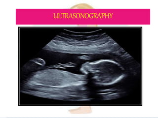
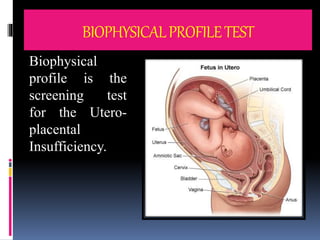
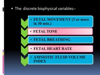
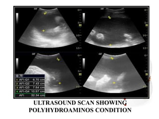
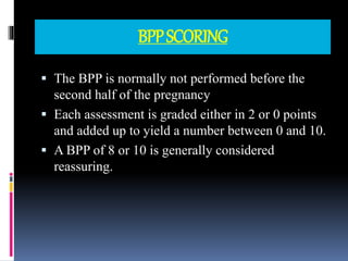
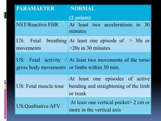
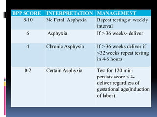
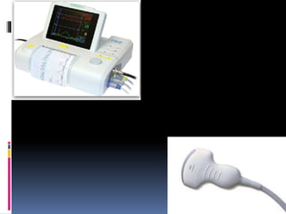
BIOPHYSICAL PROFILE
RELATED TO
MATERNAL
AND
FETAL HEALTH
PRESENTED BY-
Ms. ITISHA ROSE PRASAD
Nursing Tutor
GOV CON, Kanpur
 Pregnancy is a profound and a life changing
event, during this time the mother has to
adapt physically, socially and
psychologically to the forth coming birth of
the baby. Majority 80% of the foetal death
occurs in the ante partum period.
CAUSESOFFETALDEATH
 Thus screening of a mother and baby is a
major part of care for all the pregnancies
so that the treatment can be available at
a stage where the outcome is possible.
ULTRASONOGRAPHY
 Ultrasonographic examination of the fetus in the
early pregnancy (10-14 weeks) can detect the fetal
anomalies.
 Ultrasound is the sound waves beyond the audible
range of frequency greater than 2MHz
 The commonly used frequency range in obstetrics
is 3-5 MHz for abdominal transducer and for
vaginal transducer is 5-7MHz
FETAL/MATERNAL UTEROPLACENTAL
 Diagnosis of pregnancy
 Diagnosis of multiple
pregnancy
 To confirm cardiac
activity and fetal
viability
 Assessment of growth
 Diagnosis of ectopic
pregnancy
 During fetoscopy,
cordocentesis,
amniocentesis.
 Localization of the
placenta
 Diagnosis of
abruption placenta
 Diagnosis of molar
pregnancy
 Diagnosis of uterine
malformations.
 Assessment of liquor
volume.
 Chronicity in multiple
pregnancy
TANSABDOMINALULTRASOUNDSCAN
TRANSVAGINALULTRASOUNDSCAN-
ULTRASOUNDIMAGESINCLINCALPRACTICE
 B-mode–two dimensional (2-D) images
 M-mode is used to study the moving organs
e.g., fetal heart. This results in a wavy pattern
in the presence of motion
 Doppler ultrasound and Pulse Wave
ultrasound- It is primarily used to demonstrate
the presence, direction and velocity of blood
flow.
 Three Dimensional (3D) images.
STANDARAD ULTRASOUND
IMAGES
B mode- 2D images
2D and 3D Images Compared
ULTRSOUND MARKERS FOR FETALAND
MATERNALANOMALIES
 Nuchal Translucency- Increased fetal nuchal
skin thickness (in first trimester) > 3 mm by
TVS is a strong marker for chromosomal
anomalies ( trisomy 21, 18, 13, triploidy and
Turners syndrome)
Ultrasound
images
depicting
Nuchal
translucency
 Multiple Pregnancy- identification of
two gestational sacs indicates twin birth
in 52-63% of cases
 Anembryonic Pregnancy (blighted ovum) –
pregnancy in which the embryo never
develops or if develops it gets reabsorbed but
the gestational sac normal appearing.
 Ectopic pregnancy- TVS can detect 90% of
tubal ectopic Pregnancy. Color Doppler can
identify the placental shape (ring of fire
pattern) and enhanced blood flow pattern
outside the uterine cavity. Presence of
echogenic fluid in the pouch of Douglas is
suggestive of ectopic pregnancy.
 Hyaditiform mole- snowstorm appearance in
the scan is suggestive of hyaditiform mole
Safety of ultrasound
 Ultrasound is the essential tool in the
management of almost every pregnancy.The
effects of ultrasound on tissues are-
temperature elevation, formation of
microtubules, cavitations. However till date
there is no evidence about the harmful
effects of ultrasound still it should be
judiciously used especially the Doppler mode
and its casual use should be avoided.
BIOPHYSICALPROFILETEST
Biophysical
profile is the
screening test
for the Utero-
placental
Insufficiency.
 The discrete biophysical variables:-
• FETAL MOVEMENT (3 or more
in 30 min.)
• FETAL TONE
• FETAL BREATHING
• FETAL HEART RATE
• AMNIOTIC FLUID VOLUME
INDEX
 FETAL MOVEMENTS- There should be three
or more gross body movements in 30 minutes of
observation.
 FETAL TONE: There should be at least one
episode of motion of a limb from a position of
flexion to extension and rapid return to its
position.
 FETAL BREATHING MOVEMENTS- . There
should be at least 30 seconds of sustained fetal
breathing movements in 30 minutes of
observation
 FETAL REACTIVITY (fetal heart rate)- There
should be two or more fetal heart accelerations of
at least 15 beats per minute, within 40 minutes of
observation. These should last at least 15 seconds
and be associated with fetal movement.
AMNIOTIC FLUID INDEX (AFI)
 It is a quantitative estimate of amniotic fluid and
an indicator of fetal well being.
 Maternal abdomen is divided into quadrants taking
the umbilicus, symphysis pubis, and the fundus as
the reference points.
 Amniotic fluid index (AFI) is the sum of vertical
pockets from four quadrants of uterine cavity.
 With the ultrasound the largest vertical pocket in
each quadrant is measured.
 RESULT-
 An AFI between 8-18 cm is considered
normal
 An AFI less than 5-6 cm is considered as
oligohydroaminos
 AFI greater than or between 20-24 cm is
considered as polyhydroamnios.
ULTRASOUND SCAN SHOWING
POLYHYDROAMINOS CONDITION
BPPSCORING
 The BPP is normally not performed before the
second half of the pregnancy
 Each assessment is graded either in 2 or 0 points
and added up to yield a number between 0 and 10.
 A BPP of 8 or 10 is generally considered
reassuring.
PARAMAETER NORMAL
(2 points)
NST/Reactive FHR At least two accelerations in 30
minutes
US: Fetal breathing
movements
At least one episode of > 30s or
>20s in 30 minutes
US: Fetal activity /
gross body movements
At least two movements of the torso
or limbs within 30 min.
US: Fetal muscle tone
At least one episodes of active
bending and straightening of the limb
or trunk
US:Qualitative AFV
At least one vertical pocket> 2 cm or
more in the vertical axis
BPP SCORE INTERPRETATION MANAGEMENT
8-10 No Fetal Asphyxia Repeat testing at weekly
interval
6 Asphyxia If > 36 weeks- deliver
4 Chronic Asphyxia If > 36 weeks deliver if
<32 weeks repeat testing
in 4-6 hours
0-2 Certain Asphyxia Test for 120 min-
persists score < 4-
deliver regardless of
gestational age(induction
of labor)
FETALMOVEMENTCOUNT
The fetal movement count should be performed
daily starting at 28 weeks of pregnancy.
 Cardif ‘count 10’ formula:
 The patients count fetal movement starting at
9 am.
 The counting comes to an end as soon as the
10 movements are perceived.
Patient is instructed to report the physician if-
Less than 10 movements occur during 12
hours on the 2 successive days.
 No movements are perceived even after 12
hours in a single day.
Cardif ‘count 10’formula:
 Daily Fetal Movement Count(DFMC):
 Three counts each of one hour duration
(morning ,noon and evening ) are
recommended.
 The total counts multiplied by 4 gives daily
(12 hours) fetal movements counts.
 Patient reports to the physician if-
 There is diminished number of “kicks” to
less than 10 in 12 hour
 Or less than 3 in each hour, this is
indicative of fetal compromise
 Maternal perception of fetal movement is
reduced with-
 Fetal sleep,
 Fetal anomalies(CNS)
 Anterior placenta
 Hydramions
 Obesity
 Drugs(narcotics)
 Hypoxia.
 Maternal hypoglycemia is associated with
increased fetal movements.
NONSTRESSTEST
 A test that monitors the heart rate in response to
the fetal movement in order to assess the integrity
of fetal Central Nervous System and Cardio
Vascular System.
 This screening test is valuable to identify the fetal
wellness rather than the fetal illness.
PURPOSES
 To assess the fetal ability to cope with
continuation of a high risk pregnancy.
 To determine the projected ability of a fetus to
withstand the stress of labour.
 Previous cessarian section, placenta previa or
preterm labour.)
indications
 Maternal Indication-
 Post dated Pregnancy
 Rh Sensitization
 Maternal Age 35 or more
 Chronic Renal Disease
 Hypertension
 Sickle Cell Disease
 Diabetes
 Premature Rupture of Membrane
 History of Still Birth
 Vaginal Bleeding
INDICATIONS
 Fetal Indication-
 Decreased Fetal Movement
 Intrauterine Growth Retardation
 Fetal evaluation after Amniocentesis.
 Oligohydroaminos/Polyhydroaminos.
ARTICLES
 ARTICLES-
 Electronic Fetal heart Monitor
 Ultrasound Transducer
 Tocotransducer
 Monitor Strip
 Ultrasound Gel
 Belts to hold the transducers in place.
POSITION OF THE MOTHER
PREPARATIONOFTHEMOTHER
 Explain the procedure to the patient.
 Make sure that woman has eaten food and ask the
mother to empty her bladder.
 Place the mother in the semi fowler’s position.
 Maintain privacy.
PROCEEDURE
 Perform an abdominal palpation(Leopod’s
Maneuver)
 Confirm the presence of fetal heart tones with
fetoscope and note the area of maximum intensity.
 Apply the gel to the ultrasound transducer.
 Place the ultrasound transducer on the fetal back.
Move the transducer until clear, audible fetal heart
tones are heard and the signal lights are flashing
steadily.
 Secure the device in place with a belt.
CONTI...
 Run the Monitor and evaluate the quality of tracing
to determine if it is adequate for interpretation.
 Ask the mother to press the hand button every time
she feels the fetal movement.
 Run the monitor and obtain trcaing for every 20
minutes.
 On completion, put off the monitor and take out the
strip of paper.
 Remove the abdominal straps and wipe off the gel
from the abdominal transducer.
 Make the woman comfortable and give relevant
instructions.
RESULTS
 Reactive Non Stress Test (normal/negative)
For the test to be negative the result requires two
or more FHR accelerations of atleast 15 beats per
minutes, lasting at least 15 seconds from the
beginning of the acceleration to the end, in
association with fetal movement during a 20
minutes period.
RESULTS
 Non reactive Non Stress test (abnormal)
No acceleration or aceleration of less than
15 beats per minute or lasting less than 15
seconds in duration occur during a 40
minutes observation
VIBROACOUSTICSTIMULATION(VAS)
 It is the application of a vibrator sound stimulus to
the abdomen of a pregnant woman to induce FHR
(fetal heart rate) acceleration.
 It is used during a Non Stress Test. It is used to
change the fetal sleep state from quite (non-
REM) to active (REM)
 A reactive NST after VAS indicates a reactive
fetus.
CONTRACTIONSTRESSTEST
 A Contraction Stress Test is performed
during pregnancy to verify whether or
not the unborn baby’s heart is strong
enough to withstand labour.
indication
Intrauterine growth Restriction
Post maturity
Hypertensive Disorders of Pregnancy
Diabetes
contraindication
Hydroaminos
Previous history of caesarean section
Complications likely to produce preterm
labour
APH
Multiple Pregnancy
Positionofthemother
 The mother should be either in
semi-fowlers or
 left lateral position to prevent supine
hypotension.
PREPARATIONOFTHEMOTHER-
 Explain the procedure to the mother.
 Instruct the mother to empty the bladder to
promote comfort and avoid disruption.
 Place the mother in the comfortable position.
 Monitor the vital signs (esp. BP) for baseline
recording.
Methodsofcontraction
Nipple
Stimulation
Test
Oxytocin
Challenge
Test
NIPPLESTIMULATIONTEST
 This test involves stimulation of the nipples
(by rubbing, gently pulling), which causes the
posterior pituitary to release the hormone
oxytocin, which in turn, causes contractions.
 This method avoids the risks, discomfort and
expenses associated with intravenous infusion
of oxytocin.
Conti..
 At the beginning of the test, warm wash clothes
are applied to the breasts. A lubricating jelly is
applied to prevent soreness.
 Stimulation is initially unilateral.
 If the contractions are inadequate (fewer than 3
contractions in the first 10 minutes), then the
woman simultaneously stimulates both the nipples
for another 10 minutes.
 If still inadequate, intravenous oxytocin is used.
OXYTOCINCHALLENGETEST
 The OCT involves application of the fetal monitor
to record fetal heart rate and contraction activity.
 A dilute of IV solution of oxytocin is
administered to the mother until contractions are
occurring at a frequency of at least 3 in 10 minute
periods and lasting at least 30 seconds. When
sufficient information is obtained to evaluate the
test, the medication is turned off.
 Both the monitoring and the intravenous solution
without oxytocin in it are continued until the
contractions have diminished to the baseline
results
 Negative Contraction Stress Test-
A negative CST is the one in which no late
decelerations occur with contractions as
frequent as 3 in 10 minutes period. It is
associated with good fetal outcome.
 Positive Result-
A positive CST is one which there have
been repeated late decelerations of the fetal
heart rate patterns during the test. It is
associated with increased incidence of IUD,
fetal distress in labor and low APGAR
score.
CARDIOTOCOGRAPHY
 Cardiotocography is technical means of
recording (-graphy) the fetal heart rate (-
cardio) and the uterine contractions (-taco)
during pregnancy, typically in the third
trimester.
 It is a non invasive procedure whereby an
ultrasound transducer is strapped to the
abdomen at a point where the FHS heard is
maximum intensity.
EXTERNALFETALMONITOR
 It can be used for continuous or intermittent
monitoring. The fetal heart rate and the
activity of the uterine muscle are detected by
two transducers placed on the mother’s
abdomen (one above the fetal heart, to
monitor heart rate and the other at
the fudus of the uterus to measure frequency
of contractions).
 The heart ultrasonic sensor, similar to a
Doppler fetal monitor, detects motion of the
fetal heart rate. The pressure sensitive
contraction transducer called a
tocodynometer (toco) measures the tension
of the maternal abdominal wall.
InternalCardioitocography
 It uses an electronic transducer connected
directly to the fetal scalp. A wire electrode is
attached to the fetal scalp through the cervical
opening and is connected to the monitor. This
type of electrode is sometimes called a spiral
or scalp electrode
 The interpretation of the CTG requires description
of the following-
Uterine activity
Baseline fetal heart rate
Baseline FHR variability
Presence of accelerations
Periodic or episodic decelerations
 Uterine Activity-
Frequency
Duration
Intensity
Resting tone
Interval
Result-
Normal- contractions less than or equal to 5 in 10
minutes , averaged over a 30 min. .
Tachysystole- more than 5 contractions in 10 min,
averaged over a 30 min.
Baseline Fetal Heart Rate-
Normally FHR ranges between 120 beats per min. and
160 beats per min
Baseline FHR Variability-
Minute variations in the length of each beat due to
electrical activity in fetal heart, is baseline variability.This
causes the tracing to be jagged rather than a smooth line.
Periodic or episodic decelerations-
fetoscopy
 Fetoscopy is an endoscopic procedure during
pregnancy to allow access to the fetus, the
amniotic cavity, the umbilical cord and the
fetal side of the placenta. It uses an instrument
called fetoscope to evaluate or treat fetus
during pregnancy
Types
 External Fetoscope- it is ususally helpful to
detect the fetal heart sound after 18 weeks of
gestation.
 INTERNAL FETOSCOPE-It is a fibre optic
endoscope, which is inserted into the uterus either
transabdominally or transcervically to visualize
the fetus
RISK-
 Infection to fetus/mother
 Premature rupture of amniotic membranes
 Fetal death
 Miscarriage
 Excessive Bleeding
MAGNETICRESONANCEIMAGING
FETALINDICATIONS
FETALANATOMICAL
SURVEY
FETAL BIOMETRY
FETAL ESTIMATION
EVALUATION OF
COMPLEX
ABNORMALITIES
MATERNALINDICATIONS
ANGIOGRAPHY
DETECTION OF
THROMBOSIS
EVALUATION OF
MATERNAL TUMORS
CONTRAINDICATIONS
 Internal Cardiac Pacemaker
 Implanted Defibrillator
 Implants or other implants in the body
REFERENCES
 Bhaskar Nima. Midwifery and Obstetrical Nursing. 2nd
Edition. Banglore: Emmess Medical Publisher; 2015 Pp-
147-156
 Dutta DC. Textbook of obstetrics. 7th Edition. New Delhi:
jaypee brothers Medical Publishers (P) ltd; 2013. Pp-108-
110, 501,644-650
 Jacob Annamma. A Comprehensive textbook of Midwifery
and Gynecological Nursing. 4th Edition. ;2015. Pp- 180
 Manocha Sneh Lata. Procedure and Practises in Midwifery.
1st Edition. New Delhi: Kumar Publishing House; 2011 Pp-
301-303
 www.ncbi.nlm.nih.gov/pubmed/18253968
 www.webmd.com/baby/biophysical-profil-bpp#/
 http://emedicine.medscape.com/article/40554-overview#a2
 www.surgeryencyclopedia.com/Ce-Fi/fetoscopy.html
THANK YOU

Biophysical profile ppt.pptx

  • 1.
    BIOPHYSICAL PROFILE RELATED TO MATERNAL AND FETALHEALTH PRESENTED BY- Ms. ITISHA ROSE PRASAD Nursing Tutor GOV CON, Kanpur
  • 2.
     Pregnancy isa profound and a life changing event, during this time the mother has to adapt physically, socially and psychologically to the forth coming birth of the baby. Majority 80% of the foetal death occurs in the ante partum period.
  • 3.
  • 4.
     Thus screeningof a mother and baby is a major part of care for all the pregnancies so that the treatment can be available at a stage where the outcome is possible.
  • 5.
  • 6.
     Ultrasonographic examinationof the fetus in the early pregnancy (10-14 weeks) can detect the fetal anomalies.  Ultrasound is the sound waves beyond the audible range of frequency greater than 2MHz  The commonly used frequency range in obstetrics is 3-5 MHz for abdominal transducer and for vaginal transducer is 5-7MHz
  • 7.
    FETAL/MATERNAL UTEROPLACENTAL  Diagnosisof pregnancy  Diagnosis of multiple pregnancy  To confirm cardiac activity and fetal viability  Assessment of growth  Diagnosis of ectopic pregnancy  During fetoscopy, cordocentesis, amniocentesis.  Localization of the placenta  Diagnosis of abruption placenta  Diagnosis of molar pregnancy  Diagnosis of uterine malformations.  Assessment of liquor volume.  Chronicity in multiple pregnancy
  • 8.
  • 9.
  • 10.
    ULTRASOUNDIMAGESINCLINCALPRACTICE  B-mode–two dimensional(2-D) images  M-mode is used to study the moving organs e.g., fetal heart. This results in a wavy pattern in the presence of motion  Doppler ultrasound and Pulse Wave ultrasound- It is primarily used to demonstrate the presence, direction and velocity of blood flow.  Three Dimensional (3D) images.
  • 11.
  • 12.
    2D and 3DImages Compared
  • 15.
    ULTRSOUND MARKERS FORFETALAND MATERNALANOMALIES  Nuchal Translucency- Increased fetal nuchal skin thickness (in first trimester) > 3 mm by TVS is a strong marker for chromosomal anomalies ( trisomy 21, 18, 13, triploidy and Turners syndrome)
  • 16.
  • 17.
     Multiple Pregnancy-identification of two gestational sacs indicates twin birth in 52-63% of cases
  • 19.
     Anembryonic Pregnancy(blighted ovum) – pregnancy in which the embryo never develops or if develops it gets reabsorbed but the gestational sac normal appearing.
  • 21.
     Ectopic pregnancy-TVS can detect 90% of tubal ectopic Pregnancy. Color Doppler can identify the placental shape (ring of fire pattern) and enhanced blood flow pattern outside the uterine cavity. Presence of echogenic fluid in the pouch of Douglas is suggestive of ectopic pregnancy.
  • 22.
     Hyaditiform mole-snowstorm appearance in the scan is suggestive of hyaditiform mole
  • 23.
    Safety of ultrasound Ultrasound is the essential tool in the management of almost every pregnancy.The effects of ultrasound on tissues are- temperature elevation, formation of microtubules, cavitations. However till date there is no evidence about the harmful effects of ultrasound still it should be judiciously used especially the Doppler mode and its casual use should be avoided.
  • 24.
    BIOPHYSICALPROFILETEST Biophysical profile is the screeningtest for the Utero- placental Insufficiency.
  • 25.
     The discretebiophysical variables:- • FETAL MOVEMENT (3 or more in 30 min.) • FETAL TONE • FETAL BREATHING • FETAL HEART RATE • AMNIOTIC FLUID VOLUME INDEX
  • 26.
     FETAL MOVEMENTS-There should be three or more gross body movements in 30 minutes of observation.  FETAL TONE: There should be at least one episode of motion of a limb from a position of flexion to extension and rapid return to its position.
  • 27.
     FETAL BREATHINGMOVEMENTS- . There should be at least 30 seconds of sustained fetal breathing movements in 30 minutes of observation  FETAL REACTIVITY (fetal heart rate)- There should be two or more fetal heart accelerations of at least 15 beats per minute, within 40 minutes of observation. These should last at least 15 seconds and be associated with fetal movement.
  • 28.
    AMNIOTIC FLUID INDEX(AFI)  It is a quantitative estimate of amniotic fluid and an indicator of fetal well being.  Maternal abdomen is divided into quadrants taking the umbilicus, symphysis pubis, and the fundus as the reference points.
  • 29.
     Amniotic fluidindex (AFI) is the sum of vertical pockets from four quadrants of uterine cavity.  With the ultrasound the largest vertical pocket in each quadrant is measured.
  • 30.
     RESULT-  AnAFI between 8-18 cm is considered normal  An AFI less than 5-6 cm is considered as oligohydroaminos  AFI greater than or between 20-24 cm is considered as polyhydroamnios.
  • 31.
  • 32.
    BPPSCORING  The BPPis normally not performed before the second half of the pregnancy  Each assessment is graded either in 2 or 0 points and added up to yield a number between 0 and 10.  A BPP of 8 or 10 is generally considered reassuring.
  • 33.
    PARAMAETER NORMAL (2 points) NST/ReactiveFHR At least two accelerations in 30 minutes US: Fetal breathing movements At least one episode of > 30s or >20s in 30 minutes US: Fetal activity / gross body movements At least two movements of the torso or limbs within 30 min. US: Fetal muscle tone At least one episodes of active bending and straightening of the limb or trunk US:Qualitative AFV At least one vertical pocket> 2 cm or more in the vertical axis
  • 34.
    BPP SCORE INTERPRETATIONMANAGEMENT 8-10 No Fetal Asphyxia Repeat testing at weekly interval 6 Asphyxia If > 36 weeks- deliver 4 Chronic Asphyxia If > 36 weeks deliver if <32 weeks repeat testing in 4-6 hours 0-2 Certain Asphyxia Test for 120 min- persists score < 4- deliver regardless of gestational age(induction of labor)
  • 35.
  • 36.
    The fetal movementcount should be performed daily starting at 28 weeks of pregnancy.  Cardif ‘count 10’ formula:  The patients count fetal movement starting at 9 am.  The counting comes to an end as soon as the 10 movements are perceived. Patient is instructed to report the physician if- Less than 10 movements occur during 12 hours on the 2 successive days.  No movements are perceived even after 12 hours in a single day.
  • 37.
  • 38.
     Daily FetalMovement Count(DFMC):  Three counts each of one hour duration (morning ,noon and evening ) are recommended.  The total counts multiplied by 4 gives daily (12 hours) fetal movements counts.  Patient reports to the physician if-  There is diminished number of “kicks” to less than 10 in 12 hour  Or less than 3 in each hour, this is indicative of fetal compromise
  • 39.
     Maternal perceptionof fetal movement is reduced with-  Fetal sleep,  Fetal anomalies(CNS)  Anterior placenta  Hydramions  Obesity  Drugs(narcotics)  Hypoxia.  Maternal hypoglycemia is associated with increased fetal movements.
  • 40.
  • 41.
     A testthat monitors the heart rate in response to the fetal movement in order to assess the integrity of fetal Central Nervous System and Cardio Vascular System.  This screening test is valuable to identify the fetal wellness rather than the fetal illness.
  • 42.
    PURPOSES  To assessthe fetal ability to cope with continuation of a high risk pregnancy.  To determine the projected ability of a fetus to withstand the stress of labour.  Previous cessarian section, placenta previa or preterm labour.)
  • 43.
    indications  Maternal Indication- Post dated Pregnancy  Rh Sensitization  Maternal Age 35 or more  Chronic Renal Disease  Hypertension  Sickle Cell Disease  Diabetes  Premature Rupture of Membrane  History of Still Birth  Vaginal Bleeding
  • 44.
    INDICATIONS  Fetal Indication- Decreased Fetal Movement  Intrauterine Growth Retardation  Fetal evaluation after Amniocentesis.  Oligohydroaminos/Polyhydroaminos.
  • 45.
    ARTICLES  ARTICLES-  ElectronicFetal heart Monitor  Ultrasound Transducer  Tocotransducer  Monitor Strip  Ultrasound Gel  Belts to hold the transducers in place.
  • 47.
  • 48.
    PREPARATIONOFTHEMOTHER  Explain theprocedure to the patient.  Make sure that woman has eaten food and ask the mother to empty her bladder.  Place the mother in the semi fowler’s position.  Maintain privacy.
  • 49.
    PROCEEDURE  Perform anabdominal palpation(Leopod’s Maneuver)  Confirm the presence of fetal heart tones with fetoscope and note the area of maximum intensity.  Apply the gel to the ultrasound transducer.  Place the ultrasound transducer on the fetal back. Move the transducer until clear, audible fetal heart tones are heard and the signal lights are flashing steadily.  Secure the device in place with a belt.
  • 50.
    CONTI...  Run theMonitor and evaluate the quality of tracing to determine if it is adequate for interpretation.  Ask the mother to press the hand button every time she feels the fetal movement.  Run the monitor and obtain trcaing for every 20 minutes.  On completion, put off the monitor and take out the strip of paper.  Remove the abdominal straps and wipe off the gel from the abdominal transducer.  Make the woman comfortable and give relevant instructions.
  • 51.
    RESULTS  Reactive NonStress Test (normal/negative) For the test to be negative the result requires two or more FHR accelerations of atleast 15 beats per minutes, lasting at least 15 seconds from the beginning of the acceleration to the end, in association with fetal movement during a 20 minutes period.
  • 53.
    RESULTS  Non reactiveNon Stress test (abnormal) No acceleration or aceleration of less than 15 beats per minute or lasting less than 15 seconds in duration occur during a 40 minutes observation
  • 56.
  • 57.
     It isthe application of a vibrator sound stimulus to the abdomen of a pregnant woman to induce FHR (fetal heart rate) acceleration.  It is used during a Non Stress Test. It is used to change the fetal sleep state from quite (non- REM) to active (REM)  A reactive NST after VAS indicates a reactive fetus.
  • 58.
    CONTRACTIONSTRESSTEST  A ContractionStress Test is performed during pregnancy to verify whether or not the unborn baby’s heart is strong enough to withstand labour.
  • 59.
    indication Intrauterine growth Restriction Postmaturity Hypertensive Disorders of Pregnancy Diabetes
  • 60.
    contraindication Hydroaminos Previous history ofcaesarean section Complications likely to produce preterm labour APH Multiple Pregnancy
  • 61.
    Positionofthemother  The mothershould be either in semi-fowlers or  left lateral position to prevent supine hypotension.
  • 62.
    PREPARATIONOFTHEMOTHER-  Explain theprocedure to the mother.  Instruct the mother to empty the bladder to promote comfort and avoid disruption.  Place the mother in the comfortable position.  Monitor the vital signs (esp. BP) for baseline recording.
  • 63.
  • 64.
    NIPPLESTIMULATIONTEST  This testinvolves stimulation of the nipples (by rubbing, gently pulling), which causes the posterior pituitary to release the hormone oxytocin, which in turn, causes contractions.  This method avoids the risks, discomfort and expenses associated with intravenous infusion of oxytocin.
  • 65.
    Conti..  At thebeginning of the test, warm wash clothes are applied to the breasts. A lubricating jelly is applied to prevent soreness.  Stimulation is initially unilateral.  If the contractions are inadequate (fewer than 3 contractions in the first 10 minutes), then the woman simultaneously stimulates both the nipples for another 10 minutes.  If still inadequate, intravenous oxytocin is used.
  • 67.
    OXYTOCINCHALLENGETEST  The OCTinvolves application of the fetal monitor to record fetal heart rate and contraction activity.  A dilute of IV solution of oxytocin is administered to the mother until contractions are occurring at a frequency of at least 3 in 10 minute periods and lasting at least 30 seconds. When sufficient information is obtained to evaluate the test, the medication is turned off.  Both the monitoring and the intravenous solution without oxytocin in it are continued until the contractions have diminished to the baseline
  • 69.
    results  Negative ContractionStress Test- A negative CST is the one in which no late decelerations occur with contractions as frequent as 3 in 10 minutes period. It is associated with good fetal outcome.
  • 70.
     Positive Result- Apositive CST is one which there have been repeated late decelerations of the fetal heart rate patterns during the test. It is associated with increased incidence of IUD, fetal distress in labor and low APGAR score.
  • 72.
    CARDIOTOCOGRAPHY  Cardiotocography istechnical means of recording (-graphy) the fetal heart rate (- cardio) and the uterine contractions (-taco) during pregnancy, typically in the third trimester.  It is a non invasive procedure whereby an ultrasound transducer is strapped to the abdomen at a point where the FHS heard is maximum intensity.
  • 73.
  • 74.
     It canbe used for continuous or intermittent monitoring. The fetal heart rate and the activity of the uterine muscle are detected by two transducers placed on the mother’s abdomen (one above the fetal heart, to monitor heart rate and the other at the fudus of the uterus to measure frequency of contractions).  The heart ultrasonic sensor, similar to a Doppler fetal monitor, detects motion of the fetal heart rate. The pressure sensitive contraction transducer called a tocodynometer (toco) measures the tension of the maternal abdominal wall.
  • 75.
  • 76.
     It usesan electronic transducer connected directly to the fetal scalp. A wire electrode is attached to the fetal scalp through the cervical opening and is connected to the monitor. This type of electrode is sometimes called a spiral or scalp electrode
  • 78.
     The interpretationof the CTG requires description of the following- Uterine activity Baseline fetal heart rate Baseline FHR variability Presence of accelerations Periodic or episodic decelerations
  • 79.
     Uterine Activity- Frequency Duration Intensity Restingtone Interval Result- Normal- contractions less than or equal to 5 in 10 minutes , averaged over a 30 min. . Tachysystole- more than 5 contractions in 10 min, averaged over a 30 min.
  • 81.
    Baseline Fetal HeartRate- Normally FHR ranges between 120 beats per min. and 160 beats per min
  • 82.
    Baseline FHR Variability- Minutevariations in the length of each beat due to electrical activity in fetal heart, is baseline variability.This causes the tracing to be jagged rather than a smooth line.
  • 83.
    Periodic or episodicdecelerations-
  • 84.
  • 85.
     Fetoscopy isan endoscopic procedure during pregnancy to allow access to the fetus, the amniotic cavity, the umbilical cord and the fetal side of the placenta. It uses an instrument called fetoscope to evaluate or treat fetus during pregnancy
  • 86.
    Types  External Fetoscope-it is ususally helpful to detect the fetal heart sound after 18 weeks of gestation.
  • 87.
     INTERNAL FETOSCOPE-Itis a fibre optic endoscope, which is inserted into the uterus either transabdominally or transcervically to visualize the fetus
  • 88.
    RISK-  Infection tofetus/mother  Premature rupture of amniotic membranes  Fetal death  Miscarriage  Excessive Bleeding
  • 89.
  • 90.
  • 91.
  • 92.
    CONTRAINDICATIONS  Internal CardiacPacemaker  Implanted Defibrillator  Implants or other implants in the body
  • 93.
    REFERENCES  Bhaskar Nima.Midwifery and Obstetrical Nursing. 2nd Edition. Banglore: Emmess Medical Publisher; 2015 Pp- 147-156  Dutta DC. Textbook of obstetrics. 7th Edition. New Delhi: jaypee brothers Medical Publishers (P) ltd; 2013. Pp-108- 110, 501,644-650  Jacob Annamma. A Comprehensive textbook of Midwifery and Gynecological Nursing. 4th Edition. ;2015. Pp- 180  Manocha Sneh Lata. Procedure and Practises in Midwifery. 1st Edition. New Delhi: Kumar Publishing House; 2011 Pp- 301-303  www.ncbi.nlm.nih.gov/pubmed/18253968  www.webmd.com/baby/biophysical-profil-bpp#/  http://emedicine.medscape.com/article/40554-overview#a2  www.surgeryencyclopedia.com/Ce-Fi/fetoscopy.html
  • 94.