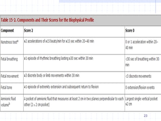
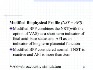
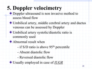
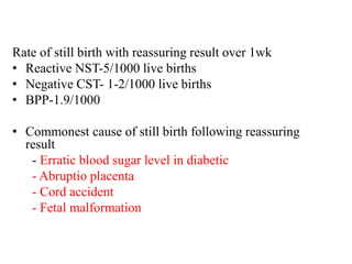
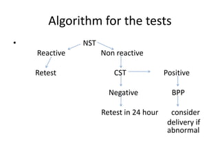
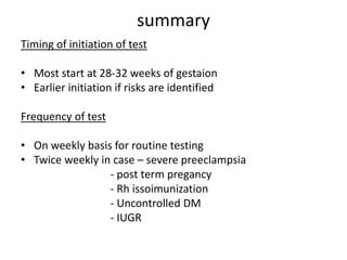
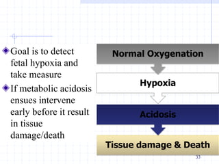
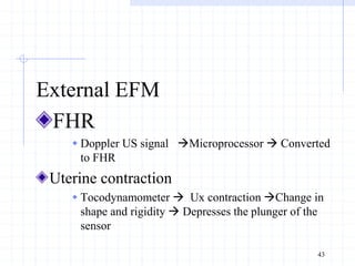
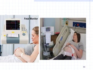
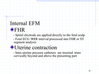
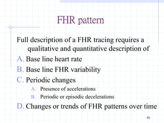
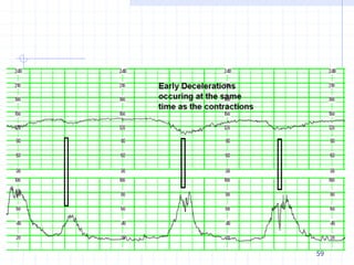
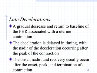
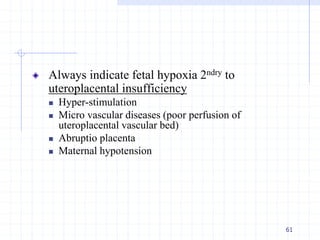
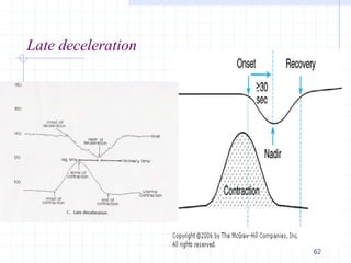
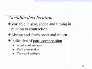
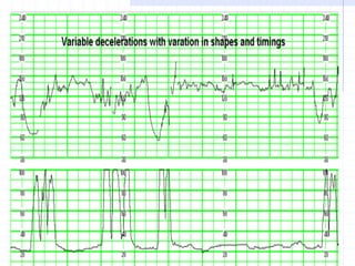
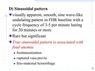
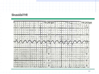
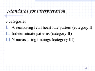
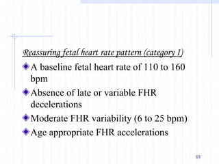
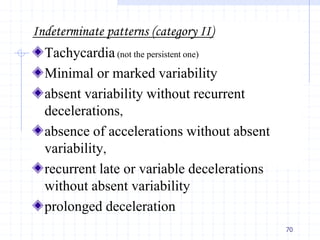
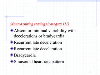
Antepartum and intrapartum
fetal surveillance
Ambo University, CMHS
For 3rd year HO students
Dr. Rabirra Waktola
June 2020
Contents
Antepartum fetal surveillance
 Definition
 Indications
 Testing/surveillance modalities
Intrapartum fetal surveillance
 Definition
 Parameters for intrapartum fetal surveillance
2
Objectives
to know some modalities of antepartum and
intrapartum surveillance
to have basic understanding on how to
do/when to do commonly used fetal
surveillance
to know how to interpret the results of the
fetal surveillance
3
Antepartum fetal surveillance
4
Definition
Assessment of the wellbeing of the
fetus/fetuses during pregnancy especially
after the fetus is considered viable
The goal is to identify those fetuses at risk
of intra uterine neurologic injury or death so
that these adverse outcomes can be
prevented.
5
Rationale
Fetal hypoxia and acidosis represent the
final common pathway to fetal injury and
death
Fetus with hypoxia will respond in a series
of detectable physiologically adaptive or
decompensatory signs
6
Indications
Uteroplacental insufficiency
 post term, suspected IUGR, DM, hypertension,
multiple pregnancy, previous still birth
Fetal compromise by other test
 Decreased fetal movement, Oligohydraminos
Routine surveillance
7
Testing modalities
Fetal movement count
Non stress testing
Contraction stress testing
Biophysical profile
Doppler velocimetry
8
1. Fetal movement count
Fetal movements are first perceived by the
mother at 17-20 weeks and becomes more
sophisticated and complicated by the end of
pregnancy
The logic behind fetal movement testing is
that fetal movements decrease in response
to hypoxia
9
Mother appreciate 80% of fetal movement,
but beyond 36 weeks only 16% of
movements are perceived by the mother
An important determinant of fetal activity is
sleep-wake cycle
Sleep cyclicity varies from as low as 20 to
as much as 75 minutes
10
Methods of FM counting
1.The Cardiff method
<10 movement over 12 hours is alarming
NB:we usually use this method in our wards in admitted patients and
there should be at least 10 movements in 12 hr duration
2.Sadovsky method
-mother count FM 3x daily each for 30min –
-normal is more than 4 movements
-<3 movement/one hour is alarming
3. Rayburn method
- count once per day for 60minutes between 7 and
11 PM after dinner
-normal is more than 4 movements
- <3 movement/1hr for two consecutive days is
alarming
11
Factor that decrease FM
Placental location
Length of fetal movement
Amniotic fluid volume
Fetal anomalies
Fetal state (if sleeping or not)
Maternal activity
Obesity
Medication use
Fetal jeopardy
12
2. Non stress testing
Almost all fetal movement (85-90%) are
accompanied by acceleration of FHR in
normal fetus
Acceleration
 If G.A >32 weeks the peak of acceleration is 15 beats per
minute or more above the baseline fetal heart rate but
acceleration last 15 seconds or more but less than 2 minutes
 If G.A< 32 weeks accelerations are defined as having peak
10 beats per minute or more above the baseline for 10
seconds or longer
13
Technique (it needs cardiotocography)
 Mother in left lateral position and continuous
electronic fetal heart monitoring conducted for
40 minutes.
 Mother provided with indicator to press
whenever she feels fetal movements.
 Tracing of fetal heart pattern made throughout
and checked for alterations associated with fetal
movement.
14
Interpretation
Reactive Non Stress Test
 Two or more accelerations that peak at 15 bpm or more
above the baseline each lasting 15 seconds or more,
within 20 minutes, with or without fetal movement
discernible by the mother
Non Reactive Non Stress Test
 Lacks sufficient fetal heart rate acceleration over a 40
minutes period
15
Factors causing Non-reactivity
Fetal sleep
Fetal anomalies-Neurologic/Cardiac
CNS depressants
Beta-blockers
Maternal smoking
Prematurity
Sepsis
Fetal jeopardy
16
3. Contraction stress test
The premises behind CST is that uterine
contractions transiently restrict oxygen
delivery to the fetus and hypoxic fetus will
demonstrate recurrent late decelerations
There are two methods of contraction stress
test
 Intravenous oxytocin
 Nipple stimulation
• NB:Read the intapartal fetal surveillance part of this slide
for the definition of decelerations 17
Fetal heart rate and uterine contraction are
recorded simultaneously with an external
monitor (cardiotocography or CTG)
18
Contraindication of CST
Patient at high risk for premature labor
- Premature ROM
- Multiple gestation
- Cervical incompetence
Condition in which uterine contraction is
dangerous
- Placenta previa
- Previous classical C/S
- Previous uterine surgery 19
Interpretation
 Negative -80% of test
 No late deceleration appearing anywhere on the
tracing with adequate uterine contraction (3
contraction in 10 min, lasting for 40-60sec)
 Negative means the fetal condition is good
 Positive -3-5% of test
 Late deceleration that are consistent and persistent
and occurring in at least >50% of contraction
without excessive uterine contraction
20
Suspicious (equivocal) -5% of tests
 Inconsistent late deceleration
Hyperstimulated -5% of cases
 Uterine contraction closer than every 2min or lasting
for >90sec
Unsatisfactory- 5% of cases
 Quality of tracing is inadequate for interpretation or
adequate uterine contraction can not be achieved
21
4. Biophysical profile
Components
1.Non stress test
2.Fetal breathing movements
3.Fetal movements
4.Fetal tone
5. Determination of the amniotic fluid volume
Other than non stress test the other components (2-5)
needs ultrasound
22
23
 Those fetal biophysical activities that
appear earliest in fetal developments are the
last to disappear with fetal hypoxia
 Fetal tone, fetal movement, fetal breathing
and FHR control develop in this sequence
 So, NST and FBM are the two parameter
that are affected initially
24
Interpretation
BPP of 10/10 OR 8/10 with normal AFV(amniotic fluid
volume) OR 8/8 NST not done
 Reassuring BPP, non asphyxiated fetus
 repeat test weekly (twice weekly in diabetes and post term)
BPP of 8/10 with abnormal AFV
- Have risk of chronic asphyxia
- Terminate pregnancy if term and
oligohydramnios
- Repeat test 1-2xweekly if not term
BPP of 6/10
- Possible fetal Asphyxia, terminate if term
- Repeat test after 4-6hour , deliver if <6
25
BPP of 4/10
 probable fetal asphyxia
 repeat the test after 4-6 hour , deliver if <6
BPP of 0-2/10
 Almost certain fetal asphyxia
 Deliver
26
Modified Biophysical Profile (NST + AFI)
Modified BPP combines the NST(with the
option of VAS) as a short term indicator of
fetal acid-base status and AFI as an
indicator of long term placental function
Modified BPP considered normal if NST is
reactive and AFI is more than 5cm
VAS-vibroacoustic stimulation 27
5. Doppler velocimetry
Doppler ultrasound is non invasive method to
assess blood flow
Umbilical artery, middle cerebral artery and ductus
venosus can be assessed by Doppler
Umbilical artery systolic/diastolic ratio is
commonly used
Abnormal result when
- if S/D ratio is above 95th percentile
- Absent diastolic flow
- Reversed diastolic flow
Usually employed in case of IUGR
28
Rate of still birth with reassuring result over 1wk
• Reactive NST-5/1000 live births
• Negative CST- 1-2/1000 live births
• BPP-1.9/1000
• Commonest cause of still birth following reassuring
result
- Erratic blood sugar level in diabetic
- Abruptio placenta
- Cord accident
- Fetal malformation
Algorithm for the tests
• NST
Reactive Non reactive
Retest CST Positive
Negative BPP
Retest in 24 hour consider
delivery if
abnormal
summary
Timing of initiation of test
• Most start at 28-32 weeks of gestaion
• Earlier initiation if risks are identified
Frequency of test
• On weekly basis for routine testing
• Twice weekly in case – severe preeclampsia
- post term pregancy
- Rh issoimunization
- Uncontrolled DM
- IUGR
Intrapartum fetal surveillance
32
Goal is to detect
fetal hypoxia and
take measure
If metabolic acidosis
ensues intervene
early before it result
in tissue
damage/death
33
Tissue damage & Death
Acidosis
Hypoxia
Normal Oxygenation
Fetal defense mechanisms against asphyxia
34
Hemoglobin F-
higher oxygen
affinity
Large placental
surface area for
exchange with
sufficient reserve-
nearly 11 meters
square
Low fetal partial
pressure of oxygen
creating a large
diffusion gradient-
30 mmH2O
Special fetal
circulation with
preferential flow of
oxygenated blood to
vital organs
Large uterine blood
flow with sufficient
reserve
Parameters used in assessing
intrapartum fetal well being
Fetal heart beat monitoring
Meconium staining of amniotic fluid
Fetal scalp blood sampling
Umbilical cord blood sampling
35
Risk factors for intrapartum asphyxia
Majority- cannot be predicted or are not
associated with the known risk factors
Pregnancies known to be associated with a
higher risk of intrapartum asphyxia and
require increased vigilance include
36
37
1. FHR monitoring
FHR monitoring in real sense is fetal brain
monitoring
Fetal brain responds to hypoxia by altering
FHR(fetal heart rate)
38
Methods of monitoring FHR
Intermittent auscultation
 Pinnard stethoscope/fetoscope
 Doppler US device
Continuous fetal heart monitoring
 Electronic fetal heart monitoring (cardiotocography)
39
Doppler US device
40
Pinnard stethoscope
Intermittent auscultation
Auscultation for period of 60sec following contraction
In high risk
 Every 15 min in 1st stage
 Every 5 min in 2nd stage
In low risk
 Every 30 min in 1st stage
 Every 15 min in 2nd stage
Advantage: Available, easy to use, inexpensive and
effective if done in consistent manner
Disadvantage: Labor intensive’ may not detect some
abnormal FHR patterns, sometimes difficult (obesity)
41
Electronic fetal monitoring (CTG)
Continuous recording & graphical
representation of FHR & uterine
contractions
EFM-can either be performed externally or
internally
42
External EFM
FHR
 Doppler US signal Microprocessor  Converted
to FHR
Uterine contraction
 Tocodynamometer  Ux contraction Change in
shape and rigidity  Depresses the plunger of the
sensor
43
44
Internal EFM
FHR
- Spiral electrode are applied directly to the fetal scalp
- Fetal ECG RR interval processed into FHR or ST
segment analysis
Uterine contraction
- Intra uterine pressure catheters are inserted trans
cervically beyond and above the presenting part
45
FHR pattern
Full description of a FHR tracing requires a
qualitative and quantitative description of
A. Base line heart rate
B. Base line FHR variability
C. Periodic changes
A. Presence of accelerations
B. Periodic or episodic decelerations
D. Changes or trends of FHR patterns over time
46
A.Base line FHR
Base line FHR - is the approximate mean
FHR during a 10 minute segment
 Normal: 120-159 beats per minute (bpm)
 Tachycardia - baseline rate above 160 bpm
- Mild 160-180bpm
- Severe ≥ 181
 Brady cardia-
 Base line FHR < 120 bpm
 mild 100-119bpm
 moderate 80-100bpm
 severe<80bpm
47
Fetal tachycardia
48
>160
BPM
Most are
not
suggestive
of fetal
jeopardy
Associated
with
 Fetal hypoxia
 Maternal fever
 Hyperthyroidism
 Maternal or fetal
anemia
 Drugs:Atropine
 Chorioamnionitis
 Fetal tachyarrhythmia
 Prematurity
Fetal bradycardia causes
Maternal hypothermia
Complete heart block
Severe asphyxia
49
B) Baseline Beat-to-beat variability
is the difference in heart rate from one beat
to the next
important index of cardiovascular function
and appears to be regulated largely by the
autonomic nervous system
50
Beat-to-beat variability
51
52
Causes of decreased/absent variability
 Hypoxia and acidosis
 Fetal anomalies
 Tachycardia
 Extreme prematurity
 Previous neurologic insult
 Fetal sleep cycles
 Drugs (CNS depressants)
53
C) Periodic changes
changes of FHR for brief duration with return to
baseline in response and relation to contraction
Decelerations (Early, late, variable, prolonged)
Accelerations
54
Acceleration
An abrupt increase in the FHR.
 Before 32 weeks of gestation, accelerations should last
≥10 sec and peak ≥10 bpm above baseline.
 After 32 weeks gestation, accelerations should
last ≥15 sec and peak ≥15 bpm above baseline.
A prolonged acceleration is ≥2 minutes but less
than 10 minutes. An acceleration of 10 minutes or
more is considered a change in baseline
Physiologic response to fetal movement
55
56
Decelerations (decrease in heart rate)
Early Decelerations
Late Decelerations
Variable Decelerations
prolonged Decelerations
57
Early Decelerations
A gradual decrease and return to baseline of the
FHR associated with a uterine contraction
The nadir of the FHR and the peak of the
contraction occur at the same time
Caused by fetal head compression during uterine
contraction, resulting in vagal stimulation and
slowing of the heart rate
not associated with fetal distress and thus are
reassuring
58
59
Late Decelerations
A gradual decrease and return to baseline of
the FHR associated with a uterine
contraction
The deceleration is delayed in timing, with
the nadir of the deceleration occurring after
the peak of the contraction
The onset, nadir, and recovery usually occur
after the onset, peak, and termination of a
contraction 60
Always indicate fetal hypoxia 2ndry to
uteroplacental insufficiency
 Hyper-stimulation
 Micro vascular diseases (poor perfusion of
uteroplacental vascular bed)
 Abruptio placenta
 Maternal hypotension
61
Late deceleration
62
Variable deceleration
Variable in size, shape and timing in
relation to contraction
Abrupt and sharp onset and return
Indicative of cord compression
 occult cord prolapse
 Cord presentation
 True cord prolapse
63
64
Prolonged deceleration
Lasts 2min-10min
Cause can be any of the mechanisms
previously described
65
D) Sinusoidal pattern
visually apparent, smooth, sine wave-like
undulating pattern in FHR baseline with a
cycle frequency of 3-5 per minute lasting
for 20 minutes or more
Rare but significant
True sinusoidal pattern is associated with
fetal anemia
 Isoimmunization
 ruptured vasa previa
 feto-maternal hemorrhage
67
Standards for interpretation
3 categories
I. A reassuring fetal heart rate pattern (category I)
II. Indeterminate patterns (category II)
III.Nonreassuring tracings (category III)
68
Reassuring fetal heart rate pattern (category I)
A baseline fetal heart rate of 110 to 160
bpm
Absence of late or variable FHR
decelerations
Moderate FHR variability (6 to 25 bpm)
Age appropriate FHR accelerations
69
Indeterminate patterns (category II)
Tachycardia(not the persistent one)
Minimal or marked variability
absent variability without recurrent
decelerations,
absence of accelerations without absent
variability,
recurrent late or variable decelerations
without absent variability
prolonged deceleration
70
Nonreassuring tracings (category III)
Absent or minimal variability with
decelerations or bradycardia
Recurrent late deceleration
Bradycardia
Sinusoidal heart rate pattern
71
2. Meconium staining of amniotic fluid
Hypoxic insult – vagal response –
meconium passage
Can occur in absence of significant or
sustained hypoxia
Has 3 grades
 Grade I-lightly meconium stained
 Grade II-between grade I and II
 Grade III-stained doughy thick meconium
72
3. Fetal scalp blood sampling
not of much practical significance in the
diagnosis of fetal asphyxia
Fetal scalp blood pH<7.2 indicates acidosis
where >/=7.2 is normal
73
4. Umbilical cord blood sampling
After delivery a sample of cord blood is
taken and analyzed for Po2, PCO2 and PH,
HCO= .
Useful to assess presence of asphyxia
retrospectively and also manage the neonate
74
Management of a non-reassuring fetal status
75
Resuscitation Definitive treatment
•Put the mother in the left lateral
position
•Oxygen by face mask at a rate of
5L/min
•Rehydrate with IV fluids
•Discontinue oxytocin
•Amnioinfusion – warm saline into the
uterine cavity through the intrauterine
catheter
•Tocolysis if hypertonus is diagnosed
•Monitor FHR further and observe for
improvements
•Expedited delivery depending on the
stage of labor
•Instrumental delivery
•Caesarean delivery
•Breech Extraction
76

Fetal surveillance 2020

  • 1.
    Antepartum and intrapartum fetalsurveillance Ambo University, CMHS For 3rd year HO students Dr. Rabirra Waktola June 2020
  • 2.
    Contents Antepartum fetal surveillance Definition  Indications  Testing/surveillance modalities Intrapartum fetal surveillance  Definition  Parameters for intrapartum fetal surveillance 2
  • 3.
    Objectives to know somemodalities of antepartum and intrapartum surveillance to have basic understanding on how to do/when to do commonly used fetal surveillance to know how to interpret the results of the fetal surveillance 3
  • 4.
  • 5.
    Definition Assessment of thewellbeing of the fetus/fetuses during pregnancy especially after the fetus is considered viable The goal is to identify those fetuses at risk of intra uterine neurologic injury or death so that these adverse outcomes can be prevented. 5
  • 6.
    Rationale Fetal hypoxia andacidosis represent the final common pathway to fetal injury and death Fetus with hypoxia will respond in a series of detectable physiologically adaptive or decompensatory signs 6
  • 7.
    Indications Uteroplacental insufficiency  postterm, suspected IUGR, DM, hypertension, multiple pregnancy, previous still birth Fetal compromise by other test  Decreased fetal movement, Oligohydraminos Routine surveillance 7
  • 8.
    Testing modalities Fetal movementcount Non stress testing Contraction stress testing Biophysical profile Doppler velocimetry 8
  • 9.
    1. Fetal movementcount Fetal movements are first perceived by the mother at 17-20 weeks and becomes more sophisticated and complicated by the end of pregnancy The logic behind fetal movement testing is that fetal movements decrease in response to hypoxia 9
  • 10.
    Mother appreciate 80%of fetal movement, but beyond 36 weeks only 16% of movements are perceived by the mother An important determinant of fetal activity is sleep-wake cycle Sleep cyclicity varies from as low as 20 to as much as 75 minutes 10
  • 11.
    Methods of FMcounting 1.The Cardiff method <10 movement over 12 hours is alarming NB:we usually use this method in our wards in admitted patients and there should be at least 10 movements in 12 hr duration 2.Sadovsky method -mother count FM 3x daily each for 30min – -normal is more than 4 movements -<3 movement/one hour is alarming 3. Rayburn method - count once per day for 60minutes between 7 and 11 PM after dinner -normal is more than 4 movements - <3 movement/1hr for two consecutive days is alarming 11
  • 12.
    Factor that decreaseFM Placental location Length of fetal movement Amniotic fluid volume Fetal anomalies Fetal state (if sleeping or not) Maternal activity Obesity Medication use Fetal jeopardy 12
  • 13.
    2. Non stresstesting Almost all fetal movement (85-90%) are accompanied by acceleration of FHR in normal fetus Acceleration  If G.A >32 weeks the peak of acceleration is 15 beats per minute or more above the baseline fetal heart rate but acceleration last 15 seconds or more but less than 2 minutes  If G.A< 32 weeks accelerations are defined as having peak 10 beats per minute or more above the baseline for 10 seconds or longer 13
  • 14.
    Technique (it needscardiotocography)  Mother in left lateral position and continuous electronic fetal heart monitoring conducted for 40 minutes.  Mother provided with indicator to press whenever she feels fetal movements.  Tracing of fetal heart pattern made throughout and checked for alterations associated with fetal movement. 14
  • 15.
    Interpretation Reactive Non StressTest  Two or more accelerations that peak at 15 bpm or more above the baseline each lasting 15 seconds or more, within 20 minutes, with or without fetal movement discernible by the mother Non Reactive Non Stress Test  Lacks sufficient fetal heart rate acceleration over a 40 minutes period 15
  • 16.
    Factors causing Non-reactivity Fetalsleep Fetal anomalies-Neurologic/Cardiac CNS depressants Beta-blockers Maternal smoking Prematurity Sepsis Fetal jeopardy 16
  • 17.
    3. Contraction stresstest The premises behind CST is that uterine contractions transiently restrict oxygen delivery to the fetus and hypoxic fetus will demonstrate recurrent late decelerations There are two methods of contraction stress test  Intravenous oxytocin  Nipple stimulation • NB:Read the intapartal fetal surveillance part of this slide for the definition of decelerations 17
  • 18.
    Fetal heart rateand uterine contraction are recorded simultaneously with an external monitor (cardiotocography or CTG) 18
  • 19.
    Contraindication of CST Patientat high risk for premature labor - Premature ROM - Multiple gestation - Cervical incompetence Condition in which uterine contraction is dangerous - Placenta previa - Previous classical C/S - Previous uterine surgery 19
  • 20.
    Interpretation  Negative -80%of test  No late deceleration appearing anywhere on the tracing with adequate uterine contraction (3 contraction in 10 min, lasting for 40-60sec)  Negative means the fetal condition is good  Positive -3-5% of test  Late deceleration that are consistent and persistent and occurring in at least >50% of contraction without excessive uterine contraction 20
  • 21.
    Suspicious (equivocal) -5%of tests  Inconsistent late deceleration Hyperstimulated -5% of cases  Uterine contraction closer than every 2min or lasting for >90sec Unsatisfactory- 5% of cases  Quality of tracing is inadequate for interpretation or adequate uterine contraction can not be achieved 21
  • 22.
    4. Biophysical profile Components 1.Nonstress test 2.Fetal breathing movements 3.Fetal movements 4.Fetal tone 5. Determination of the amniotic fluid volume Other than non stress test the other components (2-5) needs ultrasound 22
  • 23.
  • 24.
     Those fetalbiophysical activities that appear earliest in fetal developments are the last to disappear with fetal hypoxia  Fetal tone, fetal movement, fetal breathing and FHR control develop in this sequence  So, NST and FBM are the two parameter that are affected initially 24
  • 25.
    Interpretation BPP of 10/10OR 8/10 with normal AFV(amniotic fluid volume) OR 8/8 NST not done  Reassuring BPP, non asphyxiated fetus  repeat test weekly (twice weekly in diabetes and post term) BPP of 8/10 with abnormal AFV - Have risk of chronic asphyxia - Terminate pregnancy if term and oligohydramnios - Repeat test 1-2xweekly if not term BPP of 6/10 - Possible fetal Asphyxia, terminate if term - Repeat test after 4-6hour , deliver if <6 25
  • 26.
    BPP of 4/10 probable fetal asphyxia  repeat the test after 4-6 hour , deliver if <6 BPP of 0-2/10  Almost certain fetal asphyxia  Deliver 26
  • 27.
    Modified Biophysical Profile(NST + AFI) Modified BPP combines the NST(with the option of VAS) as a short term indicator of fetal acid-base status and AFI as an indicator of long term placental function Modified BPP considered normal if NST is reactive and AFI is more than 5cm VAS-vibroacoustic stimulation 27
  • 28.
    5. Doppler velocimetry Dopplerultrasound is non invasive method to assess blood flow Umbilical artery, middle cerebral artery and ductus venosus can be assessed by Doppler Umbilical artery systolic/diastolic ratio is commonly used Abnormal result when - if S/D ratio is above 95th percentile - Absent diastolic flow - Reversed diastolic flow Usually employed in case of IUGR 28
  • 29.
    Rate of stillbirth with reassuring result over 1wk • Reactive NST-5/1000 live births • Negative CST- 1-2/1000 live births • BPP-1.9/1000 • Commonest cause of still birth following reassuring result - Erratic blood sugar level in diabetic - Abruptio placenta - Cord accident - Fetal malformation
  • 30.
    Algorithm for thetests • NST Reactive Non reactive Retest CST Positive Negative BPP Retest in 24 hour consider delivery if abnormal
  • 31.
    summary Timing of initiationof test • Most start at 28-32 weeks of gestaion • Earlier initiation if risks are identified Frequency of test • On weekly basis for routine testing • Twice weekly in case – severe preeclampsia - post term pregancy - Rh issoimunization - Uncontrolled DM - IUGR
  • 32.
  • 33.
    Goal is todetect fetal hypoxia and take measure If metabolic acidosis ensues intervene early before it result in tissue damage/death 33 Tissue damage & Death Acidosis Hypoxia Normal Oxygenation
  • 34.
    Fetal defense mechanismsagainst asphyxia 34 Hemoglobin F- higher oxygen affinity Large placental surface area for exchange with sufficient reserve- nearly 11 meters square Low fetal partial pressure of oxygen creating a large diffusion gradient- 30 mmH2O Special fetal circulation with preferential flow of oxygenated blood to vital organs Large uterine blood flow with sufficient reserve
  • 35.
    Parameters used inassessing intrapartum fetal well being Fetal heart beat monitoring Meconium staining of amniotic fluid Fetal scalp blood sampling Umbilical cord blood sampling 35
  • 36.
    Risk factors forintrapartum asphyxia Majority- cannot be predicted or are not associated with the known risk factors Pregnancies known to be associated with a higher risk of intrapartum asphyxia and require increased vigilance include 36
  • 37.
  • 38.
    1. FHR monitoring FHRmonitoring in real sense is fetal brain monitoring Fetal brain responds to hypoxia by altering FHR(fetal heart rate) 38
  • 39.
    Methods of monitoringFHR Intermittent auscultation  Pinnard stethoscope/fetoscope  Doppler US device Continuous fetal heart monitoring  Electronic fetal heart monitoring (cardiotocography) 39
  • 40.
  • 41.
    Intermittent auscultation Auscultation forperiod of 60sec following contraction In high risk  Every 15 min in 1st stage  Every 5 min in 2nd stage In low risk  Every 30 min in 1st stage  Every 15 min in 2nd stage Advantage: Available, easy to use, inexpensive and effective if done in consistent manner Disadvantage: Labor intensive’ may not detect some abnormal FHR patterns, sometimes difficult (obesity) 41
  • 42.
    Electronic fetal monitoring(CTG) Continuous recording & graphical representation of FHR & uterine contractions EFM-can either be performed externally or internally 42
  • 43.
    External EFM FHR  DopplerUS signal Microprocessor  Converted to FHR Uterine contraction  Tocodynamometer  Ux contraction Change in shape and rigidity  Depresses the plunger of the sensor 43
  • 44.
  • 45.
    Internal EFM FHR - Spiralelectrode are applied directly to the fetal scalp - Fetal ECG RR interval processed into FHR or ST segment analysis Uterine contraction - Intra uterine pressure catheters are inserted trans cervically beyond and above the presenting part 45
  • 46.
    FHR pattern Full descriptionof a FHR tracing requires a qualitative and quantitative description of A. Base line heart rate B. Base line FHR variability C. Periodic changes A. Presence of accelerations B. Periodic or episodic decelerations D. Changes or trends of FHR patterns over time 46
  • 47.
    A.Base line FHR Baseline FHR - is the approximate mean FHR during a 10 minute segment  Normal: 120-159 beats per minute (bpm)  Tachycardia - baseline rate above 160 bpm - Mild 160-180bpm - Severe ≥ 181  Brady cardia-  Base line FHR < 120 bpm  mild 100-119bpm  moderate 80-100bpm  severe<80bpm 47
  • 48.
    Fetal tachycardia 48 >160 BPM Most are not suggestive offetal jeopardy Associated with  Fetal hypoxia  Maternal fever  Hyperthyroidism  Maternal or fetal anemia  Drugs:Atropine  Chorioamnionitis  Fetal tachyarrhythmia  Prematurity
  • 49.
    Fetal bradycardia causes Maternalhypothermia Complete heart block Severe asphyxia 49
  • 50.
    B) Baseline Beat-to-beatvariability is the difference in heart rate from one beat to the next important index of cardiovascular function and appears to be regulated largely by the autonomic nervous system 50
  • 51.
  • 52.
  • 53.
    Causes of decreased/absentvariability  Hypoxia and acidosis  Fetal anomalies  Tachycardia  Extreme prematurity  Previous neurologic insult  Fetal sleep cycles  Drugs (CNS depressants) 53
  • 54.
    C) Periodic changes changesof FHR for brief duration with return to baseline in response and relation to contraction Decelerations (Early, late, variable, prolonged) Accelerations 54
  • 55.
    Acceleration An abrupt increasein the FHR.  Before 32 weeks of gestation, accelerations should last ≥10 sec and peak ≥10 bpm above baseline.  After 32 weeks gestation, accelerations should last ≥15 sec and peak ≥15 bpm above baseline. A prolonged acceleration is ≥2 minutes but less than 10 minutes. An acceleration of 10 minutes or more is considered a change in baseline Physiologic response to fetal movement 55
  • 56.
  • 57.
    Decelerations (decrease inheart rate) Early Decelerations Late Decelerations Variable Decelerations prolonged Decelerations 57
  • 58.
    Early Decelerations A gradualdecrease and return to baseline of the FHR associated with a uterine contraction The nadir of the FHR and the peak of the contraction occur at the same time Caused by fetal head compression during uterine contraction, resulting in vagal stimulation and slowing of the heart rate not associated with fetal distress and thus are reassuring 58
  • 59.
  • 60.
    Late Decelerations A gradualdecrease and return to baseline of the FHR associated with a uterine contraction The deceleration is delayed in timing, with the nadir of the deceleration occurring after the peak of the contraction The onset, nadir, and recovery usually occur after the onset, peak, and termination of a contraction 60
  • 61.
    Always indicate fetalhypoxia 2ndry to uteroplacental insufficiency  Hyper-stimulation  Micro vascular diseases (poor perfusion of uteroplacental vascular bed)  Abruptio placenta  Maternal hypotension 61
  • 62.
  • 63.
    Variable deceleration Variable insize, shape and timing in relation to contraction Abrupt and sharp onset and return Indicative of cord compression  occult cord prolapse  Cord presentation  True cord prolapse 63
  • 64.
  • 65.
    Prolonged deceleration Lasts 2min-10min Causecan be any of the mechanisms previously described 65
  • 66.
    D) Sinusoidal pattern visuallyapparent, smooth, sine wave-like undulating pattern in FHR baseline with a cycle frequency of 3-5 per minute lasting for 20 minutes or more Rare but significant True sinusoidal pattern is associated with fetal anemia  Isoimmunization  ruptured vasa previa  feto-maternal hemorrhage
  • 67.
  • 68.
    Standards for interpretation 3categories I. A reassuring fetal heart rate pattern (category I) II. Indeterminate patterns (category II) III.Nonreassuring tracings (category III) 68
  • 69.
    Reassuring fetal heartrate pattern (category I) A baseline fetal heart rate of 110 to 160 bpm Absence of late or variable FHR decelerations Moderate FHR variability (6 to 25 bpm) Age appropriate FHR accelerations 69
  • 70.
    Indeterminate patterns (categoryII) Tachycardia(not the persistent one) Minimal or marked variability absent variability without recurrent decelerations, absence of accelerations without absent variability, recurrent late or variable decelerations without absent variability prolonged deceleration 70
  • 71.
    Nonreassuring tracings (categoryIII) Absent or minimal variability with decelerations or bradycardia Recurrent late deceleration Recurrent late deceleration Bradycardia Sinusoidal heart rate pattern 71
  • 72.
    2. Meconium stainingof amniotic fluid Hypoxic insult – vagal response – meconium passage Can occur in absence of significant or sustained hypoxia Has 3 grades  Grade I-lightly meconium stained  Grade II-between grade I and II  Grade III-stained doughy thick meconium 72
  • 73.
    3. Fetal scalpblood sampling not of much practical significance in the diagnosis of fetal asphyxia Fetal scalp blood pH<7.2 indicates acidosis where >/=7.2 is normal 73
  • 74.
    4. Umbilical cordblood sampling After delivery a sample of cord blood is taken and analyzed for Po2, PCO2 and PH, HCO= . Useful to assess presence of asphyxia retrospectively and also manage the neonate 74
  • 75.
    Management of anon-reassuring fetal status 75 Resuscitation Definitive treatment •Put the mother in the left lateral position •Oxygen by face mask at a rate of 5L/min •Rehydrate with IV fluids •Discontinue oxytocin •Amnioinfusion – warm saline into the uterine cavity through the intrauterine catheter •Tocolysis if hypertonus is diagnosed •Monitor FHR further and observe for improvements •Expedited delivery depending on the stage of labor •Instrumental delivery •Caesarean delivery •Breech Extraction
  • 76.