SlideShare a Scribd company logo
1 of 188
Anatomy Revision Session 2
slides are available online at
Slideshare.net/muradalshehry
After this session
The Upper Limb
Anatomy Revision Session 1
Objectives of this session
• To be able to Identify the muscles and their functions of the upper
limb.
• Know relations of muscles to important nerves.
• There will be a few pop Quiz…. Stay focused ☺
Orientation and Movement REVISION
adduction
abduction
flexionextension
circumduction
rotation
elevation
depression
supination
pronation
protraction
retraction
Orientation and Movement REVISION
Flexor digitorum superficialis
• Medial epicondyle of humerus and coronoid
process of ulna to middle phalanges (lateral) of
digits 2-5 (i.e. all except thumb)
• Flexion of metacarpophalangeal (MCP) and
proximal interphalangeal (PIP) joint
• Median nerve (C7,C8,T1)
Flexor digitorum profundus
• Ulna and interosseous membrane to distal
phalanges digits 2-5 (palmar)
• Flexion of MCP, PIP and distal interphalangeal
joint (DIP) of digits 2-5
• Ring and little fingers = ulnar nerve (C8,T1)
• Index and middle fingers = median nerve (C8,T1
anterior interosseous)
Flexor digitorum muscles:
Superficialis Splits in two, To Permit Profundus Passing through.
Flexor pollicis longus
• Radius and interosseous membrane to base of distal
phalanx of thumb (palmar)
• Flexion of MCP and IP of thumb, wrist
• Median nerve (C8, T1 anterior interosseous)
Pronator quadratus
• Distal anterior ulna to distal anterior radius
• Pronation and help to stabilise distal radioulnar joint
• Median nerve (C8, T1 anterior introsseous)
Anterior forearm muscles:
superficial group There are five, like five digits of your hand.
Place your thumb into your palm, then lay that hand palm down on your other arm.
PFPF [pass/fail, pass/fail]:
Pronator teres
Flexor carpi radialis
Palmaris longus
Flexor carpi ulnaris
Your thumb below your 4 fingers shows the muscle which is deep to the other
four: Flexor digitorum superficialis.
Median nerve
• C6-T1 fibres, medial and lateral cords
• Major nerve of anterior compartment of
forearm, THROUGH carpal tunnel
• Main nerve of thumb side of hand
Ulnar nerve and artery
• C7-T1 fibres from medial cord, medial
epicondyle of humerus, pisiform ABOVE
carpal tunnel
• Branch brachial artery, interosseous
branches, little finger side, superficial
palmar arch
median
ulnar
radial
ant. interosseous
anterior
interosseous
Median nerve passes between the heads of pronator teres
Anconeus
• Lateral epicondyle of humerus to
olecranon of ulna
• Extension of the elbow
• Radial nerve (C7-C8)
Brachioradialis
• Distal humerus (supra-epicondylar ridge) to radius
proximal to styloid process
• Puts the forearm into the semi-prone position,
flexion of elbow
• Radial nerve (C5, C6, C7) (before division into deep
and superficial)
Posterior compartment of the forearm
• Common extensor origin = lateral
epicondyle of the humerus
• Extensors of the digits, wrist
• Abductor of the thumb
• Supinator
• Radial nerve
Extensor carpi radialis longus and brevis
• Extensor carpi radialis longus from distal humerus (supra-
epicondylar ridge) to base of 2nd metacarpal
• Wrist extension and abduction
• Radial nerve (C6,C7) (pre-division)
• Extensor carpi radialis brevis from lateral epicondyle of humerus to
base of 3rd metacarpal
• Wrist extension and abduction
• Radial nerve (C7,C8) (deep = posterior interosseous)
extensor
carpi
radialis
longus
extensor
carpi
radialis
brevis
Extensor carpi ulnaris
• Lateral epicondyle of humerus (small slip from ulna) to
base of 5th metacarpal
• Wrist extension and adduction
• Radial nerve (posterior interosseous) (C7, C8)
Extensor digitorum
• Lateral epicondyle of humerus to dorsal expansion of digits 2-5 (NOT thumb)
• Extension of principally MCP, but also PIP and DIP extension of digits 2-5
• Radial nerve (posterior interosseous) (C7,C8)
Extensor digiti minimi
• Lateral epicondyle of the humerus to dorsal expansion of 5th digit (little
finger)
• MCP, PIP, DIP extension of little finger
• Radial nerve (posterior interosseous) (C7,C8)
Extensor indices
• Posterior surface of ulna (and interosseous membrane) to
dorsal expansion of digit 2 (index finger)
• MCP, PIP, DIP extension of index finger
• Wrist extension
• Radial nerve (posterior interosseous) (C7,C8)
Extensor pollicis longus
• ‘Pollicis’ = thumb
• Ulna and interosseous membrane to base of distal
phalanx of thumb
• CMC, MCP and IP extension of thumb
• Radial nerve (posterior interosseous) (C7, C8)
Extensor pollicis brevis
• Radius and interosseous membrane to base of
proximal phalanx of thumb
• Carpometacarpal and MCP thumb joint extension
• Radial nerve (posterior interosseous) (C7,C8)
Abductor pollicis longus
• Radius, interosseous membrane and ulna to base of 1st
metacarpal
• Abducts thumb and extends it at carpometacarpal joint
• Radial nerve (posterior interosseous) (C7,C8)
Supinator
• Crest of ulna, lateral epicondyle of humerus, radial collateral and annular
ligament of radius to lateral radius (proximal third)
• Supination of forearm (rotates radius)
• Radial nerve (posterior interosseous)(C7,C8)
Radial nerve passes through the
lamina of supinator
posterior
interosseous
anterior
interosseous
deep
branch of
radial
common
interosseous
superficial
artery
artery
artery
The Hand
Flexor retinaculum
• Roof of carpal tunnel
• Medial = pisiform, hook of hamate
• Lateral = tubercle of scaphoid and
ridge on trapezium
• Septum from trapezium – flexor carpi
radialis
• Attach thenar and hypothenar
structures
Carpal tunnel
Carpal tunnel syndrome causes MEDIAN TRAP:
Myxoedema
Edema premenstrually
Diabetes
Idiopathic
Agromegaly
Neoplasm
Trauma
Rheumatoid arthritis
Amyloidosis
Pregnancy
Bones and joints
carpals
metacarpals
proximal
phalanx
middle
phalanx
distal phalanx
interphalangeal (IP)
joints = synovial hinge
metacarpophalangeal
(MCP) joints = synovial
condyloid
carpometacarpal (CMC)
joints = synovial saddle
(thumb), plane (digits)
intercarpal = synovial plane
Palmar aponeurosis
aponeurosis
palmaris longus
Palmaris brevis
• Palmar aponeurosis and
flexor retinaculum to
dermis
• Grip
• Ulnar nerve (superficial
C8,T1)
Intrinsic muscles of the hand
abductor pollicis
brevis = intrinsic
muscle
tendon of flexor
digitorum superficialis =
extrinsic muscle
Intrinsic muscles – thenar eminence
• Abductor pollicis brevis = scaphoid
and trapezium to base proximal
phalanx of thumb – CMC joint
abduction – median nerve (recurrent
C8,T1)
• Flexor pollicis brevis = flexor
retinaculum, capitate, trapezium to
base proximal phalanx of thumb –
CMC joint flexion – Median
(recurrent) and some texts also ulnar
nerve (C8,T1)
• Opponens pollicis = trapezium to first
metacarpal – CMC joint opposition –
median nerve (recurrent C8,T1)
abductor
pollicis
brevis
flexor
pollicis
brevis
opponens
pollicis
Adductor pollicis • Transverse head from 3rd metacarpal;
oblique head from capitate and 2nd/3rd
metacarpals to base of proximal phalanx of
thumb
• Adduction of CMC joint of thumb
• Flexion of MCP joint of thumb
• Ulnar nerve (deep C8,T1)
transverse
head
oblique
head
Hypothenar eminence
• Abductor digiti minimi = pisiform
bone to base of 5th proximal
phalanx and dorsal digital
expansion – abduction and flexion
MCP joint little finger – ulnar nerve
(deep C8,T1)
• Flexor digiti minimi = hamate,
flexor retinaculum to base of 5th
proximal phalanx – Flex MCP joint
of little finger – ulnar nerve (deep
C8,T1)
• Opponens digiti minimi = hamate
and flexor retinaculum to 5th
metacarpal – weak opposition of
little finger – ulnar nerve (deep
C8,T1)
abductor digiti
minimi
flexor digiti
minimi
opponens
digiti minimi
Lumbricals
• 4 small muscles from tendons of
flexor digitorum profundus
• 1st (index) and 2nd (middle)
unipennate
• 3rd (ring) and 4th (little) usually
bipennate
• Insert into dorsal digital expansion
• MCP joint flexion and IP joint
extension for digits 2-4
• 1st and 2nd = median nerve (digital
C8,T1)
• 3rd and 4th = ulnar nerve (deep
C8,T1)
1
2
3
4
lumbrical
Lumbricals action
Lumbricals action Lumbrical action is to hold a pea, that is
to flex the metacarpophalangeal joint and extend the interphalangeal
joints.
When look at hand in this position, can see this makes an "L" shape,
since L
is for Lumbrical.
Palmer interossei
• Between metacarpal bones
• 1st from 2nd metacarpal to base of 2nd
proximal phalanx and dorsal
expansion
• 2nd from 4th metacarpal to base of 4th
proximal phalanx and dorsal
expansion
• 3rd from 5th metacarpal to base of 5th
proximal phalanx and dorsal
expansion
• Adduction towards middle finger (flex
MCP, extend IP)
• Ulnar nerve (deep C8,T1)
12
3
Dorsal interossei • From sides of 2 adjacent metacarpals, eg, 1st
dorsal interossei from 1st and 2nd metacarpals
• 1st inserts into 2nd proximal phalanx and dorsal
digital expansion
• 2nd and 3rd inserts into the 3rd proximal phalanx
and dorsal digital expansion
• 4th inserts into 4th proximal phalanx and dorsal
digital expansion
• Abduction (MCP flexion, IP extension)
• Ulnar nerve (deep C8,T1)
123
4
Interossei muscles
: actions of dorsal vs. palmar in hand "PAd
and DAb":
The Palmar Adduct and the Dorsal Abduct.
· Use your hand to dab with a pad.
Intrinsic muscles of hand (palmar surface)
"A OF A OF A":
· Thenar, lateral to medial:
Abductor pollicis longus
Opponens pollicis
Flexor pollicis brevis
Adductor pollicis.
· Hypothenar, lateral to medial:
Opponens digiti minimi
Flexor digiti minimi
Abductor digiti minimi
Synovial sheaths
• Synovial membrane plus fluid
• Around tendons at wrist,
palm and into digits
Cutaneous supply
median
ulnar ulnar
radial
Cutaneous upper limb
ANT POST
Carpal bones
"Stop Letting Those People Touch The Cadaver's Hand
Proximal row, lateral-to-medial: Scaphoid Lunate Triquetrum Pisiform
Distal row, lateral-to-medial: Trapezium Trapezoid Capitate Hamate
Or
"Happy Cat Tom Took Pie To Little Sister":
Hamate
Capitate
Trapezoid
Trapezium
Pisiform
Triquital
Lunate
Scaphoid
Lower Limb
Gluteal Region and Hip Joint
Outline
• Innominate (Hip) Bone
• Proximal Femur
• Muscles of the gluteal region
• Nerves and Vessels of the gluteal region
• Hip Joint
Obturator
Foramen
Iliac crest
Iliac fossa
ASIS
AIIS
PSIS
PIIS
Arcuate Line
Ischial Tuberosity
Ischial Spine
Lesser Sciatic
Notch
Greater Sciatic
Notch
Pubis
Innominate:
Medial view
Innominate:
Lateral view
Iliac crest
ASIS
AIIS
PSIS
PIIS
Ischial Tuberosity
Ischial Spine
Lesser Sciatic
Notch
Greater Sciatic
Notch
Pubis
Posterior
Gluteal Line
Anterior
Gluteal Line
Inferior
Gluteal Line
Acetabulum
Pubic Tubercle
Ischium
Greater and Lesser Sciatic Foramina
Muscles of Gluteal Region
Lateral Rotators of Hip
• Gluteus Maximus
(extensor)
• Piriformis
• Obturator internus
• Gemelli
• Quadratus femoris
Medial Rotators and Abductors
of Hip
• Gluteus Medius
• Gluteus Minimus
Support
• Tensor fascia lata
Gluteus Maximus
• Sacrum, S/T ligament and ilium
behind posterior gluteal line 
Iliotibial tract (3/4) and gluteal
tuberosity of femur (1/4)
• Extends hip; assists lateral
rotation
• Inferior gluteal nerve
• (L5, S1, 2)
Tensor Fascia Lata
• Ant. Iliac Crest and ASIS 
Iliotibial tract
• Tenses fascia lata and
iliotibial tract; Supports
femur on tibia during
standing
• Superior Gluteal nerve
• (L4, 5, S1)
Gluteus
Maximus
Iliotibialtract
Gluteus Medius
• Ilium between posterior and
anterior gluteal lines 
greater trochanter (lateral
surface)
• Abducts and medially rotates
hip
• Superior gluteal nerve
• (L4, 5, S1)
Gluteus Minimus
• Ilium between anterior and
inferior gluteal lines 
greater trochanter (anterior
surface)
• Abducts and medially
rotates hip
• Superior gluteal nerve
• (L4, 5, S1)
Piriformis
• Anterior Sacrum and S/T lig.
Greater Trochanter (superior
border)
• Passes through greater sciatic
foramen
• Laterally rotates hip
• Anterior rami of L5, S1, 2
Obturator internus
• Pelvic surface of Obturator mem.
and surrounding bones 
Trochanteric Fossa of greater
trochanter
• Passes through lesser sciatic foramen
• Laterally rotates hip
• Nerve to obturator internus (L5,
S1)
Gemelli
• Superior: Ischial spine
• Inferior: Ischial tuberosity
Blend with tendon of obturator
internus (Trochanteric fossa)
• Laterally rotate hip
• Superior: Nerve to obturator
internus
• Inferior: Nerve to quadratus
femoris
Quadratus femoris
• Ischial tuberosity Quadrate
tubercle on intertrochanteric crest
of femur
• Laterally rotates hip
• Nerve to quadratus femoris (L5, S1)
Obturator externus
• Outer surface of obturator mem. 
Trochanteric fossa
• Laterally rotates hip
• Obturator nerve
• L3, 4
Anterior
Lateral Rotators
"Play Golf Or Go On Quacking":
· From top to bottom:
Piriformis
Gemellus superior
Obturator internus
Gemellus inferior
Obturator externus
Quadratus femoris
· Alternatively: "P-GO-GO-Q".
Sciatic nerve
• L4, 5, S1, 2, 3
• Tibial always leaves below
piriformis
• Common Peroneal may leave
below, above or through piriformis
Gluteal vessels and nerves
• Superior gluteal artery and
nerve
• Inferior gluteal artery and
nerve
• Piriformis
Posterior cutaneous nerve of thigh
• Lies on posterior aspect of
sciatic nerve
• (S1, 2, 3)
• Inferior Clunial nerves to skin of
inferior half of buttock
Cutaneous Nerves – Gluteal and Posterior Thigh
Superior Clunial
(posterior rami L1,2,3)
Middle Clunial
(posterior rami S1,2,3)
Posterior Cuntaneous n. of Thigh
with Inferior Clunial branches
Hip Joint
• Articular surfaces
• Capsule attachments
• Ligaments
• Relations of the Joint
Articular Surfaces
Capsule attachments
Iliofemoral ligament
• AIIS → Intertrochanteric line
• Upper and lower bands
Limits:
• Extension
• Lateral rotation
• Adduction (upper)
• Abduction (lower)
Pubofemoral ligament
• Iliopubic eminence → Lower part of intertrochanteric
line
Limits:
• Extension
• Lateral rotation
• Abduction
Ischiofemoral ligament
• Posterior aspect of acetabulum →
Greater trochanter
Limits:
• Extension
• Medial rotation
• Adduction
Blood Supply to the Hip Joint
Anterior and Medial Thigh
Outline
• Compartments of the Thigh
• Features of the femur
• Muscles of Anterior Compartment
• Muscles of Medial Compartment
• Adductor Canal
• Nerves
• Arteries
Compartments of Thigh
Flex Hip; Extend Knee
Anterior Compartment
Subcutaneous tissue
Fascia Lata
Medial Compartment
Posterior
Compartment
Great Saphenous vein
Intermuscular Septum
Iliotibial tract
Extend Hip; Flex Knee
Adduct Hip
Proximal femur
(anterior) Head
Greater trochanter
Intertrochanteric line
Lesser trochanter
Shaft
Fovea
Neck
Proximal femur
(posterior)
Greater trochanter
Trochanteric Fossa
Intertrochanteric crest
Lesser trochanter
Pectineal line
Gluteal tuberosity
Anterior Thigh
Hip Flexors
• Iliacus
• Psoas major
• Pectineus
• Sartorius
Knee Extensors
• Quadriceps
• Rectus femoris
• Vastus medialis
• Vastus lateralis
• Vastus intermedius
Hip Flexors – Iliopsoas
• Iliacus
• Iliac crest, fossa, ala of sacrum, ant. sacroiliac
lig. → Psoas tendon, lesser trochanter
• Femoral nerve (L2, 3)
• Flex hip
• Psoas major
• T12-L5, IV discs, lumbar transverse processes
→ Lesser trochanter
• Anterior rami L1-3
• Flex hip
Hip Flexors – Pectineus
• Pectineus
• Superior ramus of pubis →
Pectineal line of femur
• Femoral nerve (L2, 3) [occassionally br.
from obturator]
• Adduct and flex hip; assist with medial
rotation
Hip Flexors – Sartorious
• Sartorious
• ASIS
→ Superior part of medial surface Tibia
• Femoral nerve (L2, 3)
• Flex, abduct, laterally rotate hip; flex knee
Knee Extensors – Quadriceps
• Rectus femoris
• AIIS, ilium above acetabulum
• Vastus lateralis
• Gr. Trochanter & Lat. Linea Aspera
• Vastus medialis
• Intertrochanteric line & Med. Linea Aspera
• Vastus intermedius
• Ant. and Lat. Shaft of femur
• → Quadriceps tendon then tibial
tuberosity via Patellar lig.
• Extend knee (rectus fem. also flexes hip)
• Femoral nerve (L2,3,4)
Rectus
Femoris
Vastus
Lateralis
Vastus
Medialis
Vastus
Intermedius
Anterior Thigh
Rectus Femoris
Vastus Lateralis
Vastus Medialis
Sartorious
Iliopsoas
Pectineus
Femoral nerve
• Passes deep to inguinal ligament, medial to
ASIS, on tendon of iliopsoas m.
• Muscular branches: anterior thigh
• Articular branches: hip and knee
• Cutaneous branches: anteromedial thigh
• Saphenous nerve is terminal cutaneous br.
– anteromedial knee, leg and foot
Iliacus
Rectus
femoris
Vasti
muscles
Pectineus
Medial Thigh
Hip Adductors
• Gracilis
• Adductor Longus
• Adductor Brevis
• Adductor Magnus
• Obturator Externus
Hip Adductors – Gracilis
• Gracilis
• Body and Inf. ramus of Pubis →Superior
Medial surface Tibia
• Obturator (L2, 3)
• Adducts hip; flexes knee
Hip Adductors – Adductor Longus
• Adductor Longus
• Body pubis → Middle ⅓ Linea aspera
• Obturator (L2,3,4)
• Adducts thigh
Hip Adductors – Adductor Brevis
• Adductor Brevis
• Body and Inf. ramus of Pubis →Pectineal Line
and proximal Linea aspera
• Obturator (L2,3,4)
• Adducts thigh (may assist flexion)
Hip Adductors – Adductor Magnus
• Adductor Magnus
• Ischiopubic ramus and Ischial tuberosity
→Linea aspera, Med. Supracondylar line,
Adductor tubercle
• Obturator (L2,3,4); Hamstring part –
tibial part Sciatic n. (L4)
• Adducts thigh; Adductor part flexes and
hamstring part extends thigh
Hip Adductors – Obturator Externus
• Obturator Externus
• Margins obturator foramen and
Obturator membrane →Trochanteric
fossa
• Obturator (L3,4)
• Laterally rotates thigh
Medial Thigh
Adductor
Longus
Gracilis
Adductor
Brevis
Adductor
Magnus
Obturator
Externus
Adductor
Magnus
Obturator Nerve
Obturator externus
Adductor Brevis
Adductor Longus
Adductor Magnus
Gracilis
• Anterior divisions L2-4
• Runs along lateral wall of pelvis to the
obturator canal
• Anterior and Posterior branches
• Muscular branches to medial thigh
• Anterior branch – cutaneous to middle part of
medial thigh Obturator
Externus
Adductor
Brevis
Posterior
Branch
Anterior
Branch
Posterior
Branch
Anterior
Branch
Adductor Canal
• Apex of Femoral Triangle  Adductor
Hiatus (in adductor magnus)
• Underlies the distal half of sartorius m.
• Femoral vessels travel in canal and pass
through Hiatus to reach Popliteal fossa
• Saphenous nerve runs in canal then
passes between sartorius and gracilis to
supply skin of anteromedial knee, leg
and foot
Femoral a. and v.
entering adductor canal
Saphenous n.
Sartorius forming roof of
adductor canal
Arterial Supply
Femoral artery – Continuation of Ext. Iliac a.
• Femoral triangle → Adductor canal → Adductor hiatus to become Popliteal
a.
• Supplies anterior and anteromedial thigh
Profunda femoris → br. of Femoral
• Runs posterior to Adductor Longus → 3/4 perforating br.s through Add.
Magnus to supply mm in med., post. and lat. part of ant. Compartment
• Also gives Medial and Lateral Circumflex femoral br.s
Obturator artery (from Int. Iliac a.)
• Through obturator foramen to medial compartment → ant. and post. br.s
• Anterior branch: muscles of medial compartment
• Posterior branch: muscles attached to ischial tuberosity
Femoral
Profunda
Femoris
External
Iliac
Commom
Iliac
Lateral Circumflex
Femoral
Femoral
Profunda
FemorisMedial Circumflex
Femoral
Perforating
Branches
Popliteal
Obturator
Adductor
Magnus
Adductor
Hiatus
Openings for
Perforating br.s
Anterior Posterior
Cruciate
Anastomosis
Posterior thigh and popliteal
fossa
Outline
• Muscles of posterior thigh
• Sciatic nerve
• Popliteal fossa
• Roof
• Boundaries
• Contents
• Floor
• Popliteal artery and branches
104
Posterior Thigh
• Hamstrings
• Semitendinousus
• Semimembranosus
• Biceps Femoris
• Extend hip, Flex knee
Semitendinosus
• Semitendinosus
• Ischial tuberosity Medial
surface of superior tibia
• Tibial division of Sciatic
• L5,S1,S2
Semimembranosus
• Semimembranosus
• Ischial tuberosity 
Posterior surface of
Medial Condyle of tibia
• Tibial division of Sciatic
• L5,S1,S2
Biceps Femoris
• Biceps femoris
• Long head: Ischial tuberosity
• Short head: Linea aspera and Lat.
Supracondylar line of femur
Head of Fibula
• Long head = Tibial division of
Sciatic (L5,S1,S2)
• Short head = Common peroneal
division of Sciatic (L5,S1,S2)
Rotation of Knee
• Knee Flexed:
• Lateral rotation by:
• Biceps Femoris
• Medial rotation by:
• Semimembranosus
• Semitendinosus
• Gracilis
• Sartorius
Sciatic Nerve
• L4-S3
• Deep to long head of
biceps femoris
• Tibial and Common
Peroneal (fibular)
Divisions
Tibial
nerve
Common
Peroneal
nerve
Right
Popliteal Fossa – Roof
• Deep fascia
• Pierced by:
• Sural nerve
• Short saphenous vein
Sural nerve
Short Saphenous
Vein
Popliteal Fossa – Boundaries
• Superiorly: Diverging tendons of Hamstrings
• Biceps femoris laterally
• Semimembranosus and Semitendinosus
medially
• Inferiorly: Medial and Lateral heads of
Gastrocnemius
Popliteal Fossa – Contents
• Popliteal artery
• Popliteal vein
• Terminal branches of Sciatic
• Tibial nerve
• Common peroneal nerve
Medial
& Deep
Lateral &
Superficial
Popliteal artery
Popliteal vein
Tibial nerve
Common
Peroneal nerve
• medial to lateral arrangment "Serve And Volley Next Ball":
Semimembranosus/ Semitendonosus
Artery
Vein
Nerve
Biceps femoris
· Lateral and medial heads of Gastrocnemius are inferior borders.
Popliteus
Joint capsule
Femur
Popliteal Fossa – Floor
• Popliteal surface of
femur
• Capsule of Knee Jt.
• Popliteus muscle
Popliteal Fossa – Floor
• Oblique popliteal ligament
• Expansion of Semimembranosus
• Reinforces knee joint capsule
• Middle Genicular vessels
• Pierce joint capsule to supply cruciate
ligaments
Popliteal Artery
• Continuation of femoral
• Adductor hiatus →Inferior
border of Popliteus
• 5 genicular branches
• Terminal branches:
• Anterior Tibial artery
• Posterior Tibial artery
Medial Superior
Genicular artery
Lateral Superior
Genicular artery
Medial Inferior
Genicular artery
Lateral Inferior
Genicular artery
Anterior and
Lateral Leg
Dorsum of Foot
Outline
• Compartments of the leg
• Interosseous membrane and superior tibiofibular joint
• Muscles of Lateral Compartment
• Muscles of Anterior Compartment
• Extensor Retinacula
• Dorsum of Foot
• Peroneal nerves
• Arteries of the Leg and Dorsum of Foot
Compartments of the Leg
Interosseous membrane
• Provides a surface for muscle
attachment
• Fibres pass inferolaterally from Tibia
to Fibula
• Helps resist the downward pull of
muscles attached to the fibula
Superior tibiofibular joint
Anterior and Lateral Compartments
Anterior Compartment:
4 Muscles
Extensors of ankle jt. and digits
Tendons anterior to ankle jt.
Lateral Compartment
2 Muscles
Evertors of the foot
Tendons posterior to lateral
malleolus
Lateral Compartment
Peroneus Longus:
Head and upper ⅔ of lat. Fibula
 base of 1st Metatarsal and Medial
Cuneiform
Superficial Peroneal Nerve (L5,S1,S2)
Lateral Compartment
Peroneus Brevis:
Inf. ⅔ of lat. Fibula
 Tuberosity on Lat. side of Base of
5th Metatarsal
Superficial Peroneal Nerve (L5,S1,S2)
Lateral Compartment
Peroneus Longus Tendon
Peroneus Brevis Tendon
Peroneal Trochlea
of Calcaneus
Cuboid
5th Metatarsal
Calcaneus
Talus
Anterior compartment
• Tibialis anterior
• Extensor digitorum longus
• Extensor hallucis longus
• Peroneus tertius
• All supplied by Deep Peroneal
Nerve
• L4, L5
Tibialis Anterior
Lat. condyle of Tibia, Sup. ½ of Lat.
Tibial surface, Interosseous mem.
→ Med. and Inf. surfaces of
Medial Cuneiform and base of 1st
metatarsal
Dorsiflexes ankle; Inverts foot
R
R
Extensor Digitorum Longus
Lat. Condyle Tibia, Sup. ¾ of med
surface Fibula, Interosseous membrane
Middle and distal Phalanges of
Lateral 4 digits
Extends lateral 4 digits; Dorsiflexes
ankle
R
Inf. ⅓ anterior Fibula and
Interosseous membrane
Dorsum of base of 5th
Metatarsal
Dorsiflexes ankle; Assists
in eversion of foot
Peroneus Tertius
R
Extensor Hallucis Longus
Middle ant. surface Fibula,
Interosseous membrane
Dorsal aspect of distal Phalanx
of Hallux
Extends Hallux; Dorsiflexes ankle
Extensor Retinacula
Superior Extensor Retinaculum
Ant. Border of Tibia  Lower end of Fibula
Inferior Extensor Retinaculum
(Y-shaped)
Stem attached to Calcaneus laterally
Upper Limb attached to Medial Malleolus
Lower Limb passes round medial border
of foot to blend with dense fascia over
abductor hallucis
Anterior muscles of leg
"The Hospitals Are Not Dirty Places":
T: Tibialis anterior
H: extensor Hallucis longus
A: anterior tibial Artery
N: deep fibular Nerve
D: extensor Digitorum longus
P: Peronius tertius [aka fibularis tertius]
Dorsum
of Foot
Muscles of Dorsum of Foot
Superior surface of Calcaneus,
Inferior Extensor Retinaculum
Extensor Hallucis Brevis
Base of Proximal Phalanx of Hallux
Extensor Digitorum Brevis
 Long extensor tendons of toes 2-4
Deep Peroneal nerve
L5/S1
Superficial peroneal nerve
• Branch of common peroneal
between peroneus longus and
neck of fibula
• Lateral compartment of leg
supplying muscles and continuing
as cutaneous nerve
• Cutaneous innervation to distal
anterior surface of leg and most
of dorsum of foot
Deep Peroneal Nerve
• Branch of common peroneal
between peroneus longus and neck
of fibula
• Passes through extensor digitorum
longus and travels with anterior tibial
a. supplying all anterior
compartment mm.
• Crosses the ankle jt. to supply ext.
digitorum brevis and ext. hallucis
brevis
• Cutaneous innervation to skin
between digits 1 and 2
Cutaneous Innervation
Anterior Cutaneous
branches of Femoral
Saphenous nerve
(from Femoral)Lateral Sural
Cutaneous nerve
Superficial Peroneal
nerve
Deep Peroneal nerve
Lateral Dorsal Cutaneous
nerve of foot
(Termination of Sural n.)
Arteries of the Leg
• Popliteal a. gives anterior and
posterior tibial aa.
• Anterior tibial a. – through
interosseous membrane and
descends on this
• Posterior tibial a. – gives the
fibular a. that provides
perforating br.s to lateral
compartment
Anterior
Tibial a.
Posterior
Tibial a.
Fibular a.
• Dorsalis Pedis artery is a
continuaton of Anterior Tibial a.
• Gives Deep Plantar artery to sole of
the foot
• Gives Arcuate artery which runs
across metatarsals to anastomose
with Lateral Tarsal a.
• Metatarsal and digital aa.
Arteries of Dorsum of Foot
Anterior
Tibial a.
Dorsalis
Pedis a.
Deep
Plantar a.
Arcuate a.
Lateral
Tarsal a.
Posterior Leg and Ankle Joint
Outline
• Posterior aspect of the bones of the leg
• Superficial and deep muscles of the posterior compartment
• Flexor retinaculum
• Posterior arteries
• Tibial nerve
• Inferior Tibiofibular joint
• Ankle joint
Posterior Aspect of Leg Bones
Popliteal area
Soleal line
Apex
Head
Neck
Medial Malleolus Lateral Malleolus
Tibia Fibula
Fibula
Groove for Peroneus longus and brevis tendons
The FibuLA is LAteral.
Muscles of the Posterior Compartment
• 7 muscles
• 3 superficial; 4 deep
• Superficial muscles all insert on posterior
surface of calcaneous via
Tendocalcaneous
• Tendons of deep muscles pass behind
medial malleolus to plantar surface of foot
Superficial Muscles - Gastrocnemius
Medial Head:
Popliteal surface of femur
sup. to medial condyle
Lateral Head:
Lateral aspect of lateral
condyle of femur
Tibial nerve (S1, 2)
Plantarflexes ankle;
Flexes knee
Right
Superficial Muscles - Plantaris
Lateral supracondylar
line of femur (proximal to
lateral head of gastrocnemius)
Tibial nerve (S1, 2)
Assists Plantarflexion
of ankle
Right
Superficial Muscles - Soleus
Soleal line of Tibia
Upper ⅓ posterior Fibula
Tendinous arch between
bony attachments
Tibial nerve (S1, 2)
Plantarflexes ankle joint
Right
Deep Muscles - Popliteus
Pit for popliteus (lateral condyle
of femur) and Lateral meniscus
→Popliteal area of tibia
(above soleal line)
Tibial n. (L4, 5, S1)
Unlocks knee joint by laterally
rotating femur on fixed tibia
Right
Deep Muscles – Flexor Digitorum Longus
Posterior Surface of Tibia
→Base of distal Phalanx of
digits 2-4
Tibial nerve (L5, S1, 2)
Flexes lateral 4 digits
Weak plantarflexor of ankle
Right
Deep Muscles – Flexor Hallucis Longus
Posterior Surface of Fibula
→Base of distal Phalanx of
Hallux
Tibial nerve (L5, S1, 2)
Flexes Hallux
Weak plantarflexor of ankle
Right
Deep Muscles – Tibialis Posterior
Posterior Surface of Tibia and
Fibula, Interosseous membrane
→Tuberosity of Navicular,
Cuneiforms, Cuboid,
Sustentaculum tali of Calcaneus,
base of 2nd, 3rd, and 4th
Metatarsals
Tibial nerve (L4, 5)
Plantar flexes ankle
Inverts foot
Right
Inversion vs. Eversion Muscles in leg
Second letter rule for inversion/eversion:
· Eversion muscles:
pErineus longus
pErineus brevis
pErineus terius
· Inversion muscles:
tIbialis anterior
tIbialis posterior
Plantarflexion vs. Dorsiflexion
Plantar flexion
occurs when you squish a Plant with your foot.
Dorsiflexion
Look at your toe nails
Tendons of Deep Muscles
Tibialis Posterior tendon passes deep to Flexor
Digitorum Longus
Groove posterior to the medial malleolus
Flexor Hallucis Longus grooves the posterior
surface of the lower end of the tibia, the
posterior surface of the talus
Medial to lateral arrangement of tendons at
ankle joint: Tibialis Posterior, Flexor Digitorum
Longus and Flexor Hallucis Longus
(Tom, Dick and Harry)
TP
FDL
FHL
Flexor Retinaculum
Medial Malleolus→ Calcaneous
Posterior tibial vessels
and tibial nerve found
between FDL and FHL
tendons
Right
Tarsal tunnel: contents
"Tiny Dogs Are Not Hunters“ Or :
From superior to inferior:
T: Tibialis posterior
F: flexor Digitorum longus
A: posterior tibial Artery
N: tibial Nerve
H: flexor Hallucis longus
Arteries of Posterior Leg
Posterior Tibial Artery: terminal
branch of popliteal artery
Between heads of gastrocnemius
and deep to soleus to run on
surface of TP and FDL
Gives Fibular artery as a branch
Terminates as Medial and Lateral
Plantar arteries in the foot
Fibular artery: runs medial to
fibula, usually within FHL
Tibial a.
Fibular a.
Cutaneous Innervation
Posterior Cutaneous
nerve of Thigh
Lateral Sural
Cutaneous nerve
Medial Sural
Cutaneous nerve
(Sural Nerve)
Saphenous nerve
(from Femoral)
Tibial nerve
• Passes between the heads of
gastrocnemius and deep to
tendinous arch of soleus, with Post.
Tibial a.
• Lies between flexor hallucis longus
and flexor digitorum longus at the
ankle jt.
• Gives terminal branches that
supply the sole of the foot: Medial
and Lateral Plantar nn
Inferior tibiofibular joint
Ankle joint
Distal ends of Tibia
and Fibula form:
Malleolar Mortise
Trochlea of Talus
Ankle joint
Range of movement
Capsule and Ligaments
Attaches around articular margins
Anteriorly extends onto neck of
talus
Weak anteriorly and posteriorly
Supported laterally and medially
by strong Collateral Ligaments
Anterior and posterior views with
weak parts of capsule removed
Lateral Collateral Ligaments
Anterior Talofibular → neck of talus
Posterior Talofibular → lateral tubercle of talus
Calcaneofibular → lateral surface of calcaneous
Lateral Collateral Ligaments
Calcaneofibular
Ant. talofibular
Posterior
talofibular Post. talofibular
Right
Right
Medial Collateral Ligament (Deltoid)
Fans out from Medial
malleolus and has 4
parts:
1) Anterior Tibiotalar
2) Tibionavicular
3) Tibiocalcaneal
4) Posterior Tibiotalar
1
2
3
4
3 4
Plantar Surface of Foot
Outline
• Bones of the foot
• Deep fascia
• The four layers of the foot
• Plantar arteries
• Plantar nerves
• Cutaneous nerves of the foot
Foot – Dorsal View
Phalanges
Metatarsals
Tarsals
Navicular
Talus
Calcaneus
Cuneiforms
(medial, intermediate, lateral)
Foot – Ventral View
Phalanges
Metatarsals
Tarsals
Navicular
Sustentaculum
Tali
Cuboid
Tubercle of
Calcaneus
Cuneiforms
Sesamoid bones
within Flexor
Hallucis Brevis
tendon
Head of Talus
The Tarsal Bones
“The Circus Needs More Interesting Little Clowns”.
• T: Talus
• C: Calcaneus
• N: Navicular
• M: Medial cuneiform
• I: Intermediate cuneiform
• L: Lateral cuneiform
• C: Cuboid Sustentaculum
Tali
Cuboid
Tubercle of
Calcaneus
Cuneiforms
Head of Talus
Deep Fascia & Plantar Aponeurosis
Deep Fascia:
Thick central portion
Weaker medially and laterally
Plantar Aponeurosis:
Superficial Ligament formed by central
portion of deep fascia
Proximal attachment to Calcaneus
Divides into 5 bands distally – continuous
with fibrous digital sheaths
Vertical intermuscular septae:
Medial, Central and Lateral compartments
Compartments of the Foot
Lateral Compartment
Muscles of the little toe:
Abductor Digiti Minimi
Flexor Digiti Minimi Brevis
Medial Compartment
Muscles of the great toe:
Abductor Hallucis
Flexor Hallucis Brevis
Central Compartment
Flexor Digitorum Brevis
Muscles associated with tendon of FDL
(lumbricals and quadratus plantae)
Adductor Hallucis
Interossei
Plantar and Dorsal interossei
Muscles of Dorsum of Foot
Extensor Digitorum Brevis
Extensor Hallucis Brevis
Foot – Layer 1
Abductor Hallucis (1)
Med. Tubercle of Calcaneus;
Flex. Retinaculum
→ Med. Base proximal Phalynx
Flexor Digitorum Brevis (2)
Medial Tubercle of Calcaneus
→ Middle Phalanges of lateral
4 toes
Abductor Digiti Minimi (3)
Tubercle of Calcaneus
→ Lat. Base proximal Phalynx
1
2
1
2
3
3
Foot – Layer 2
Long flexor tendons to the toes:
FDL and FHL
Lumbricals *
Tendons of FDL
→ Med. side of dorsal tendon
expansion
Quadratus Plantae (QP)
Med. and Lat. surfaces of
Calcaneus
→ Lateral side of FDL tendon
* * *
*
QPQP
FDL
FDL
* * **
Layers 1 & 2
Foot – Layer 3
Flexor Hallucis Brevis
Cuboid and Lat. Cuneiform
→ Both sides base Proximal
Phalanx
Adductor Hallucis
Transverse head: Plantar ligament
of MTPJoints
Oblique head: Base metatarsals 2&4
→ Lat. side base Proximal Phalanx
Flexor Digiti Minimi Brevis*
Base 5th Metatarsal
→ Base Proximal Phalanx
FHB
FHB
Ad H
Ad H
Ad H
*
*
Foot – Layer 4
Interossei
3x Plantar Interossei
Bases and Med. side of Metatarsals 3-5 (unipennate)
→ Med. side of proximal phalanx of 3rd -5th digit
PAD – Plantar ADduct
4x Dorsal Interossei
Adjacent sides of Metatarsals 1-5 (bipennate)
→ 1st on med. side proximal phalanx of 2nd digit
→2nd-4th on lat. side proximal phalanx of 2nd -4th digits
DAB – Dorsal ABduct
Plantar interossei
1
2
3
Dorsal Interossei
1
1
2
2
3
3
4
4
Layer 4
Tendons:
Peroneus Longus *
Tibialis Posterior (TP)
Long
Plantar
*
*
*
*
TP
TP
TP
TP
Layers 3&4
Plantar Nerves
Terminal branches of Tibial
nerve deep to flexor retinaculum
Enter foot deep to Abductor
Hallucis
Medial Plantar Nerve:
Between Abductor Hallucis and
FDB
Lateral Plantar Nerve:
Between Layers 1&2
Deep and Superficial branches
Deep branch btwn Layers 3&4 Tibial
nerve
Sural
nerve
Medial Plantar Nerve:
Abductor Hallucis
Flexor digitorum brevis
1st Lumbrical
Flexor hallucis brevis
Lateral Plantar Nerve:
All other muscles
S2, 3
Innervation of Muscles of Foot
Medial
Plantar n.
Lateral
Plantar n.
Cutaneous Innervation of the Foot
Superficial
Peroneal
Deep
Peroneal
Saphenous
Saphenous
Dorsal Lateral
Cutaneous of Foot
Dorsal Lateral
Cutaneous of Foot
Medial
Plantar
Lateral
Plantar
Calcaneal
Branches
Plantar Arteries
Terminal branches of Posterior Tibial
artery deep to flexor retinaculum
Enter foot deep to Abductor Hallucis
Medial Plantar artery:
Muscles of hallux and overlying skin
Occassionally: Superficial Plantar Arch
Lateral Plantar artery:
Initially btwn Layers 1 and 2
Deep Plantar Arch btwn Layers 3 and 4
Deep Plantar a. from Dorsalis Pedis
Plantar Metatarsal and Digital aa.
Medial
Plantar a.
Lateral
Plantar a.
Superficial
Plantar Arch
Deep
Plantar Arch

More Related Content

What's hot

What's hot (20)

Anatomy - Back of forearm
Anatomy - Back of forearmAnatomy - Back of forearm
Anatomy - Back of forearm
 
Popliteal fossa
Popliteal fossa   Popliteal fossa
Popliteal fossa
 
Muscles of upper
Muscles of upperMuscles of upper
Muscles of upper
 
Anatomy of posterior compartment of the arm
Anatomy of  posterior compartment of the armAnatomy of  posterior compartment of the arm
Anatomy of posterior compartment of the arm
 
9. posterior compartment of the forearm
9. posterior compartment of the forearm9. posterior compartment of the forearm
9. posterior compartment of the forearm
 
Hand anatomy
Hand anatomyHand anatomy
Hand anatomy
 
Forearm anatomy complete
Forearm anatomy completeForearm anatomy complete
Forearm anatomy complete
 
The hand(2)
The hand(2)The hand(2)
The hand(2)
 
Anatomy of Hand
Anatomy of HandAnatomy of Hand
Anatomy of Hand
 
The anatomy of pectoral region
The  anatomy of pectoral regionThe  anatomy of pectoral region
The anatomy of pectoral region
 
Anatomy of anterior compartment of arm
Anatomy of anterior compartment of armAnatomy of anterior compartment of arm
Anatomy of anterior compartment of arm
 
Slideshow: Anterior Leg
Slideshow: Anterior LegSlideshow: Anterior Leg
Slideshow: Anterior Leg
 
8. lateral and back of the leg
8. lateral and back of the leg8. lateral and back of the leg
8. lateral and back of the leg
 
THIGH ANATOMY.pptx
THIGH ANATOMY.pptxTHIGH ANATOMY.pptx
THIGH ANATOMY.pptx
 
Arches of foot
Arches of footArches of foot
Arches of foot
 
Upper limb bones-joints-muscles-Dr.B.B.Gosai
Upper limb bones-joints-muscles-Dr.B.B.GosaiUpper limb bones-joints-muscles-Dr.B.B.Gosai
Upper limb bones-joints-muscles-Dr.B.B.Gosai
 
Presentation pons BRAIN
Presentation pons BRAINPresentation pons BRAIN
Presentation pons BRAIN
 
Sole of foot
Sole of footSole of foot
Sole of foot
 
Sole of the foot
Sole of the footSole of the foot
Sole of the foot
 
Slideshow: Lateral Leg
Slideshow: Lateral LegSlideshow: Lateral Leg
Slideshow: Lateral Leg
 

Viewers also liked

210 upper limb rs updated
210 upper limb rs updated210 upper limb rs updated
210 upper limb rs updated
AHS_anatomy2
 
Anatomy upper limb scapulohumeral 24112010
Anatomy upper limb scapulohumeral  24112010Anatomy upper limb scapulohumeral  24112010
Anatomy upper limb scapulohumeral 24112010
Lawrence James
 
Forearm And Hand 5
Forearm And Hand 5Forearm And Hand 5
Forearm And Hand 5
jo Han
 
1.13.10 kubal thoracic outlet syndrome
1.13.10 kubal thoracic outlet syndrome1.13.10 kubal thoracic outlet syndrome
1.13.10 kubal thoracic outlet syndrome
urhgghex
 
Management of primary bone tumours
Management of primary bone tumoursManagement of primary bone tumours
Management of primary bone tumours
NOHD, Kano, Nigeria
 
muscle s of upper limb
muscle s of upper limbmuscle s of upper limb
muscle s of upper limb
Naresh Odnala
 

Viewers also liked (20)

Upper Limb
Upper LimbUpper Limb
Upper Limb
 
210 upper limb rs updated
210 upper limb rs updated210 upper limb rs updated
210 upper limb rs updated
 
Anatomy of upper limb rehan
Anatomy of upper limb rehanAnatomy of upper limb rehan
Anatomy of upper limb rehan
 
Seminar clinical anatomy of upper limb joints and muscles
Seminar clinical anatomy of upper limb joints and musclesSeminar clinical anatomy of upper limb joints and muscles
Seminar clinical anatomy of upper limb joints and muscles
 
12 whole upper limb
12 whole upper limb12 whole upper limb
12 whole upper limb
 
Blood supply of upper limb by Dr-Ismail Khan
Blood supply of upper limb by Dr-Ismail KhanBlood supply of upper limb by Dr-Ismail Khan
Blood supply of upper limb by Dr-Ismail Khan
 
Bone and Muscles of the Hand
Bone and Muscles of the HandBone and Muscles of the Hand
Bone and Muscles of the Hand
 
A&P Chapter1
A&P  Chapter1A&P  Chapter1
A&P Chapter1
 
Anatomy of the arm (detailed)
Anatomy of the arm (detailed)Anatomy of the arm (detailed)
Anatomy of the arm (detailed)
 
Anatomy atlas
Anatomy atlasAnatomy atlas
Anatomy atlas
 
Brachial plexus & applied
Brachial plexus & appliedBrachial plexus & applied
Brachial plexus & applied
 
Introduction to Anatomy (Anatomical Terms)
Introduction to Anatomy (Anatomical Terms)Introduction to Anatomy (Anatomical Terms)
Introduction to Anatomy (Anatomical Terms)
 
Topic 4 joint
Topic 4 jointTopic 4 joint
Topic 4 joint
 
UPPER LIMB ULTRASOUND ANATOMY for Medical Students
UPPER LIMB ULTRASOUND ANATOMY for Medical StudentsUPPER LIMB ULTRASOUND ANATOMY for Medical Students
UPPER LIMB ULTRASOUND ANATOMY for Medical Students
 
Anatomy upper limb scapulohumeral 24112010
Anatomy upper limb scapulohumeral  24112010Anatomy upper limb scapulohumeral  24112010
Anatomy upper limb scapulohumeral 24112010
 
Forearm And Hand 5
Forearm And Hand 5Forearm And Hand 5
Forearm And Hand 5
 
1.13.10 kubal thoracic outlet syndrome
1.13.10 kubal thoracic outlet syndrome1.13.10 kubal thoracic outlet syndrome
1.13.10 kubal thoracic outlet syndrome
 
Management of primary bone tumours
Management of primary bone tumoursManagement of primary bone tumours
Management of primary bone tumours
 
muscle s of upper limb
muscle s of upper limbmuscle s of upper limb
muscle s of upper limb
 
Painful Conditions of the Shoulder by: Dr. Craig Arntz
Painful Conditions of the Shoulder by: Dr. Craig Arntz Painful Conditions of the Shoulder by: Dr. Craig Arntz
Painful Conditions of the Shoulder by: Dr. Craig Arntz
 

Similar to Anatomy revision 2

Hand anatomy new
Hand anatomy newHand anatomy new
Hand anatomy new
Qazi Manaan
 
Muscles of posterior compartment of forearm (1).pptx
Muscles of posterior compartment of forearm (1).pptxMuscles of posterior compartment of forearm (1).pptx
Muscles of posterior compartment of forearm (1).pptx
JamalAhmad50
 

Similar to Anatomy revision 2 (20)

Median Nerve .pptx
Median Nerve .pptxMedian Nerve .pptx
Median Nerve .pptx
 
Median nerve
Median nerveMedian nerve
Median nerve
 
Median nerve injuries
Median nerve injuriesMedian nerve injuries
Median nerve injuries
 
Median nerve injury
Median nerve injuryMedian nerve injury
Median nerve injury
 
Median nerve injury
Median nerve injuryMedian nerve injury
Median nerve injury
 
4c5ff848-18ff-493d-be9e-b2c0e33c3e12.pdf
4c5ff848-18ff-493d-be9e-b2c0e33c3e12.pdf4c5ff848-18ff-493d-be9e-b2c0e33c3e12.pdf
4c5ff848-18ff-493d-be9e-b2c0e33c3e12.pdf
 
Median nerve
Median nerveMedian nerve
Median nerve
 
Muscular anatomy of upper limb, MRI Anatomy
Muscular anatomy of upper limb, MRI AnatomyMuscular anatomy of upper limb, MRI Anatomy
Muscular anatomy of upper limb, MRI Anatomy
 
740_Hand.ppt
740_Hand.ppt740_Hand.ppt
740_Hand.ppt
 
median nerve injuries.pptx
median nerve injuries.pptxmedian nerve injuries.pptx
median nerve injuries.pptx
 
Hand anatomy new
Hand anatomy newHand anatomy new
Hand anatomy new
 
ULNAR AND MEDIAN NERVE INJURY.pptx
ULNAR AND MEDIAN NERVE INJURY.pptxULNAR AND MEDIAN NERVE INJURY.pptx
ULNAR AND MEDIAN NERVE INJURY.pptx
 
Brachium to hand musculature
Brachium to hand musculatureBrachium to hand musculature
Brachium to hand musculature
 
Muscles of posterior compartment of forearm (1).pptx
Muscles of posterior compartment of forearm (1).pptxMuscles of posterior compartment of forearm (1).pptx
Muscles of posterior compartment of forearm (1).pptx
 
Post leprotic hand reconstroction
Post leprotic hand reconstroctionPost leprotic hand reconstroction
Post leprotic hand reconstroction
 
Radial, ulnar and median nerve injuries
Radial, ulnar and median nerve injuriesRadial, ulnar and median nerve injuries
Radial, ulnar and median nerve injuries
 
Claw hand
Claw handClaw hand
Claw hand
 
Wristandhand.pptx
Wristandhand.pptxWristandhand.pptx
Wristandhand.pptx
 
Median nerve palsy
Median nerve palsy Median nerve palsy
Median nerve palsy
 
Median nerve palsy final
Median nerve palsy finalMedian nerve palsy final
Median nerve palsy final
 

Recently uploaded

Spellings Wk 4 and Wk 5 for Grade 4 at CAPS
Spellings Wk 4 and Wk 5 for Grade 4 at CAPSSpellings Wk 4 and Wk 5 for Grade 4 at CAPS
Spellings Wk 4 and Wk 5 for Grade 4 at CAPS
AnaAcapella
 
QUATER-1-PE-HEALTH-LC2- this is just a sample of unpacked lesson
QUATER-1-PE-HEALTH-LC2- this is just a sample of unpacked lessonQUATER-1-PE-HEALTH-LC2- this is just a sample of unpacked lesson
QUATER-1-PE-HEALTH-LC2- this is just a sample of unpacked lesson
httgc7rh9c
 

Recently uploaded (20)

Understanding Accommodations and Modifications
Understanding  Accommodations and ModificationsUnderstanding  Accommodations and Modifications
Understanding Accommodations and Modifications
 
How to Manage Call for Tendor in Odoo 17
How to Manage Call for Tendor in Odoo 17How to Manage Call for Tendor in Odoo 17
How to Manage Call for Tendor in Odoo 17
 
HMCS Max Bernays Pre-Deployment Brief (May 2024).pptx
HMCS Max Bernays Pre-Deployment Brief (May 2024).pptxHMCS Max Bernays Pre-Deployment Brief (May 2024).pptx
HMCS Max Bernays Pre-Deployment Brief (May 2024).pptx
 
Play hard learn harder: The Serious Business of Play
Play hard learn harder:  The Serious Business of PlayPlay hard learn harder:  The Serious Business of Play
Play hard learn harder: The Serious Business of Play
 
Spellings Wk 4 and Wk 5 for Grade 4 at CAPS
Spellings Wk 4 and Wk 5 for Grade 4 at CAPSSpellings Wk 4 and Wk 5 for Grade 4 at CAPS
Spellings Wk 4 and Wk 5 for Grade 4 at CAPS
 
Economic Importance Of Fungi In Food Additives
Economic Importance Of Fungi In Food AdditivesEconomic Importance Of Fungi In Food Additives
Economic Importance Of Fungi In Food Additives
 
dusjagr & nano talk on open tools for agriculture research and learning
dusjagr & nano talk on open tools for agriculture research and learningdusjagr & nano talk on open tools for agriculture research and learning
dusjagr & nano talk on open tools for agriculture research and learning
 
Jamworks pilot and AI at Jisc (20/03/2024)
Jamworks pilot and AI at Jisc (20/03/2024)Jamworks pilot and AI at Jisc (20/03/2024)
Jamworks pilot and AI at Jisc (20/03/2024)
 
UGC NET Paper 1 Unit 7 DATA INTERPRETATION.pdf
UGC NET Paper 1 Unit 7 DATA INTERPRETATION.pdfUGC NET Paper 1 Unit 7 DATA INTERPRETATION.pdf
UGC NET Paper 1 Unit 7 DATA INTERPRETATION.pdf
 
NO1 Top Black Magic Specialist In Lahore Black magic In Pakistan Kala Ilam Ex...
NO1 Top Black Magic Specialist In Lahore Black magic In Pakistan Kala Ilam Ex...NO1 Top Black Magic Specialist In Lahore Black magic In Pakistan Kala Ilam Ex...
NO1 Top Black Magic Specialist In Lahore Black magic In Pakistan Kala Ilam Ex...
 
Accessible Digital Futures project (20/03/2024)
Accessible Digital Futures project (20/03/2024)Accessible Digital Futures project (20/03/2024)
Accessible Digital Futures project (20/03/2024)
 
OS-operating systems- ch05 (CPU Scheduling) ...
OS-operating systems- ch05 (CPU Scheduling) ...OS-operating systems- ch05 (CPU Scheduling) ...
OS-operating systems- ch05 (CPU Scheduling) ...
 
QUATER-1-PE-HEALTH-LC2- this is just a sample of unpacked lesson
QUATER-1-PE-HEALTH-LC2- this is just a sample of unpacked lessonQUATER-1-PE-HEALTH-LC2- this is just a sample of unpacked lesson
QUATER-1-PE-HEALTH-LC2- this is just a sample of unpacked lesson
 
TỔNG ÔN TẬP THI VÀO LỚP 10 MÔN TIẾNG ANH NĂM HỌC 2023 - 2024 CÓ ĐÁP ÁN (NGỮ Â...
TỔNG ÔN TẬP THI VÀO LỚP 10 MÔN TIẾNG ANH NĂM HỌC 2023 - 2024 CÓ ĐÁP ÁN (NGỮ Â...TỔNG ÔN TẬP THI VÀO LỚP 10 MÔN TIẾNG ANH NĂM HỌC 2023 - 2024 CÓ ĐÁP ÁN (NGỮ Â...
TỔNG ÔN TẬP THI VÀO LỚP 10 MÔN TIẾNG ANH NĂM HỌC 2023 - 2024 CÓ ĐÁP ÁN (NGỮ Â...
 
Exploring_the_Narrative_Style_of_Amitav_Ghoshs_Gun_Island.pptx
Exploring_the_Narrative_Style_of_Amitav_Ghoshs_Gun_Island.pptxExploring_the_Narrative_Style_of_Amitav_Ghoshs_Gun_Island.pptx
Exploring_the_Narrative_Style_of_Amitav_Ghoshs_Gun_Island.pptx
 
FICTIONAL SALESMAN/SALESMAN SNSW 2024.pdf
FICTIONAL SALESMAN/SALESMAN SNSW 2024.pdfFICTIONAL SALESMAN/SALESMAN SNSW 2024.pdf
FICTIONAL SALESMAN/SALESMAN SNSW 2024.pdf
 
Towards a code of practice for AI in AT.pptx
Towards a code of practice for AI in AT.pptxTowards a code of practice for AI in AT.pptx
Towards a code of practice for AI in AT.pptx
 
What is 3 Way Matching Process in Odoo 17.pptx
What is 3 Way Matching Process in Odoo 17.pptxWhat is 3 Way Matching Process in Odoo 17.pptx
What is 3 Way Matching Process in Odoo 17.pptx
 
Interdisciplinary_Insights_Data_Collection_Methods.pptx
Interdisciplinary_Insights_Data_Collection_Methods.pptxInterdisciplinary_Insights_Data_Collection_Methods.pptx
Interdisciplinary_Insights_Data_Collection_Methods.pptx
 
How to setup Pycharm environment for Odoo 17.pptx
How to setup Pycharm environment for Odoo 17.pptxHow to setup Pycharm environment for Odoo 17.pptx
How to setup Pycharm environment for Odoo 17.pptx
 

Anatomy revision 2

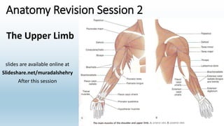
  • 1. Anatomy Revision Session 2 slides are available online at Slideshare.net/muradalshehry After this session The Upper Limb
  • 2. Anatomy Revision Session 1 Objectives of this session • To be able to Identify the muscles and their functions of the upper limb. • Know relations of muscles to important nerves. • There will be a few pop Quiz…. Stay focused ☺
  • 3. Orientation and Movement REVISION adduction abduction flexionextension circumduction rotation
  • 5. Flexor digitorum superficialis • Medial epicondyle of humerus and coronoid process of ulna to middle phalanges (lateral) of digits 2-5 (i.e. all except thumb) • Flexion of metacarpophalangeal (MCP) and proximal interphalangeal (PIP) joint • Median nerve (C7,C8,T1)
  • 6. Flexor digitorum profundus • Ulna and interosseous membrane to distal phalanges digits 2-5 (palmar) • Flexion of MCP, PIP and distal interphalangeal joint (DIP) of digits 2-5 • Ring and little fingers = ulnar nerve (C8,T1) • Index and middle fingers = median nerve (C8,T1 anterior interosseous)
  • 7. Flexor digitorum muscles: Superficialis Splits in two, To Permit Profundus Passing through.
  • 8. Flexor pollicis longus • Radius and interosseous membrane to base of distal phalanx of thumb (palmar) • Flexion of MCP and IP of thumb, wrist • Median nerve (C8, T1 anterior interosseous)
  • 9. Pronator quadratus • Distal anterior ulna to distal anterior radius • Pronation and help to stabilise distal radioulnar joint • Median nerve (C8, T1 anterior introsseous)
  • 10. Anterior forearm muscles: superficial group There are five, like five digits of your hand. Place your thumb into your palm, then lay that hand palm down on your other arm. PFPF [pass/fail, pass/fail]: Pronator teres Flexor carpi radialis Palmaris longus Flexor carpi ulnaris Your thumb below your 4 fingers shows the muscle which is deep to the other four: Flexor digitorum superficialis.
  • 11. Median nerve • C6-T1 fibres, medial and lateral cords • Major nerve of anterior compartment of forearm, THROUGH carpal tunnel • Main nerve of thumb side of hand
  • 12. Ulnar nerve and artery • C7-T1 fibres from medial cord, medial epicondyle of humerus, pisiform ABOVE carpal tunnel • Branch brachial artery, interosseous branches, little finger side, superficial palmar arch
  • 14. Anconeus • Lateral epicondyle of humerus to olecranon of ulna • Extension of the elbow • Radial nerve (C7-C8)
  • 15. Brachioradialis • Distal humerus (supra-epicondylar ridge) to radius proximal to styloid process • Puts the forearm into the semi-prone position, flexion of elbow • Radial nerve (C5, C6, C7) (before division into deep and superficial)
  • 16. Posterior compartment of the forearm • Common extensor origin = lateral epicondyle of the humerus • Extensors of the digits, wrist • Abductor of the thumb • Supinator • Radial nerve
  • 17. Extensor carpi radialis longus and brevis • Extensor carpi radialis longus from distal humerus (supra- epicondylar ridge) to base of 2nd metacarpal • Wrist extension and abduction • Radial nerve (C6,C7) (pre-division) • Extensor carpi radialis brevis from lateral epicondyle of humerus to base of 3rd metacarpal • Wrist extension and abduction • Radial nerve (C7,C8) (deep = posterior interosseous) extensor carpi radialis longus extensor carpi radialis brevis
  • 18. Extensor carpi ulnaris • Lateral epicondyle of humerus (small slip from ulna) to base of 5th metacarpal • Wrist extension and adduction • Radial nerve (posterior interosseous) (C7, C8)
  • 19. Extensor digitorum • Lateral epicondyle of humerus to dorsal expansion of digits 2-5 (NOT thumb) • Extension of principally MCP, but also PIP and DIP extension of digits 2-5 • Radial nerve (posterior interosseous) (C7,C8)
  • 20. Extensor digiti minimi • Lateral epicondyle of the humerus to dorsal expansion of 5th digit (little finger) • MCP, PIP, DIP extension of little finger • Radial nerve (posterior interosseous) (C7,C8)
  • 21. Extensor indices • Posterior surface of ulna (and interosseous membrane) to dorsal expansion of digit 2 (index finger) • MCP, PIP, DIP extension of index finger • Wrist extension • Radial nerve (posterior interosseous) (C7,C8)
  • 22. Extensor pollicis longus • ‘Pollicis’ = thumb • Ulna and interosseous membrane to base of distal phalanx of thumb • CMC, MCP and IP extension of thumb • Radial nerve (posterior interosseous) (C7, C8)
  • 23. Extensor pollicis brevis • Radius and interosseous membrane to base of proximal phalanx of thumb • Carpometacarpal and MCP thumb joint extension • Radial nerve (posterior interosseous) (C7,C8)
  • 24. Abductor pollicis longus • Radius, interosseous membrane and ulna to base of 1st metacarpal • Abducts thumb and extends it at carpometacarpal joint • Radial nerve (posterior interosseous) (C7,C8)
  • 25. Supinator • Crest of ulna, lateral epicondyle of humerus, radial collateral and annular ligament of radius to lateral radius (proximal third) • Supination of forearm (rotates radius) • Radial nerve (posterior interosseous)(C7,C8)
  • 26. Radial nerve passes through the lamina of supinator
  • 29. Flexor retinaculum • Roof of carpal tunnel • Medial = pisiform, hook of hamate • Lateral = tubercle of scaphoid and ridge on trapezium • Septum from trapezium – flexor carpi radialis • Attach thenar and hypothenar structures
  • 31. Carpal tunnel syndrome causes MEDIAN TRAP: Myxoedema Edema premenstrually Diabetes Idiopathic Agromegaly Neoplasm Trauma Rheumatoid arthritis Amyloidosis Pregnancy
  • 32. Bones and joints carpals metacarpals proximal phalanx middle phalanx distal phalanx interphalangeal (IP) joints = synovial hinge metacarpophalangeal (MCP) joints = synovial condyloid carpometacarpal (CMC) joints = synovial saddle (thumb), plane (digits) intercarpal = synovial plane
  • 34. Palmaris brevis • Palmar aponeurosis and flexor retinaculum to dermis • Grip • Ulnar nerve (superficial C8,T1)
  • 35. Intrinsic muscles of the hand abductor pollicis brevis = intrinsic muscle tendon of flexor digitorum superficialis = extrinsic muscle
  • 36. Intrinsic muscles – thenar eminence • Abductor pollicis brevis = scaphoid and trapezium to base proximal phalanx of thumb – CMC joint abduction – median nerve (recurrent C8,T1) • Flexor pollicis brevis = flexor retinaculum, capitate, trapezium to base proximal phalanx of thumb – CMC joint flexion – Median (recurrent) and some texts also ulnar nerve (C8,T1) • Opponens pollicis = trapezium to first metacarpal – CMC joint opposition – median nerve (recurrent C8,T1) abductor pollicis brevis flexor pollicis brevis opponens pollicis
  • 37. Adductor pollicis • Transverse head from 3rd metacarpal; oblique head from capitate and 2nd/3rd metacarpals to base of proximal phalanx of thumb • Adduction of CMC joint of thumb • Flexion of MCP joint of thumb • Ulnar nerve (deep C8,T1) transverse head oblique head
  • 38. Hypothenar eminence • Abductor digiti minimi = pisiform bone to base of 5th proximal phalanx and dorsal digital expansion – abduction and flexion MCP joint little finger – ulnar nerve (deep C8,T1) • Flexor digiti minimi = hamate, flexor retinaculum to base of 5th proximal phalanx – Flex MCP joint of little finger – ulnar nerve (deep C8,T1) • Opponens digiti minimi = hamate and flexor retinaculum to 5th metacarpal – weak opposition of little finger – ulnar nerve (deep C8,T1) abductor digiti minimi flexor digiti minimi opponens digiti minimi
  • 39. Lumbricals • 4 small muscles from tendons of flexor digitorum profundus • 1st (index) and 2nd (middle) unipennate • 3rd (ring) and 4th (little) usually bipennate • Insert into dorsal digital expansion • MCP joint flexion and IP joint extension for digits 2-4 • 1st and 2nd = median nerve (digital C8,T1) • 3rd and 4th = ulnar nerve (deep C8,T1) 1 2 3 4 lumbrical
  • 40. Lumbricals action Lumbricals action Lumbrical action is to hold a pea, that is to flex the metacarpophalangeal joint and extend the interphalangeal joints. When look at hand in this position, can see this makes an "L" shape, since L is for Lumbrical.
  • 41. Palmer interossei • Between metacarpal bones • 1st from 2nd metacarpal to base of 2nd proximal phalanx and dorsal expansion • 2nd from 4th metacarpal to base of 4th proximal phalanx and dorsal expansion • 3rd from 5th metacarpal to base of 5th proximal phalanx and dorsal expansion • Adduction towards middle finger (flex MCP, extend IP) • Ulnar nerve (deep C8,T1) 12 3
  • 42. Dorsal interossei • From sides of 2 adjacent metacarpals, eg, 1st dorsal interossei from 1st and 2nd metacarpals • 1st inserts into 2nd proximal phalanx and dorsal digital expansion • 2nd and 3rd inserts into the 3rd proximal phalanx and dorsal digital expansion • 4th inserts into 4th proximal phalanx and dorsal digital expansion • Abduction (MCP flexion, IP extension) • Ulnar nerve (deep C8,T1) 123 4
  • 43. Interossei muscles : actions of dorsal vs. palmar in hand "PAd and DAb": The Palmar Adduct and the Dorsal Abduct. · Use your hand to dab with a pad.
  • 44.
  • 45. Intrinsic muscles of hand (palmar surface) "A OF A OF A": · Thenar, lateral to medial: Abductor pollicis longus Opponens pollicis Flexor pollicis brevis Adductor pollicis. · Hypothenar, lateral to medial: Opponens digiti minimi Flexor digiti minimi Abductor digiti minimi
  • 46. Synovial sheaths • Synovial membrane plus fluid • Around tendons at wrist, palm and into digits
  • 47.
  • 50. Carpal bones "Stop Letting Those People Touch The Cadaver's Hand Proximal row, lateral-to-medial: Scaphoid Lunate Triquetrum Pisiform Distal row, lateral-to-medial: Trapezium Trapezoid Capitate Hamate Or "Happy Cat Tom Took Pie To Little Sister": Hamate Capitate Trapezoid Trapezium Pisiform Triquital Lunate Scaphoid
  • 52. Gluteal Region and Hip Joint
  • 53. Outline • Innominate (Hip) Bone • Proximal Femur • Muscles of the gluteal region • Nerves and Vessels of the gluteal region • Hip Joint
  • 54. Obturator Foramen Iliac crest Iliac fossa ASIS AIIS PSIS PIIS Arcuate Line Ischial Tuberosity Ischial Spine Lesser Sciatic Notch Greater Sciatic Notch Pubis Innominate: Medial view
  • 55. Innominate: Lateral view Iliac crest ASIS AIIS PSIS PIIS Ischial Tuberosity Ischial Spine Lesser Sciatic Notch Greater Sciatic Notch Pubis Posterior Gluteal Line Anterior Gluteal Line Inferior Gluteal Line Acetabulum Pubic Tubercle Ischium
  • 56. Greater and Lesser Sciatic Foramina
  • 57. Muscles of Gluteal Region Lateral Rotators of Hip • Gluteus Maximus (extensor) • Piriformis • Obturator internus • Gemelli • Quadratus femoris Medial Rotators and Abductors of Hip • Gluteus Medius • Gluteus Minimus Support • Tensor fascia lata
  • 58. Gluteus Maximus • Sacrum, S/T ligament and ilium behind posterior gluteal line  Iliotibial tract (3/4) and gluteal tuberosity of femur (1/4) • Extends hip; assists lateral rotation • Inferior gluteal nerve • (L5, S1, 2)
  • 59. Tensor Fascia Lata • Ant. Iliac Crest and ASIS  Iliotibial tract • Tenses fascia lata and iliotibial tract; Supports femur on tibia during standing • Superior Gluteal nerve • (L4, 5, S1) Gluteus Maximus Iliotibialtract
  • 60. Gluteus Medius • Ilium between posterior and anterior gluteal lines  greater trochanter (lateral surface) • Abducts and medially rotates hip • Superior gluteal nerve • (L4, 5, S1)
  • 61. Gluteus Minimus • Ilium between anterior and inferior gluteal lines  greater trochanter (anterior surface) • Abducts and medially rotates hip • Superior gluteal nerve • (L4, 5, S1)
  • 62. Piriformis • Anterior Sacrum and S/T lig. Greater Trochanter (superior border) • Passes through greater sciatic foramen • Laterally rotates hip • Anterior rami of L5, S1, 2
  • 63. Obturator internus • Pelvic surface of Obturator mem. and surrounding bones  Trochanteric Fossa of greater trochanter • Passes through lesser sciatic foramen • Laterally rotates hip • Nerve to obturator internus (L5, S1)
  • 64. Gemelli • Superior: Ischial spine • Inferior: Ischial tuberosity Blend with tendon of obturator internus (Trochanteric fossa) • Laterally rotate hip • Superior: Nerve to obturator internus • Inferior: Nerve to quadratus femoris
  • 65.
  • 66. Quadratus femoris • Ischial tuberosity Quadrate tubercle on intertrochanteric crest of femur • Laterally rotates hip • Nerve to quadratus femoris (L5, S1)
  • 67. Obturator externus • Outer surface of obturator mem.  Trochanteric fossa • Laterally rotates hip • Obturator nerve • L3, 4 Anterior
  • 68. Lateral Rotators "Play Golf Or Go On Quacking": · From top to bottom: Piriformis Gemellus superior Obturator internus Gemellus inferior Obturator externus Quadratus femoris · Alternatively: "P-GO-GO-Q".
  • 69. Sciatic nerve • L4, 5, S1, 2, 3 • Tibial always leaves below piriformis • Common Peroneal may leave below, above or through piriformis
  • 70. Gluteal vessels and nerves • Superior gluteal artery and nerve • Inferior gluteal artery and nerve • Piriformis
  • 71. Posterior cutaneous nerve of thigh • Lies on posterior aspect of sciatic nerve • (S1, 2, 3) • Inferior Clunial nerves to skin of inferior half of buttock
  • 72. Cutaneous Nerves – Gluteal and Posterior Thigh Superior Clunial (posterior rami L1,2,3) Middle Clunial (posterior rami S1,2,3) Posterior Cuntaneous n. of Thigh with Inferior Clunial branches
  • 73. Hip Joint • Articular surfaces • Capsule attachments • Ligaments • Relations of the Joint
  • 76. Iliofemoral ligament • AIIS → Intertrochanteric line • Upper and lower bands Limits: • Extension • Lateral rotation • Adduction (upper) • Abduction (lower)
  • 77. Pubofemoral ligament • Iliopubic eminence → Lower part of intertrochanteric line Limits: • Extension • Lateral rotation • Abduction
  • 78. Ischiofemoral ligament • Posterior aspect of acetabulum → Greater trochanter Limits: • Extension • Medial rotation • Adduction
  • 79. Blood Supply to the Hip Joint
  • 81. Outline • Compartments of the Thigh • Features of the femur • Muscles of Anterior Compartment • Muscles of Medial Compartment • Adductor Canal • Nerves • Arteries
  • 82. Compartments of Thigh Flex Hip; Extend Knee Anterior Compartment Subcutaneous tissue Fascia Lata Medial Compartment Posterior Compartment Great Saphenous vein Intermuscular Septum Iliotibial tract Extend Hip; Flex Knee Adduct Hip
  • 83. Proximal femur (anterior) Head Greater trochanter Intertrochanteric line Lesser trochanter Shaft Fovea Neck
  • 84. Proximal femur (posterior) Greater trochanter Trochanteric Fossa Intertrochanteric crest Lesser trochanter Pectineal line Gluteal tuberosity
  • 85. Anterior Thigh Hip Flexors • Iliacus • Psoas major • Pectineus • Sartorius Knee Extensors • Quadriceps • Rectus femoris • Vastus medialis • Vastus lateralis • Vastus intermedius
  • 86. Hip Flexors – Iliopsoas • Iliacus • Iliac crest, fossa, ala of sacrum, ant. sacroiliac lig. → Psoas tendon, lesser trochanter • Femoral nerve (L2, 3) • Flex hip • Psoas major • T12-L5, IV discs, lumbar transverse processes → Lesser trochanter • Anterior rami L1-3 • Flex hip
  • 87. Hip Flexors – Pectineus • Pectineus • Superior ramus of pubis → Pectineal line of femur • Femoral nerve (L2, 3) [occassionally br. from obturator] • Adduct and flex hip; assist with medial rotation
  • 88. Hip Flexors – Sartorious • Sartorious • ASIS → Superior part of medial surface Tibia • Femoral nerve (L2, 3) • Flex, abduct, laterally rotate hip; flex knee
  • 89. Knee Extensors – Quadriceps • Rectus femoris • AIIS, ilium above acetabulum • Vastus lateralis • Gr. Trochanter & Lat. Linea Aspera • Vastus medialis • Intertrochanteric line & Med. Linea Aspera • Vastus intermedius • Ant. and Lat. Shaft of femur • → Quadriceps tendon then tibial tuberosity via Patellar lig. • Extend knee (rectus fem. also flexes hip) • Femoral nerve (L2,3,4) Rectus Femoris Vastus Lateralis Vastus Medialis Vastus Intermedius
  • 90. Anterior Thigh Rectus Femoris Vastus Lateralis Vastus Medialis Sartorious Iliopsoas Pectineus
  • 91. Femoral nerve • Passes deep to inguinal ligament, medial to ASIS, on tendon of iliopsoas m. • Muscular branches: anterior thigh • Articular branches: hip and knee • Cutaneous branches: anteromedial thigh • Saphenous nerve is terminal cutaneous br. – anteromedial knee, leg and foot Iliacus Rectus femoris Vasti muscles Pectineus
  • 92. Medial Thigh Hip Adductors • Gracilis • Adductor Longus • Adductor Brevis • Adductor Magnus • Obturator Externus
  • 93. Hip Adductors – Gracilis • Gracilis • Body and Inf. ramus of Pubis →Superior Medial surface Tibia • Obturator (L2, 3) • Adducts hip; flexes knee
  • 94. Hip Adductors – Adductor Longus • Adductor Longus • Body pubis → Middle ⅓ Linea aspera • Obturator (L2,3,4) • Adducts thigh
  • 95. Hip Adductors – Adductor Brevis • Adductor Brevis • Body and Inf. ramus of Pubis →Pectineal Line and proximal Linea aspera • Obturator (L2,3,4) • Adducts thigh (may assist flexion)
  • 96. Hip Adductors – Adductor Magnus • Adductor Magnus • Ischiopubic ramus and Ischial tuberosity →Linea aspera, Med. Supracondylar line, Adductor tubercle • Obturator (L2,3,4); Hamstring part – tibial part Sciatic n. (L4) • Adducts thigh; Adductor part flexes and hamstring part extends thigh
  • 97. Hip Adductors – Obturator Externus • Obturator Externus • Margins obturator foramen and Obturator membrane →Trochanteric fossa • Obturator (L3,4) • Laterally rotates thigh
  • 99. Obturator Nerve Obturator externus Adductor Brevis Adductor Longus Adductor Magnus Gracilis • Anterior divisions L2-4 • Runs along lateral wall of pelvis to the obturator canal • Anterior and Posterior branches • Muscular branches to medial thigh • Anterior branch – cutaneous to middle part of medial thigh Obturator Externus Adductor Brevis Posterior Branch Anterior Branch Posterior Branch Anterior Branch
  • 100. Adductor Canal • Apex of Femoral Triangle  Adductor Hiatus (in adductor magnus) • Underlies the distal half of sartorius m. • Femoral vessels travel in canal and pass through Hiatus to reach Popliteal fossa • Saphenous nerve runs in canal then passes between sartorius and gracilis to supply skin of anteromedial knee, leg and foot Femoral a. and v. entering adductor canal Saphenous n. Sartorius forming roof of adductor canal
  • 101. Arterial Supply Femoral artery – Continuation of Ext. Iliac a. • Femoral triangle → Adductor canal → Adductor hiatus to become Popliteal a. • Supplies anterior and anteromedial thigh Profunda femoris → br. of Femoral • Runs posterior to Adductor Longus → 3/4 perforating br.s through Add. Magnus to supply mm in med., post. and lat. part of ant. Compartment • Also gives Medial and Lateral Circumflex femoral br.s Obturator artery (from Int. Iliac a.) • Through obturator foramen to medial compartment → ant. and post. br.s • Anterior branch: muscles of medial compartment • Posterior branch: muscles attached to ischial tuberosity
  • 103. Posterior thigh and popliteal fossa
  • 104. Outline • Muscles of posterior thigh • Sciatic nerve • Popliteal fossa • Roof • Boundaries • Contents • Floor • Popliteal artery and branches 104
  • 105. Posterior Thigh • Hamstrings • Semitendinousus • Semimembranosus • Biceps Femoris • Extend hip, Flex knee
  • 106. Semitendinosus • Semitendinosus • Ischial tuberosity Medial surface of superior tibia • Tibial division of Sciatic • L5,S1,S2
  • 107. Semimembranosus • Semimembranosus • Ischial tuberosity  Posterior surface of Medial Condyle of tibia • Tibial division of Sciatic • L5,S1,S2
  • 108. Biceps Femoris • Biceps femoris • Long head: Ischial tuberosity • Short head: Linea aspera and Lat. Supracondylar line of femur Head of Fibula • Long head = Tibial division of Sciatic (L5,S1,S2) • Short head = Common peroneal division of Sciatic (L5,S1,S2)
  • 109. Rotation of Knee • Knee Flexed: • Lateral rotation by: • Biceps Femoris • Medial rotation by: • Semimembranosus • Semitendinosus • Gracilis • Sartorius
  • 110. Sciatic Nerve • L4-S3 • Deep to long head of biceps femoris • Tibial and Common Peroneal (fibular) Divisions Tibial nerve Common Peroneal nerve Right
  • 111. Popliteal Fossa – Roof • Deep fascia • Pierced by: • Sural nerve • Short saphenous vein Sural nerve Short Saphenous Vein
  • 112. Popliteal Fossa – Boundaries • Superiorly: Diverging tendons of Hamstrings • Biceps femoris laterally • Semimembranosus and Semitendinosus medially • Inferiorly: Medial and Lateral heads of Gastrocnemius
  • 113. Popliteal Fossa – Contents • Popliteal artery • Popliteal vein • Terminal branches of Sciatic • Tibial nerve • Common peroneal nerve Medial & Deep Lateral & Superficial Popliteal artery Popliteal vein Tibial nerve Common Peroneal nerve
  • 114.
  • 115. • medial to lateral arrangment "Serve And Volley Next Ball": Semimembranosus/ Semitendonosus Artery Vein Nerve Biceps femoris · Lateral and medial heads of Gastrocnemius are inferior borders.
  • 116. Popliteus Joint capsule Femur Popliteal Fossa – Floor • Popliteal surface of femur • Capsule of Knee Jt. • Popliteus muscle
  • 117. Popliteal Fossa – Floor • Oblique popliteal ligament • Expansion of Semimembranosus • Reinforces knee joint capsule • Middle Genicular vessels • Pierce joint capsule to supply cruciate ligaments
  • 118. Popliteal Artery • Continuation of femoral • Adductor hiatus →Inferior border of Popliteus • 5 genicular branches • Terminal branches: • Anterior Tibial artery • Posterior Tibial artery Medial Superior Genicular artery Lateral Superior Genicular artery Medial Inferior Genicular artery Lateral Inferior Genicular artery
  • 120. Outline • Compartments of the leg • Interosseous membrane and superior tibiofibular joint • Muscles of Lateral Compartment • Muscles of Anterior Compartment • Extensor Retinacula • Dorsum of Foot • Peroneal nerves • Arteries of the Leg and Dorsum of Foot
  • 122. Interosseous membrane • Provides a surface for muscle attachment • Fibres pass inferolaterally from Tibia to Fibula • Helps resist the downward pull of muscles attached to the fibula
  • 124. Anterior and Lateral Compartments Anterior Compartment: 4 Muscles Extensors of ankle jt. and digits Tendons anterior to ankle jt. Lateral Compartment 2 Muscles Evertors of the foot Tendons posterior to lateral malleolus
  • 125. Lateral Compartment Peroneus Longus: Head and upper ⅔ of lat. Fibula  base of 1st Metatarsal and Medial Cuneiform Superficial Peroneal Nerve (L5,S1,S2)
  • 126. Lateral Compartment Peroneus Brevis: Inf. ⅔ of lat. Fibula  Tuberosity on Lat. side of Base of 5th Metatarsal Superficial Peroneal Nerve (L5,S1,S2)
  • 127. Lateral Compartment Peroneus Longus Tendon Peroneus Brevis Tendon Peroneal Trochlea of Calcaneus Cuboid 5th Metatarsal Calcaneus Talus
  • 128. Anterior compartment • Tibialis anterior • Extensor digitorum longus • Extensor hallucis longus • Peroneus tertius • All supplied by Deep Peroneal Nerve • L4, L5
  • 129. Tibialis Anterior Lat. condyle of Tibia, Sup. ½ of Lat. Tibial surface, Interosseous mem. → Med. and Inf. surfaces of Medial Cuneiform and base of 1st metatarsal Dorsiflexes ankle; Inverts foot R R
  • 130. Extensor Digitorum Longus Lat. Condyle Tibia, Sup. ¾ of med surface Fibula, Interosseous membrane Middle and distal Phalanges of Lateral 4 digits Extends lateral 4 digits; Dorsiflexes ankle R
  • 131. Inf. ⅓ anterior Fibula and Interosseous membrane Dorsum of base of 5th Metatarsal Dorsiflexes ankle; Assists in eversion of foot Peroneus Tertius R
  • 132. Extensor Hallucis Longus Middle ant. surface Fibula, Interosseous membrane Dorsal aspect of distal Phalanx of Hallux Extends Hallux; Dorsiflexes ankle
  • 133. Extensor Retinacula Superior Extensor Retinaculum Ant. Border of Tibia  Lower end of Fibula Inferior Extensor Retinaculum (Y-shaped) Stem attached to Calcaneus laterally Upper Limb attached to Medial Malleolus Lower Limb passes round medial border of foot to blend with dense fascia over abductor hallucis
  • 134. Anterior muscles of leg "The Hospitals Are Not Dirty Places": T: Tibialis anterior H: extensor Hallucis longus A: anterior tibial Artery N: deep fibular Nerve D: extensor Digitorum longus P: Peronius tertius [aka fibularis tertius]
  • 136. Muscles of Dorsum of Foot Superior surface of Calcaneus, Inferior Extensor Retinaculum Extensor Hallucis Brevis Base of Proximal Phalanx of Hallux Extensor Digitorum Brevis  Long extensor tendons of toes 2-4 Deep Peroneal nerve L5/S1
  • 137. Superficial peroneal nerve • Branch of common peroneal between peroneus longus and neck of fibula • Lateral compartment of leg supplying muscles and continuing as cutaneous nerve • Cutaneous innervation to distal anterior surface of leg and most of dorsum of foot
  • 138. Deep Peroneal Nerve • Branch of common peroneal between peroneus longus and neck of fibula • Passes through extensor digitorum longus and travels with anterior tibial a. supplying all anterior compartment mm. • Crosses the ankle jt. to supply ext. digitorum brevis and ext. hallucis brevis • Cutaneous innervation to skin between digits 1 and 2
  • 139. Cutaneous Innervation Anterior Cutaneous branches of Femoral Saphenous nerve (from Femoral)Lateral Sural Cutaneous nerve Superficial Peroneal nerve Deep Peroneal nerve Lateral Dorsal Cutaneous nerve of foot (Termination of Sural n.)
  • 140. Arteries of the Leg • Popliteal a. gives anterior and posterior tibial aa. • Anterior tibial a. – through interosseous membrane and descends on this • Posterior tibial a. – gives the fibular a. that provides perforating br.s to lateral compartment Anterior Tibial a. Posterior Tibial a. Fibular a.
  • 141. • Dorsalis Pedis artery is a continuaton of Anterior Tibial a. • Gives Deep Plantar artery to sole of the foot • Gives Arcuate artery which runs across metatarsals to anastomose with Lateral Tarsal a. • Metatarsal and digital aa. Arteries of Dorsum of Foot Anterior Tibial a. Dorsalis Pedis a. Deep Plantar a. Arcuate a. Lateral Tarsal a.
  • 142. Posterior Leg and Ankle Joint
  • 143. Outline • Posterior aspect of the bones of the leg • Superficial and deep muscles of the posterior compartment • Flexor retinaculum • Posterior arteries • Tibial nerve • Inferior Tibiofibular joint • Ankle joint
  • 144. Posterior Aspect of Leg Bones Popliteal area Soleal line Apex Head Neck Medial Malleolus Lateral Malleolus Tibia Fibula Fibula Groove for Peroneus longus and brevis tendons The FibuLA is LAteral.
  • 145. Muscles of the Posterior Compartment • 7 muscles • 3 superficial; 4 deep • Superficial muscles all insert on posterior surface of calcaneous via Tendocalcaneous • Tendons of deep muscles pass behind medial malleolus to plantar surface of foot
  • 146. Superficial Muscles - Gastrocnemius Medial Head: Popliteal surface of femur sup. to medial condyle Lateral Head: Lateral aspect of lateral condyle of femur Tibial nerve (S1, 2) Plantarflexes ankle; Flexes knee Right
  • 147. Superficial Muscles - Plantaris Lateral supracondylar line of femur (proximal to lateral head of gastrocnemius) Tibial nerve (S1, 2) Assists Plantarflexion of ankle Right
  • 148. Superficial Muscles - Soleus Soleal line of Tibia Upper ⅓ posterior Fibula Tendinous arch between bony attachments Tibial nerve (S1, 2) Plantarflexes ankle joint Right
  • 149. Deep Muscles - Popliteus Pit for popliteus (lateral condyle of femur) and Lateral meniscus →Popliteal area of tibia (above soleal line) Tibial n. (L4, 5, S1) Unlocks knee joint by laterally rotating femur on fixed tibia Right
  • 150. Deep Muscles – Flexor Digitorum Longus Posterior Surface of Tibia →Base of distal Phalanx of digits 2-4 Tibial nerve (L5, S1, 2) Flexes lateral 4 digits Weak plantarflexor of ankle Right
  • 151. Deep Muscles – Flexor Hallucis Longus Posterior Surface of Fibula →Base of distal Phalanx of Hallux Tibial nerve (L5, S1, 2) Flexes Hallux Weak plantarflexor of ankle Right
  • 152. Deep Muscles – Tibialis Posterior Posterior Surface of Tibia and Fibula, Interosseous membrane →Tuberosity of Navicular, Cuneiforms, Cuboid, Sustentaculum tali of Calcaneus, base of 2nd, 3rd, and 4th Metatarsals Tibial nerve (L4, 5) Plantar flexes ankle Inverts foot Right
  • 153. Inversion vs. Eversion Muscles in leg Second letter rule for inversion/eversion: · Eversion muscles: pErineus longus pErineus brevis pErineus terius · Inversion muscles: tIbialis anterior tIbialis posterior
  • 154. Plantarflexion vs. Dorsiflexion Plantar flexion occurs when you squish a Plant with your foot. Dorsiflexion Look at your toe nails
  • 155. Tendons of Deep Muscles Tibialis Posterior tendon passes deep to Flexor Digitorum Longus Groove posterior to the medial malleolus Flexor Hallucis Longus grooves the posterior surface of the lower end of the tibia, the posterior surface of the talus Medial to lateral arrangement of tendons at ankle joint: Tibialis Posterior, Flexor Digitorum Longus and Flexor Hallucis Longus (Tom, Dick and Harry) TP FDL FHL
  • 156. Flexor Retinaculum Medial Malleolus→ Calcaneous Posterior tibial vessels and tibial nerve found between FDL and FHL tendons Right
  • 157. Tarsal tunnel: contents "Tiny Dogs Are Not Hunters“ Or : From superior to inferior: T: Tibialis posterior F: flexor Digitorum longus A: posterior tibial Artery N: tibial Nerve H: flexor Hallucis longus
  • 158. Arteries of Posterior Leg Posterior Tibial Artery: terminal branch of popliteal artery Between heads of gastrocnemius and deep to soleus to run on surface of TP and FDL Gives Fibular artery as a branch Terminates as Medial and Lateral Plantar arteries in the foot Fibular artery: runs medial to fibula, usually within FHL Tibial a. Fibular a.
  • 159. Cutaneous Innervation Posterior Cutaneous nerve of Thigh Lateral Sural Cutaneous nerve Medial Sural Cutaneous nerve (Sural Nerve) Saphenous nerve (from Femoral)
  • 160. Tibial nerve • Passes between the heads of gastrocnemius and deep to tendinous arch of soleus, with Post. Tibial a. • Lies between flexor hallucis longus and flexor digitorum longus at the ankle jt. • Gives terminal branches that supply the sole of the foot: Medial and Lateral Plantar nn
  • 162. Ankle joint Distal ends of Tibia and Fibula form: Malleolar Mortise Trochlea of Talus
  • 165. Capsule and Ligaments Attaches around articular margins Anteriorly extends onto neck of talus Weak anteriorly and posteriorly Supported laterally and medially by strong Collateral Ligaments Anterior and posterior views with weak parts of capsule removed
  • 166. Lateral Collateral Ligaments Anterior Talofibular → neck of talus Posterior Talofibular → lateral tubercle of talus Calcaneofibular → lateral surface of calcaneous
  • 167. Lateral Collateral Ligaments Calcaneofibular Ant. talofibular Posterior talofibular Post. talofibular Right Right
  • 168. Medial Collateral Ligament (Deltoid) Fans out from Medial malleolus and has 4 parts: 1) Anterior Tibiotalar 2) Tibionavicular 3) Tibiocalcaneal 4) Posterior Tibiotalar 1 2 3 4 3 4
  • 170. Outline • Bones of the foot • Deep fascia • The four layers of the foot • Plantar arteries • Plantar nerves • Cutaneous nerves of the foot
  • 171. Foot – Dorsal View Phalanges Metatarsals Tarsals Navicular Talus Calcaneus Cuneiforms (medial, intermediate, lateral)
  • 172. Foot – Ventral View Phalanges Metatarsals Tarsals Navicular Sustentaculum Tali Cuboid Tubercle of Calcaneus Cuneiforms Sesamoid bones within Flexor Hallucis Brevis tendon Head of Talus
  • 173. The Tarsal Bones “The Circus Needs More Interesting Little Clowns”. • T: Talus • C: Calcaneus • N: Navicular • M: Medial cuneiform • I: Intermediate cuneiform • L: Lateral cuneiform • C: Cuboid Sustentaculum Tali Cuboid Tubercle of Calcaneus Cuneiforms Head of Talus
  • 174. Deep Fascia & Plantar Aponeurosis Deep Fascia: Thick central portion Weaker medially and laterally Plantar Aponeurosis: Superficial Ligament formed by central portion of deep fascia Proximal attachment to Calcaneus Divides into 5 bands distally – continuous with fibrous digital sheaths Vertical intermuscular septae: Medial, Central and Lateral compartments
  • 175. Compartments of the Foot Lateral Compartment Muscles of the little toe: Abductor Digiti Minimi Flexor Digiti Minimi Brevis Medial Compartment Muscles of the great toe: Abductor Hallucis Flexor Hallucis Brevis Central Compartment Flexor Digitorum Brevis Muscles associated with tendon of FDL (lumbricals and quadratus plantae) Adductor Hallucis Interossei Plantar and Dorsal interossei Muscles of Dorsum of Foot Extensor Digitorum Brevis Extensor Hallucis Brevis
  • 176. Foot – Layer 1 Abductor Hallucis (1) Med. Tubercle of Calcaneus; Flex. Retinaculum → Med. Base proximal Phalynx Flexor Digitorum Brevis (2) Medial Tubercle of Calcaneus → Middle Phalanges of lateral 4 toes Abductor Digiti Minimi (3) Tubercle of Calcaneus → Lat. Base proximal Phalynx 1 2 1 2 3 3
  • 177. Foot – Layer 2 Long flexor tendons to the toes: FDL and FHL Lumbricals * Tendons of FDL → Med. side of dorsal tendon expansion Quadratus Plantae (QP) Med. and Lat. surfaces of Calcaneus → Lateral side of FDL tendon * * * * QPQP FDL FDL * * **
  • 179. Foot – Layer 3 Flexor Hallucis Brevis Cuboid and Lat. Cuneiform → Both sides base Proximal Phalanx Adductor Hallucis Transverse head: Plantar ligament of MTPJoints Oblique head: Base metatarsals 2&4 → Lat. side base Proximal Phalanx Flexor Digiti Minimi Brevis* Base 5th Metatarsal → Base Proximal Phalanx FHB FHB Ad H Ad H Ad H * *
  • 180. Foot – Layer 4 Interossei 3x Plantar Interossei Bases and Med. side of Metatarsals 3-5 (unipennate) → Med. side of proximal phalanx of 3rd -5th digit PAD – Plantar ADduct 4x Dorsal Interossei Adjacent sides of Metatarsals 1-5 (bipennate) → 1st on med. side proximal phalanx of 2nd digit →2nd-4th on lat. side proximal phalanx of 2nd -4th digits DAB – Dorsal ABduct
  • 183. Layer 4 Tendons: Peroneus Longus * Tibialis Posterior (TP) Long Plantar * * * * TP TP TP TP
  • 185. Plantar Nerves Terminal branches of Tibial nerve deep to flexor retinaculum Enter foot deep to Abductor Hallucis Medial Plantar Nerve: Between Abductor Hallucis and FDB Lateral Plantar Nerve: Between Layers 1&2 Deep and Superficial branches Deep branch btwn Layers 3&4 Tibial nerve Sural nerve
  • 186. Medial Plantar Nerve: Abductor Hallucis Flexor digitorum brevis 1st Lumbrical Flexor hallucis brevis Lateral Plantar Nerve: All other muscles S2, 3 Innervation of Muscles of Foot Medial Plantar n. Lateral Plantar n.
  • 187. Cutaneous Innervation of the Foot Superficial Peroneal Deep Peroneal Saphenous Saphenous Dorsal Lateral Cutaneous of Foot Dorsal Lateral Cutaneous of Foot Medial Plantar Lateral Plantar Calcaneal Branches
  • 188. Plantar Arteries Terminal branches of Posterior Tibial artery deep to flexor retinaculum Enter foot deep to Abductor Hallucis Medial Plantar artery: Muscles of hallux and overlying skin Occassionally: Superficial Plantar Arch Lateral Plantar artery: Initially btwn Layers 1 and 2 Deep Plantar Arch btwn Layers 3 and 4 Deep Plantar a. from Dorsalis Pedis Plantar Metatarsal and Digital aa. Medial Plantar a. Lateral Plantar a. Superficial Plantar Arch Deep Plantar Arch