This document provides an overview of the anatomy of the hand, including:
- Surface anatomy and bony landmarks of the hand and wrist
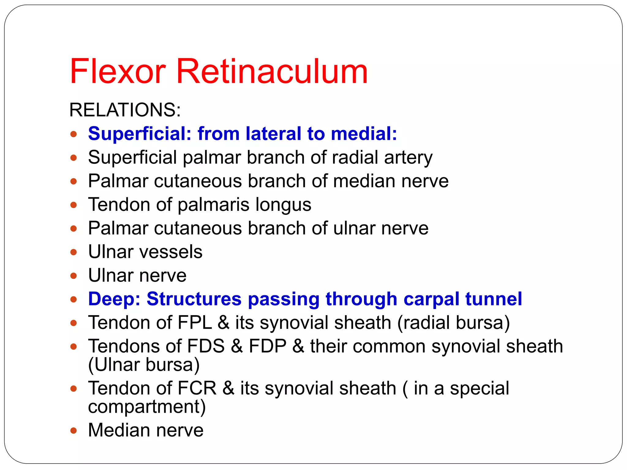
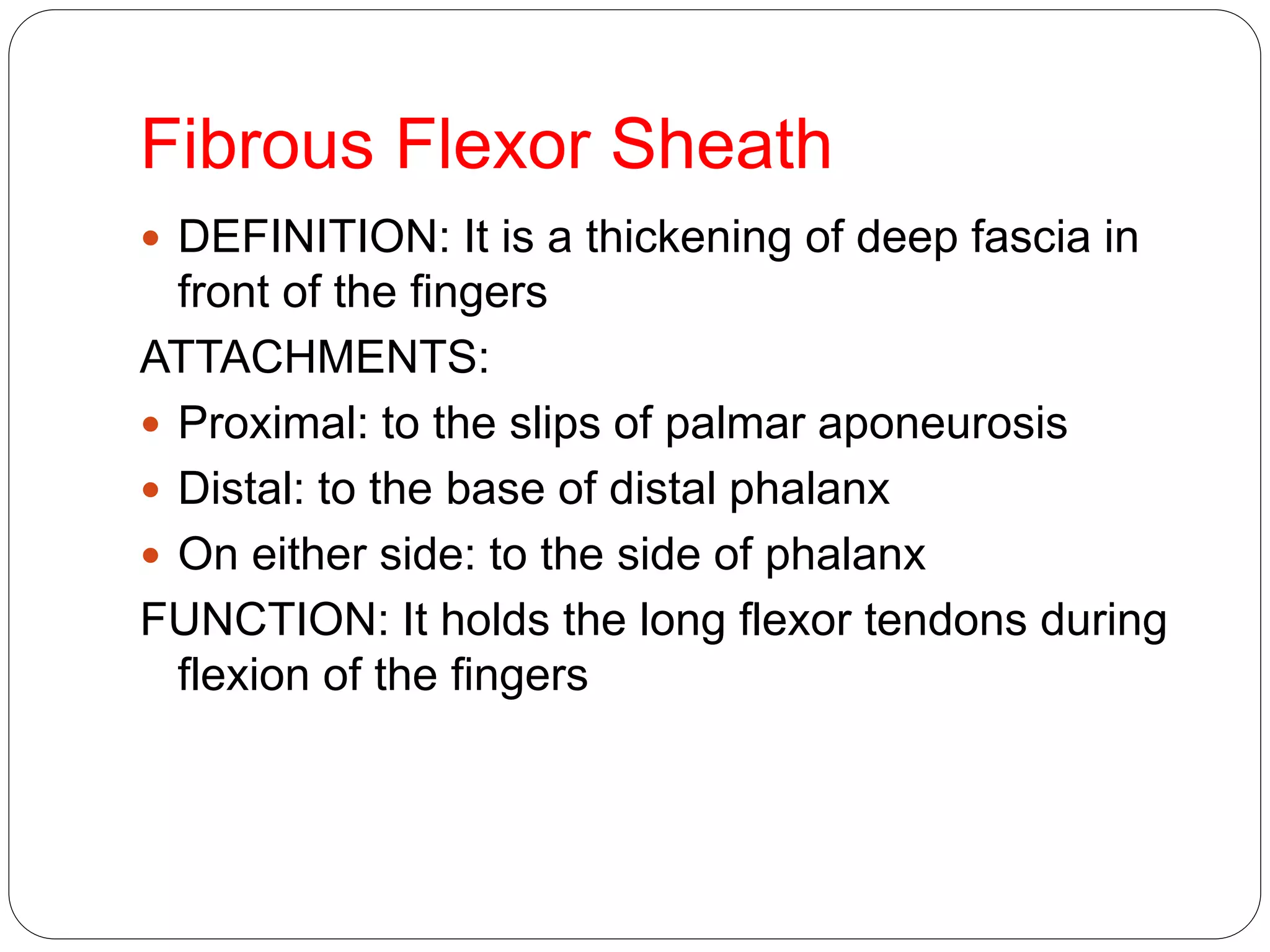
- Compartments and spaces of the hand, including the palmar aponeurosis and flexor retinaculum
- Intrinsic muscles of the hand grouped into thenar, hypothenar, lumbrical and interossei muscles
- Arterial arches including the superficial and deep palmar arches
- Nerve innervation including the median, ulnar and radial nerves
- Clinical concerns involving the hand like carpal tunnel syndrome and De Quervain's tenosynovitis are also discussed.
Application of Matrices in real life. Presentation on application of matrices
740_Hand.ppt
1. Dr. K. S. Ravi
MBBS, MD(JIPMER), MAMS
Department of Anatomy
AIIMS Rishikesh
2. Learning objectives
Surface anatomy of Hand
Bony landmarks
Compartments and Spaces of hand
Flexor Retinaculum & Structures passing below
& above FR
Intrinsic muscles of hand
Superficial & Deep Palmar Arches
Dorsum of hand & Dorsal venous Arch
Nerves in hand
Applied anatomy
13. Palmar Aponeurosis
It is a thickening of deep fascia in the middle of the palm
DESCRIPTION: It is triangular in shape:
Apex: directed proximally, continuous with tendon of
palmaris longus
Base: directed distally, divided into 4 slips for the
medial 4 fingers
Margins: send septa to metacarpal bones separating
the structures under the aponeurosis from thenar &
hypothenar muscles
FUNCTION: It protects the underlying tendons, vessels
& nerves
CLINICAL ANATOMY: DUPUYTREN’S
CONTRACTURE: shortening of the medial part of
15. Flexor Retinaculum
It is a thickening of deep fascia that lies over the
front of the carpal bones converting the carpal
groove (formed by carpal bones) into a tunnel
ATTACHMENTS:
Lateral: by 2 laminae: superficial (to tubercles of
scaphoid & trapezium) & deep (to the medial lip
of the groove on the trapezium)
Medial: to pisiform & hook of hamate
16. Flexor Retinaculum
RELATIONS:
Superficial: from lateral to medial:
Superficial palmar branch of radial artery
Palmar cutaneous branch of median nerve
Tendon of palmaris longus
Palmar cutaneous branch of ulnar nerve
Ulnar vessels
Ulnar nerve
Deep: Structures passing through carpal tunnel
Tendon of FPL & its synovial sheath (radial bursa)
Tendons of FDS & FDP & their common synovial sheath
(Ulnar bursa)
Tendon of FCR & its synovial sheath ( in a special
compartment)
Median nerve
17. Flexor Retinaculum
FUNCTION: It keeps the flexor tendons in
position during movement of wrist joint
CLINICAL ANATOMY (CARPAL TUNNEL
SYNDROME): Compression of median nerve
under the flexor retinaculum
19. Fibrous Flexor Sheath
DEFINITION: It is a thickening of deep fascia in
front of the fingers
ATTACHMENTS:
Proximal: to the slips of palmar aponeurosis
Distal: to the base of distal phalanx
On either side: to the side of phalanx
FUNCTION: It holds the long flexor tendons during
flexion of the fingers
20. Intrinsic Muscles OfHand
Situated totally within the hand
Divided into 4 groups:-
•Thenar
•Hypothenar
•Lumbricals
•Interossei muscles
21. Intrinsic Muscles
LATERAL GROUP:
FOUR THENAR MUSCLES
MEDIAL GROUP:
THREE HYPOTHENAR MUSCLES
PALMARIS BREVIS
CENTRAL GROUP:
FOUR LUMBRICALS
FOUR PALMAR INTEROSSEI
FOUR DORSAL INTEROSSEI
ALL MUSCLES ARE SUPPLIED BY C8 & T1 SPINAL
SEGMENTS THROUGH MEDIAN & ULNAR NERVES
23. The Thenar Group
•Abductor Pollicis
Brevis
•Flexor Pollicis Brevis
•Opponens Pollicis
•Adductor Pollicis
Muscles
24. ABDUCTOR POLLICIS BREVIS
Origin Scaphoid & Trapezium &
Flexor Retinaculum
Insertion Lateral side of base of
proximal phalanx of thumb
Action Abducts thumb
Innervation median nerve (C8 and
T1
25. Flexor Pollicis Brevis
Origin S-Tubercle of Trapezium
D-Capitate & Trapezoid bones
Insertion Lateral side of base
of proximal phalanx of thumb
Action Flexes thumb
InnervationRecurrent branch of
median nerve (C8 and T1)
26. Opponens Pollicis
Origin Flexor retinaculum and
Tubercles of Trapezium
Insertion Lateral side of 1st
metacarpal
Action Draws 1st metacarpal
laterally to oppose thumb toward
center of palm
Innervation Recurrent branch of
median nerve (C8 and T1)
27. Adductor Pollicis
It has 2 heads that are separated by a
gap through which the radial artery
passes
Origin: Oblique head – capitate &
bases of 2nd and 3rd metacarpals,
Transverse head – anterior surface of
body of 3rd metacarpal bone
Insertion: medial side of base of
proximal phalanx of thumb
Innervation: ulnar nerve
Action: adducts thumb towards middle
digit
28. The Hypothenar Group
Hypothenar muscles
1. Opponens Digiti
Minimi
2.Abductor Digiti Minimi
3.Flexor Digiti Minimi
Brevis
4.Palmaris Brevis
29. Opponens Digiti Minimi
Origin Hook of hamate
and flexor retinaculum
Insertion Medial border
of 5th metacarpal
Action Brings little finger
(5th digit) into opposition
with thumb
InnervationDeep branch
of ulnar nerve (C8 and T1)
30. Abductor Digiti Minimi
The most superficial of the
hypothenar muscles forming
the hypothenar eminence
Origin : Pisiform bone
Insertion: Medial side of
base of proximal phalanx of
5th digit
Action: Abducts 5th digit
31. Flexor Digiti Minimi Brevis
Origin Hook of hamate
and flexor retinaculum
Insertion Medial side of
base of proximal phalanx
of little finger
Action Flexes proximal
phalanx of little (5th) finger
Innervation ulnar
nerve
32. Palmar Brevis
It lies in the fascia deep to the
skin of the hypo thenar em
inence
A relatively unimportant mu scle
except that it covers and protects
the ulnar nerve and artery
Origi n: Flexor retinaculum and
palmar aponeurosis
Inserti on: Skin on the medial side
of the palm
Acti on: Wrinkles the skin on the
medial side of the palm and deepens
the hollow of the palm, as in cupping
of the hand, thereby aiding the grip
33. INTEROSSEI MUSCLES
Interosseous Muscles
They are located between the
metacarpal bones
Arranged in 2 layers: 4 Palmar
and 4 Dorsal
Located between bones 1. Dorsal
interossei 1 to 4
Origin: Adjacent sides of 2 metacarpal
bones
Insertion : Extensor expansion's and
bases of proximal phalanges of digit 2
to 4
Action: Abducts digits and assist
lumbricals
2. Palmar interossei 1 to 4
Origin : Palmar surfaces of 2nd, 3th , 4th
and 5th metacarpal bones
Insertion: Extensor expansion of digits
and bases of proximal phalanges of
digits 2, 4, and 5
Action: Adducts digits and assist
lumbricals
34. Lumbrical Muscles
They are named as such because
of their elongated worml i ke
form
1.Lumbri cal s 1 and 2
Ori gi n : lateral 2 tendons of flexor
digitorum profundus
2.Lumbri cal s 3 and 4
Ori gi n: medial 3 tendons of flexor
digitorum profundus
Insertion: lateral sides of extensor
expansions of digits 2 to 5
Acti on: To flex digits at MCP joints
and extend IP joints
35. ARTERIAL ARCHES IN HAND
SUPERFICIAL PALMAR
ARCH
DEEP PALMAR ARCH
Formation
Site
Surface anatomy
Branches
36. Superficial Palmar Arch
FORMATION:
Direct continuation of ulnar artery (mainly)
Superficial branch of radial artery
SITE: between palmar aponeurosis & long flexor
tendons
SURFACE ANATOMY: level with the distal border of the
fully extended thumb
BRANCHES: digital branches to the medial three & half
fingers
N.B.: Radial artery gives 2 branches that supplies the
lateral one & half fingers:
Radialis indicis: supplies lateral side of index
Princeps pollicis: supplies both sides of thumb
37. Deep Palmar Arch
FORMATION:
Direct continuation of radial artery (mainly)
Deep branch of ulnar artery
SITE: between long flexor tendons & metacarpal
bones
SURFACE ANATOMY: lies one inch proximal to
superficial palmar arch
BRANCHES:
Branches sharing in anastomosis around wrist joint
Articular & muscular branches
47. Clinical Concerns
Carpal tunnel Ganglion cyst
- The transverse carpal ligament in the wrist
puts pressure on the medial nerve
- - possible cause: overuse, hormonal
- http://www.youtube.com/watch?v=SGyKQ
chSEJ4
- Is a fluid filled cyst that develops
out of a joint.
- - possible cause: joint trauma
- http://www.youtube.com/watch?v=
mJ6oj3lkqm8
48. De Quervain’s Tenosynovitis
-irritation of the sheath around the tendon
- affects the tendons on the thumb side of the wrist
- possibly caused by repetitive actions, over use
- http://www.youtube.com/watch?v=q87zSRYHa1o
49. 1. Nerve damage that impairs the flexion of distal
interphalangeal joints of index and middle fingers also
produces which of the following conditions ?
a) Atrophy of Hypothenar eminence
b) Loss of adduction of thumb
c) Similar impairment of flexion of distal I.P joint of little finger
d) Weakness in pronation of forearm
2.A man is unable to hold a postcard between his index and
middle fingers because of an injury to which of the
following nerves ?
a) Superficial branch of ulnar nerve
b) Deep branch of ulnar nerve
c) Recurrent branch of median nerve
d) Posterior interosseous nerve
50. 3. All of the following muscles form the
boundry of the anatomical snuff box,
EXCEPT :
a) Brachioradialis
b) Abductor pollicis longus
c) Extensor pollicis longus
d) Extensor pollicis brevis
4. Which is the longest metacarpal bone ?
a) Second metacarpal
b) Third metacarpal
c) Fourth metacarpal
d) Fifth metacarpal
51. 5. After falling on his outstretched hand a patient
complains of tenderness in the space between
Extensor pollicis brevis and extensor pollicis
longus tendons. Which bone is most likely
fractured by the injury ?
a) Scaphoid
b) First metacarpal
c) Radial styloid process
d) Trapezium
6. Which is the most frequently fractured carpal
bone ?
a) Lunate
b) Scaphoid
c) Capitate
d) Trapezium
52. 7. A middle aged woman suffering from myxedema, suddenly
woke up one night by a severe bout of pain in her right wrist
and middle finger. The pain seems to move up her forearm.
After a thorough neurological check up her physician
diagnosed her condition as Carpal Tunnel Syndrome.Which
nerve is involved in this syndrome ?
a) Median nerve
b) Palmar cutaneous branch of median nerve
c) Ulnar nerve
d) Anterior interosseous nerve
8. Examination of a patient reveals paralysis of the Abductor pollicis
brevis muscle. All of the following can be sites of lesion that
resulted in this paralysis , EXCEPT :
One answer only.
a) Lower trunk of brachial plexus
b) Lateral root of median nerve
c) Medial root of median nerve
d) Recurrent branch of median nerve
53. 9. A patient exhibits weakness of Pinch grip ;
other thumb movements are normal. There is
no sensory loss in the hand. The probable cause
is damage to :
a) Posterior interosseous nerve
b) Anterior interosseous nerve
c) Deep branch of ulnar nerve
d) Median nerve proximal to flexor retinaculum
10. In carpal tunnel syndrome which of the
follwoing conditions occur due to motor deficit ?
a) Claw hand
b) Pointing index finger
c) Benediction hand
d) Simian hand
Rx PG MCQ