Necrosis is the death of cells and living tissue. There are several types of necrosis that are characterized by the microscopic appearance of dead cells. Coagulative necrosis occurs due to ischemia and results in pale, swollen tissue on gross examination and swollen eosinophilic cells on microscopy. Liquefactive necrosis occurs due to infection and results in soft, cystic lesions containing debris. Caseous necrosis is seen in tuberculosis and resembles dry cheese with granular debris. Fat necrosis affects pancreatic and breast tissue, appearing as yellowish deposits containing calcium salts. Fibrinoid necrosis involves fibrin deposition in tissues damaged by immune processes.
Introduction to necrosis by Mohamed Faizal Asan from the General Pathology department.
Necrosis is localized tissue death, followed by degradation by hydrolytic enzymes with inflammatory response.
In necrosis, lytic enzymes digest cells leading to homogeneous eosinophilic cytoplasm, and proteins undergo denaturation causing nuclear changes like pyknosis.
There are five types of necrosis: Coagulative, Liquefactive, Caseous, Fat, and Fibrinoid necrosis.
Most common necrosis type due to ischemia, affecting heart, kidney, and spleen; characterized by swelling and eosinophilia.
This type occurs due to ischemic injury and infections; it leads to soft tissue with liquid centers and cyst formation.
Combination of coagulative and liquefactive necrosis found in tuberculosis, resembling dry cheese with characteristic histological features.
Occurs in different anatomical sites and characterized by yellowish-white deposits, with calcium soap formation seen in acute pancreatitis.
Deposition of fibrin-like material in immunologic tissue injury, with gross appearance of local hemorrhage around the necrosis.
Closing remarks on necrosis, highlighting the importance of understanding its types and their implications.
What is Necrosisof a cell?
Necrosis is defined as a localised area
of death of a tissue followed by
degradation of tissue by Hydrolytic
enzymes liberated from Dead cells,It
is invariably accompanied by
inflammatory reaction
3.
What happens innecrosis?
1) Cell digestion by Lytic enzymes :
Cells become
homogeneous with eosinophilic
cytoplasm
sometimes it may also
ungergo vacuolation or dystrophic
calcification.
4.
2)Denaturation ofproteins :
Nuclear changes happens
PYKNOSIS – The nucleus shrinks
resulting in condensation of neuclear
chromatin.
KARYOLYSIS – Dissolution of the
nucleus.
KARYORRHESIS – The nucleus
frangments into granular clumps.
1.Coagulative Necrosis
Mostcommon type of necrosis
Occurs due to a focal irreversible
injury by Ischemia
Common organs
affected:Heart,Kidney,Spleen
Gross appearance:They appear
pale,swollen,yellowish and softer,On
progression they become shrunken
7.
Coagulative necrosis ofa kidney
Histological appearance :
•Conversion of normal cells
IntoTombstones.(Main Hallmark)
•Necrosed cells are swollen and become
more eosinophilic
8.
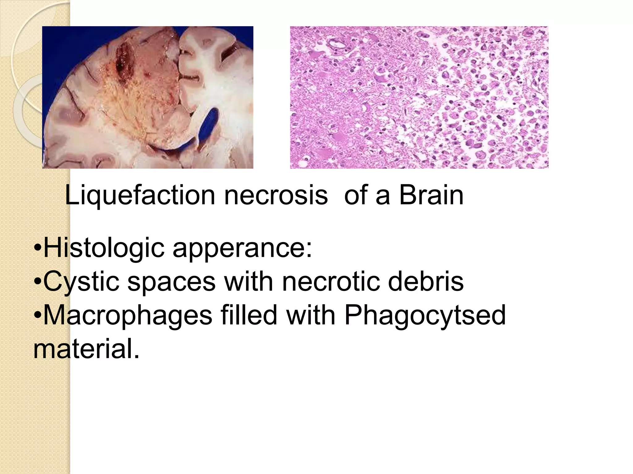
2.Liquefactive Necrosis
Alsocalled as Colliquative necrosis
Occurs due toIschemic injury and
bacterial and Fungal infection.
Common Examples:Infract
brain,Abscess cavity
Gross Appearance:Initially soft with a
liquefied centre later a cyst wall is
formed.
9.
Liquefaction necrosis ofa Brain
•Histologic apperance:
•Cystic spaces with necrotic debris
•Macrophages filled with Phagocytsed
material.
10.
Caseous Necrosis
Combinationof both Coagulative and
liquefactive necrosis
Found in centre of foci of Tuberculous
infection.
Gross Appearance:Resembles dry
cheese and are
soft,Granular,Yellowish colour.
11.
Caseous necrosis ofa TB lymph
node
Histologic Appearance:
Necrosed foci that are
Structureless,eosinophilic with
Granular debris.
Consists of Langerhans Giant cells.
12.
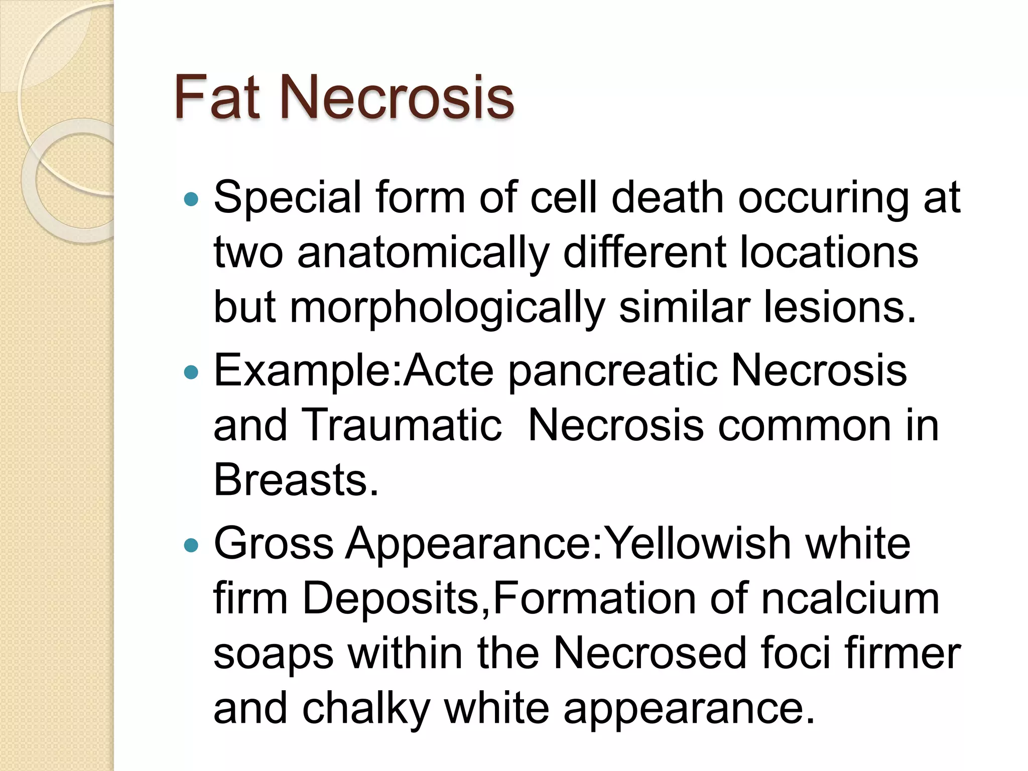
Fat Necrosis
Specialform of cell death occuring at
two anatomically different locations
but morphologically similar lesions.
Example:Acte pancreatic Necrosis
and Traumatic Necrosis common in
Breasts.
Gross Appearance:Yellowish white
firm Deposits,Formation of ncalcium
soaps within the Necrosed foci firmer
and chalky white appearance.
13.
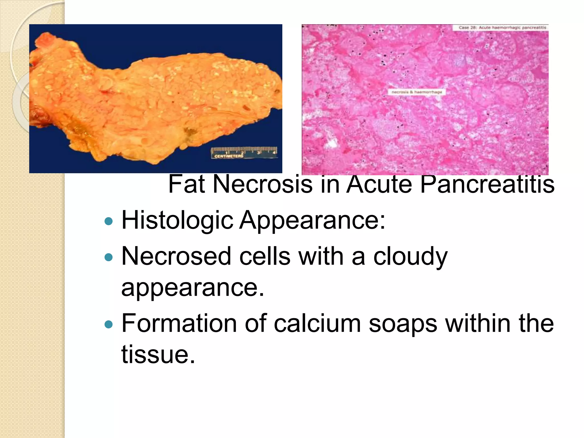
Fat Necrosis inAcute Pancreatitis
Histologic Appearance:
Necrosed cells with a cloudy
appearance.
Formation of calcium soaps within the
tissue.
14.
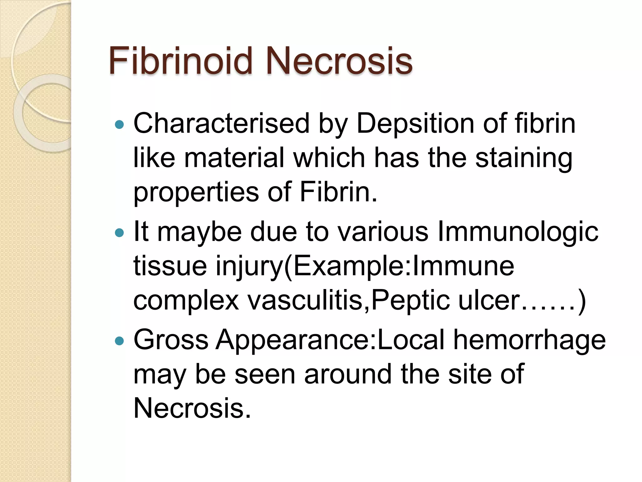
Fibrinoid Necrosis
Characterisedby Depsition of fibrin
like material which has the staining
properties of Fibrin.
It maybe due to various Immunologic
tissue injury(Example:Immune
complex vasculitis,Peptic ulcer……)
Gross Appearance:Local hemorrhage
may be seen around the site of
Necrosis.
15.
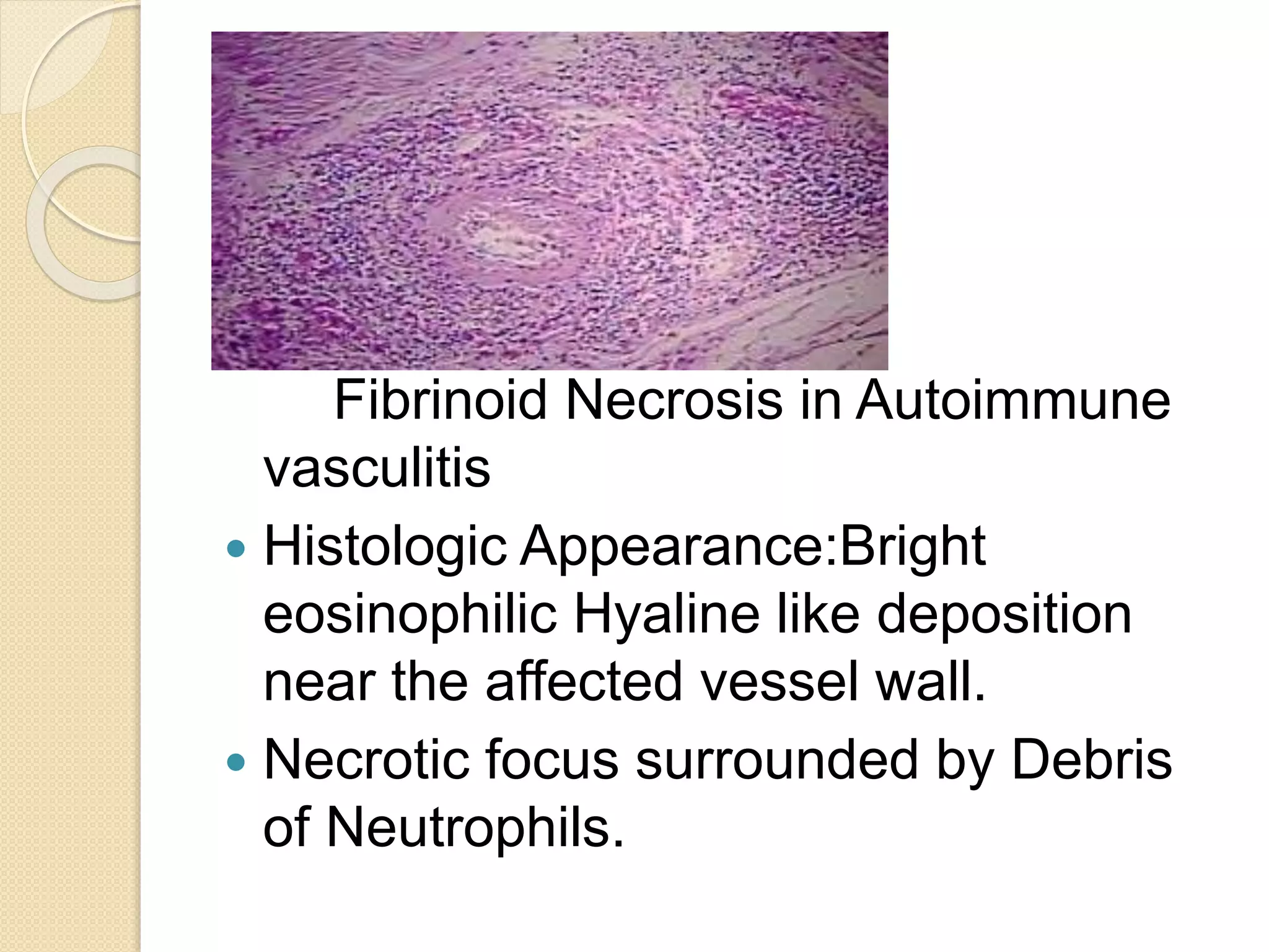
Fibrinoid Necrosis inAutoimmune
vasculitis
Histologic Appearance:Bright
eosinophilic Hyaline like deposition
near the affected vessel wall.
Necrotic focus surrounded by Debris
of Neutrophils.